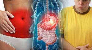
Both examinations are highly informative diagnostic methods that make it possible to make a diagnosis in difficult cases when diseases of the lower parts of the digestive system are suspected.
Intestinal ultrasound or colonoscopy expands the possibilities of medical care for patients with signs of gastrointestinal pathology. The test helps make a diagnosis when symptoms have just begun and the disease is at an early stage. In such a situation, it is possible to completely cure almost any disease.
After questioning, examining and palpating the patient, the doctor prescribes a series of laboratory tests and also gives a referral for instrumental diagnostic methods (ultrasound of the intestines or colonoscopy). The choice of method is determined by the patient’s condition and the nature of the suspected pathology.
The fundamental difference between the methods is that when performing ultrasound, internal organs are studied through the surface of the body using sensors that emit ultrasonic waves that are reflected from anatomical structures. Colonoscopy allows you to see the walls of the gastrointestinal tract from the inside through direct inspection using a video camera attached to a probe that is inserted into the intestines.
For the patient, intestinal ultrasound is a more comfortable and painless procedure, but often diagnosis is only possible using colonoscopy. The methods cannot replace each other; in complex clinical situations, both procedures are required. The doctor decides which test will be ordered first - intestinal ultrasound or colonoscopy - based on the symptoms and manifestations of the disease.
Indications
Both methods have a common purpose - examination of the gastrointestinal tract for the purpose of diagnosing inflammatory diseases, neoplasms, internal bleeding, foreign bodies, and structural anomalies of organs. The specificity of each method provides an advantage in diagnosis when a specific disease is suspected.
Common symptoms for which an intestinal ultrasound or colonoscopy may be prescribed:
- prolonged constipation, alternating constipation and diarrhea;
- the presence of bloody, mucous, purulent impurities in the stool, change in the color of stool;
- bloating, increased gastrointestinal motility, increased gas formation;
- pain of any nature, localized in the abdomen, groin, near the anus;
- intestinal colic;
- weight loss;
- suspicion of cancer or the development of metastases in the gastrointestinal tract;
- the presence of a space-occupying formation when palpating the gastrointestinal tract.
In the diagnosis of gastrointestinal diseases, the manifestations of which are localized on the mucous membrane, colonoscopy is prescribed. The study has the advantage of being able to take a biopsy for histological examination of the material, as well as perform operations to remove detected tumors and stop bleeding.
Colonoscopy is performed for the following pathologies:
- bleeding from the lower gastrointestinal tract, manifested by traces of blood in the stool;
- nonspecific ulcerative colitis;
- gastrointestinal polyps;
- cancer or benign neoplasm;
- Crohn's disease;
- intestinal obstruction of unknown etiology.
Ultrasound is prescribed as a screening method, as well as after operations on the gastrointestinal tract to monitor the healing and recovery processes. As a method for diagnosing diseases, ultrasound allows one to study parts of the gastrointestinal tract and surrounding tissues and obtain information about the boundaries of pathological foci and formations.
Intestinal ultrasound is prescribed for the following indications:
- clarification of the localization of endometriosis in the presence of appropriate symptoms;
- inflammatory diseases – appendicitis, colitis, enteritis;
- suspicion of germination of pelvic tumors in the gastrointestinal tract;
- ischemic conditions of the gastrointestinal tract;
- injuries;
- elongated sigmoid colon;
- peritonitis;
- irritable bowel syndrome;
- gastrointestinal deformations, fecal stones;
- Hirschsprung's disease.
Intestinal ultrasound is an absolutely safe and painless examination that can be performed an unlimited number of times. Colonoscopy is more traumatic and unpleasant for the person being examined.
Contraindications
When deciding which study will be performed - intestinal ultrasound or colonoscopy, the doctor makes a decision in each case individually, depending on the patient’s health condition and the presence of contraindications.
Ultrasound examination is not dangerous for healthy people and for patients with poor health. Ultrasound is a universal method with a small number of limitations that are associated with the condition of the patient’s external integument and his mental health.
Contraindications to intestinal ultrasound:
- open wounds,
- acute phase of infectious diseases;
- inappropriate behavior of the patient due to mental illness.
Unlike intestinal ultrasound, colonoscopy is an invasive procedure - it is introduced into the internal environment of the body. The examination is quite traumatic and painful.
There are a number of contraindications when colonoscopy is not performed:
- intestinal bleeding;
- bronchial asthma in exacerbation;
- high-grade arterial hypertension;
- coronary heart disease, heart attack, heart failure;
- stroke, shock;
- pregnancy;
- epilepsy and other mental illnesses;
- exacerbation of inflammatory bowel diseases.
The high information content of colonoscopy is determined by the possibility of direct examination of the mucous membrane of the gastrointestinal tract and taking material for further examination. The procedure has a number of relative contraindications: menstruation, infectious diseases, hernia, decreased blood clotting. Once these conditions are eliminated, colonoscopy becomes possible.
How to examine the intestines: Ultrasound plus additional techniques
The intestine has three sections: the large, small intestine and rectum, and the study of each of them has its own characteristics and nuances.
- Ultrasound of the colon
helps detect cancer at a very early stage. To make sure, the patient is prescribed a contrast X-ray and colonoscopy. Irrigoscopy, an X-ray examination using contrast fluid, will also be very effective. The method allows you to “see” areas that are invisible to colonoscopy and difficult to distinguish with ultrasound, for example, areas of bends or accumulations of mucus. - Ultrasound of the small intestine
is hampered by tortuosity and deep burial, as well as the accumulation of gases that distort the image on the monitor. A special curved sensor and the latest high-precision equipment help to examine the small intestine. Ultrasound evaluates wall thickness, visualization of layers, patency, wall expansion, and peristalsis. - An ultrasound of the duodenum
is performed together with an examination of the stomach. Allows 100% diagnosis of stomach ulcers, cancer, gastroduodenitis.
Depending on the area being examined, the doctor uses a sensor with certain characteristics.
Preparation
Normally, the gastrointestinal tract is filled with feces and air inclusions. This state of the organ makes it difficult to obtain informative images reflecting the condition of the walls of the gastrointestinal tract, which reduces the diagnostic value of the study. Before conducting any examination of the gastrointestinal tract, preliminary preparation is necessary.
General principles of preparation:
- For several days before diagnosis, it is necessary to follow a diet aimed at reducing the load on the gastrointestinal tract and reducing gas formation;
- limit as much as possible the consumption of legumes, cabbage, onions, mushrooms, dairy products, eggs, sweets, carbonated drinks;
- exclude alcohol;
- drink at least 2 liters of fluid per day;
- It is recommended to include lean meat, chicken, cereals, and vegetable broths in the diet;
- meals are taken in small portions 5-6 times a day;
- You must come for the study at least 6 hours after your last meal;
- if diet is not enough to reduce the amount of gases in the gastrointestinal tract, you should take activated charcoal and espumizan on the day before the examination;
- if constipation occurs, you should use laxatives;
- On the eve of the procedure, it is necessary to cleanse the large intestine with an enema.

Neglecting the preparation rules leads to insufficient cleansing of the stomach, which is a relative contraindication for the procedure. If an examination is carried out in such a condition, then there is a high probability of missing a small pathological formation, and the diagnostic value of the study will be reduced.
When visiting a doctor, you must take a referral for testing and medical documentation related to your current state of health - an advisory opinion from the attending physician with a preliminary diagnosis, laboratory test results. If an intestinal ultrasound or colonoscopy is repeated, you must have the results of previous examinations with you. The maximum amount of information about the patient’s health status helps the doctor make an effective diagnosis.
Symptoms of a stomach ulcer
The most common complaint characterizing the presence of a peptic ulcer in a patient is pain in the stomach.
Abdominal pain
The pain of a stomach ulcer is in most cases sharp, burning, aching. However, the pain can also be vague, dull or felt like a feeling of fullness in the stomach (heaviness in the abdomen), sometimes reminiscent of hunger. In people suffering from duodenal ulcers, abdominal pain occurs 1.5-3 hours after eating. Night attacks of pain often occur.
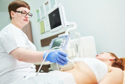
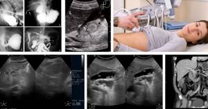
How is an ultrasound of the intestines performed?
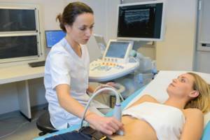
The procedure is carried out by several types of sensors with different purposes and depth of penetration of ultrasonic waves. The patient enters the doctor's office, takes off his clothes, then lies down on his back. The main method of examination is transabdominal. A transparent gel is applied to the abdomen, which is necessary to conduct the signal from the sensor to the body. The doctor examines the projection area of the gastrointestinal tract, receiving visual information on the device monitor.
Inspection of the rectum during a transabdominal examination is difficult, since the organ is deeply located and protected by the pubic bone. To diagnose possible pathology of the rectum, a transrectal sensor is used with insertion directly into the rectum. The patient is asked to turn on his side and bend his legs. In women, a transvaginal sensor is sometimes used for better access to the gastrointestinal tract.
When performing an ultrasound, a liquid can be injected into the large intestine, which allows you to examine the walls of the straightened tract. The technique allows you to evaluate peristalsis and absorption capacity of the mucous membrane.
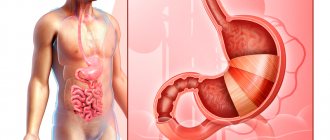
What does a stomach ultrasound show?
What does an ultrasound of the stomach show in adults:
- the presence (or absence) of gastritis or ulcers;
- malignant tumor;
- narrowing of the pylorus, if it is sufficiently pronounced;
- intestinal obstruction (this requires ultrasound examination of the entire gastrointestinal tract);
- any abnormal phenomena in the structure of the organ under study.
Decoding the result
When performing an ultrasound of the stomach, you can normally see the following:
- the organ in section usually looks like an oval ring-shaped formation with a rim, the nature of which is echo-negative, and the center is echo-positive;
- the walls of the stomach in the proximal sections have a thickness of about 5 mm, and in the pyloric section - about 7 mm;
- the presence of 5 layers in the wall of the stomach, differing in the degree of echogenicity;
- the serous membrane, which is located outside the stomach, is hyperechoic;
- the muscle sheath is hypoechoic, its thickness is about 2.5 mm;
- the submucosal membrane (with average echogenicity) measures about 3 mm;
- the mucosal plate, consisting of muscles, is usually low hypoechoic, its thickness is no more than 1 mm;
- the mucosa has a thickness of about 1.5 mm, its character is hyperechoic;
- A glass of liquid drunk by the patient is removed from the stomach in about 1/3 of an hour, and the primary withdrawal of liquid is normally about 180 seconds.
It is important to assess not only the thickness of the stomach walls, but also how uniform they are.
Can stomach cancer be seen on an ultrasound?
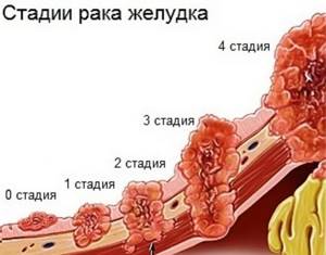
In the early stages, stomach cancer cannot be seen on ultrasound. This is due to the fact that at this stage of the disease, changes in the stomach are subtle and very difficult to identify.
In the later stages, changes in the body are already quite extensive. An ultrasound of the abdominal cavity will allow us to judge the presence of metastases and their size.
However, the sensitivity of the scanner is limited, so obvious tumors are detected only when they reach a large size.
Can a stomach ulcer be seen on an ultrasound?
Is a stomach ulcer visible on an ultrasound? The area where the ulcer is will not reflect ultrasound waves. Having noticed such a hole, the specialist will understand that the patient has an ulcer. However, such an indication on the disease screen is considered conditional. Additional examination will be required.
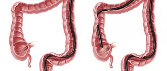
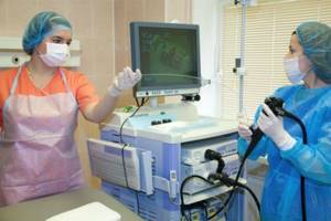
How is a colonoscopy performed?
The success of the procedure depends on the quality of the patient’s preparation and the accurate implementation of the doctor’s instructions. The examination is carried out using local or general anesthesia.
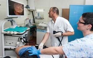
The patient undresses, lies on the table on his left side and pulls his legs to his chest. The tip of the colonoscope is inserted into the anus, to which a flexible tube is attached. The advancement of the tube into the large intestine occurs gradually, with the injection of a small amount of air, which ensures a moderate expansion of the walls of the gastrointestinal tract and easier passage of the tube.
Through the tube, a video camera and instruments can be brought to the walls of the tract to perform operations, stop bleeding, and take a biopsy. During the procedure, the mucous membrane is examined. The detected polyp can be removed using a surgical instrument inserted through a tube. The presence of a neoplasm or ulcer means taking the material with special forceps for examination in the laboratory.
The duration of the examination depends on the volume of manipulations, on average – 20-60 minutes. After the procedure, the patient may experience discomfort - accumulation of gas, the urge to defecate. The condition normalizes on its own within a few hours; to speed up the process, you can take espumizan or activated carbon.
What organs are included in an abdominal ultrasound?
An ultrasound scan of the abdominal cavity can clearly assess the condition of the following organs:
- liver, porta hepatis, lobe sizes;
- gallbladder, duct of Wirsung;
- biliary tract;
- pancreas;
- spleen;
- kidneys and adrenal glands;
- retroperitoneal space;
- lymph nodes;
- portal vein.
Unfortunately, ultrasound of the abdominal cavity does not visualize the intestine well. Diagnosis of this organ using ultrasound is not very informative, and it is better for the patient to undergo an MRI of the gastrointestinal tract, virtual colonoscopy or enterography.
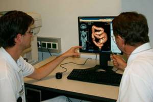
Virtual colonoscopy
The method is the construction of a model of the gastrointestinal tract with the ability to study the relief of the walls of the tract, neoplasms, defects of the mucous membrane, and developmental anomalies. The image is obtained as a result of a computed tomography scan of the abdominal cavity with preliminary expansion of the gastrointestinal tract cavity with air.
Indications and preparation for the procedure are similar to other instrumental examination methods. The advantage of virtual colonoscopy is that it is non-invasive, painless for the patient, and the ability to build a detailed three-dimensional model of the gastrointestinal tract. The method is used when an oncological process in the gastrointestinal tract and adjacent tumors of the abdominal cavity and pelvis is suspected, as well as to monitor postoperative changes in the large intestine. The examination is prescribed when an intestinal ultrasound or colonoscopy cannot be performed due to contraindications.
The disadvantage of this method is the inability to perform surgery or take material for research. Due to the use of ionizing radiation, the method has limited use in pregnant women and children.
How to prepare for an ultrasound of the stomach?
How reliable the results of the study are depends on whether gastric ultrasound is performed by specialists and on proper preparation for the gastric ultrasound procedure.
You are required to follow a diet for about two days before the procedure.
Avoid: foods that cause gas (rye bread, peas, beans, cabbage, kefir, carbonated mineral water, fresh fruits and vegetables).
- Questions often arise before an ultrasound: is it done on an empty stomach or not, is it possible to eat before an ultrasound of the stomach. The last meal on the day preceding the study should be no later than seven to eight o’clock in the evening.
- On the morning of the ultrasound of the stomach and intestines, do not eat, drink or smoke.
- Patients with severe hunger pains are allowed to drink half a glass of tea and eat a cracker.
Ultrasound diagnostic results
On the monitor during the examination, you can see and record the size, shape of the organ, the thickness of the tract walls, and the presence of pathological formations several millimeters in size.
How does pathology manifest itself on ultrasound diagnostics:
- There is normally no fluid in the abdominal cavity. The accumulation of fluid appears as an anechoic area. The presence of fluid indicates peritonitis, internal bleeding, inflammatory processes, and tumors. Based on the location of the liquid, one can judge the reasons for its appearance;
- intestinal obstruction manifests itself as increased visualization of intestinal loops filled with gas or liquid. The reason for this condition is the closure of the lumen by a foreign body or tumor;
- appendicitis is manifested by thickening of the appendix, accumulation of gases in the lumen of the gastrointestinal tract, and a small amount of fluid in the abdominal cavity;
- tumors and neoplasms are characterized by an increase in the thickness of the intestinal wall due to hypoechoic areas of round or irregular shape. Ultrasound shows the size of the tumor, the degree and depth of the lesion, and invasion into neighboring organs;
- adhesions are high-density strands, often combined with deformation of nearby sections of the tract.
The conclusion is drawn up by the ultrasound doctor and given to the patient. The document contains a detailed description of the state of the gastrointestinal tract and all pathological changes and neoplasms. The information allows the attending physician to make the correct diagnosis.
Advantages and disadvantages of ultrasound of the stomach when examining the gastrointestinal tract
The doctor prescribes an ultrasound examination of the stomach to the patient as a primary auxiliary diagnostic method.
The advantages of ultrasound are as follows:
- the exit section most susceptible to diseases is examined;
- ultrasound “sees” any foreign bodies in the cavity;
- Ultrasound accurately assesses the thickness of the walls of the organ;
- thanks to the method, venous blood flow is clearly visible;
- using diagnostics, benign and malignant tumors of minimal size are identified;
- stomach ulcers are well assessed;
- the degree of inflammation of the gastric mucosa varies;
- the method allows you to see reflux disease - the reflux of the contents of the lower sections back into the stomach;
- the organ is examined from different points and in different sections, which is impossible with x-rays;
- Ultrasound sees what is happening in the thickness of the stomach wall;
- thanks to the echo structure, ultrasound can easily distinguish a polyp from an oncological neoplasm;
- in addition to diagnosing the stomach, ultrasound diagnostics reveals concomitant pathologies of other organs (usually with gastritis, diseases of the biliary tract and pancreas develop);
- Ultrasound is performed on newborns and small children for whom it is impossible to undergo an FGDS or x-ray.
The main advantage of ultrasound over FGDS is the ability to detect forms of cancer developing in the thickness of the organ wall (infiltration forms), which cannot be detected using fibrogastroscopy.
Despite all the advantages, ultrasound has some disadvantages that do not allow the method to become widespread as an independent examination of the stomach.
The disadvantages include the following:
- Unlike endoscopic examination, ultrasound does not allow tissue samples to be taken for further study (for example, gastric juice;
- scraping of the mucous membrane, tissue biopsy);
- Ultrasound cannot assess the degree of changes in the mucous membrane;
- limitation of the areas studied (it is possible to examine only the outlet zone of the stomach).
Colonoscopy results
A healthy large intestine has a smooth, shiny, pink mucous membrane, without areas of hyperemia or edema. Cicatricial changes, diverticula, pathological narrowing or expansion of the intestinal lumen, ulcers, and polyps are normally absent.
What some diseases look like:
- with nonspecific ulcerative colitis, the mucous membrane becomes bright red, swelling, granularity, and areas of hemorrhage appear;
- intestinal diverticula look like branches of the gastrointestinal tract with an entrance diameter of up to 2 mm, at the site of the lesion the tone of the intestinal wall is increased, the folds are thickened;
- cancer looks like a heterogeneous neoplasm of irregular shape with a granular surface, surrounded by an area of edema, and bleeds when touched.
The results of examining the intestinal walls with a video camera can be obtained shortly after the procedure. Based on the results of the conclusion, the attending physician makes a diagnosis or refers the patient for further diagnostics. If a biopsy was performed, the result of the examination of the fragment will be issued within two weeks. Microscopic examination allows you to make the most accurate diagnosis.
What will an ultrasound of the abdominal organs show?
Ultrasound of OBP will show well:
- inflammatory and necrotic liver lesions;
- hepatitis;
- cysts and tumors of organs, polyps;
- formation of stones, inflammation or scarring in the gallbladder and ducts;
- cholecystitis;
- damage to the kidneys and adrenal glands;
- acute and chronic pancreatitis of the pancreas;
- enlarged lymph nodes in the abdominal cavity;
- enlarged spleen;
- abnormal arrangement of organs.
Unfortunately, ultrasound examination is not informative enough to judge 100% the malignancy and benignity of a space-occupying lesion. Therefore, if tumors are detected, the doctor will refer the patient for further examination using an MRI of the abdominal cavity with contrast.