Magnetic resonance imaging of the intestines is an imaging test that provides detailed pictures of the intestines. This method can help the doctor diagnose inflammation, bleeding, obstruction and other problems. It is non-invasive and does not use ionizing radiation. MRI uses a magnetic field to create detailed images of the intestines. The computer analyzes the images. Before the exam, oral and intravenous contrast material is administered to isolate the bowel. Medicine may also be given to reduce bowel movements (peristalsis) that may interfere with images. It is very important to tell the doctor about any health problems, recent surgeries or allergies, and whether the patient is likely to be pregnant. The magnetic field is not harmful, but may cause some medical devices to malfunction. Most orthopedic implants do not pose any risk, but you should always tell the technologist if there are any devices or metal in the body.
What does the equipment look like?
Content:
- What does the equipment look like?
- How does this procedure happen?
- Application of intestinal MRI
- How to prepare
- MRI of the intestines for pregnant women
- MRI for children
- How the procedure is performed
- What are the benefits and risks
- What are the limitations of intestinal MRI?
- MRI or colonoscopy
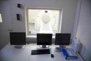
Typically, an MRI scanner consists of a large cylindrical tube surrounded by a circular magnet. The patient lies on a movable examination table that slides into the center of the magnet. Some newer MRI machines have a larger opening, which may be more comfortable for larger patients or those with claustrophobia.
Other MRI machines are open on the sides (open MRI). Some MRI scanners, called short-circuit systems, are designed so that the magnet does not completely surround the person. Open ones are especially useful for studying larger patients or those with claustrophobia. Newer open MRI units provide very high-quality images for many types of examinations. Older open MRI devices may not provide the same image quality. Some types of tests cannot be performed using open MRI. The computer workstation that processes image information is located in a separate room.
Preparation for the procedure
- Preparation for an MRI study of the intestines begins several days in advance with a gentle diet, excluding foods that lead to bloating.
- As with any other examination of the large intestine, before an MRI it must be cleansed using laxatives or enemas (unless there is an obstruction).
- The examination is carried out only on an empty stomach.
- During an MRI of the intestine with contrast, a preliminary test is done to determine the tolerance of the contrast agent.
- Before the procedure itself, metal objects are removed from the body.
How does this procedure happen?
Unlike conventional X-rays and CT scans, MRI scans do not use ionizing radiation. Instead, the radiofrequency pulses reconfigure the hydrogen atoms that naturally exist within the body while the patient is in the scanner, without causing any chemical changes in the tissue. As the hydrogen atoms return to their normal alignment, they release different amounts of energy that vary depending on the type of body tissue they come from. An MRI scanner captures this energy and creates a picture of the tissues scanned based on this information. The electric current does not come into contact with the patient. The signals are then processed by a computer to generate a series of images, each showing a thin section of the intestine. The images can then be examined from different angles by an interpreting radiologist. Often, differentiating abnormal (diseased) tissue from normal tissue is better with MRI than with other imaging techniques such as X-rays, CT, and ultrasound.
Computed tomography of the intestine: progress of the procedure
Immediately before the procedure, the patient is asked to remove all things, get rid of jewelry, watches, foreign objects, mobile phone, etc.
Then the subject is placed on the diagnostic table, the operator gives all the necessary instructions: take the required position (on the side, back, stomach). If necessary, to ensure the patient's immobility, which is necessary to obtain high-quality images, the limbs are fixed using special belts.
To perform a CT scan of the intestine, the rectum must be filled with carbon dioxide or air. For this purpose, a specialist inserts a thin tube into the patient’s anus, through which gas is injected using a bulb or pump. This event is necessary in order to maximally straighten the intestinal walls, which normally have bends and folds. Thanks to the “inflation” of the intestine, the view for the radiologist is improved and pathological processes in hidden areas are revealed.
After preparation, the patient is moved into the scanner itself, asked to hold his breath and change his body position for the next sections.
The CT scan takes an average of 15 minutes, after which the specialist removes the gas tube and allows the patient to stand up.
How to prepare
Medicines and food can be taken as usual. Some MRI exams may require the use of a contrast agent. The radiologist, technologist, or nurse should ask if the patient has any allergies, such as allergies to iodine or radiocontrast material, drugs, food, or the environment, or asthma. The contrast material most commonly used for MRI examinations contains a metal called gadolinium. Gadolinium may be used in patients allergic to iodine contrast. It is much less common for a patient to be allergic to the gadolinium-based contrast agent used for MRI than to the iodinated contrast agent used for CT. However, even if a patient is known to be allergic to gadolinium contrast, it can still be used after appropriate pretreatment. You should also tell the radiologist if you have any serious health problems or any recent surgeries. Some conditions, such as severe kidney disease, may prevent the use of gadolinium contrast for MRI. If there is a history of kidney disease or liver transplantation, blood tests will need to be done to determine whether the kidneys are functioning adequately.

Jewelry and other accessories should be left at home if possible or removed until the MRI scan. Because they can interfere with the magnetic field, metal and electronic objects are not allowed in the exam room. In addition to affecting MRI images, these objects can cause damage.
These items include:
- watches, hearing aids;
- metal zippers and pins;
- removable dentures;
- glasses, pens, pocket knives;
- piercings.
In most cases, the MRI test is safe for patients who have metal implants, except for some types. People with these implants cannot undergo this test and are prohibited from entering the area where the MRI scan takes place:
- cochlear (ear) implant;
- some types of clamps used for brain aneurysms;
- almost all cardiac defibrillators and pacemakers.
You should tell your doctor if you have medical or electronic devices in your body. These objects may interfere with examination or potentially pose a risk depending on their nature and the strength of the MRI magnet. Some implanted devices may require time after placement before they are safe for MRI scanning, usually taking no more than six weeks. Examples include, but are not limited to: artificial heart valves, implanted drug infusion ports, artificial limbs or metal joint prostheses, implanted nerve stimulators, pins, screws, plates, stents. If there is any question about their presence, x-ray diagnostics can be used to detect and identify the location of any metal objects. The magnetic field does not affect dental fillings and braces. Family members accompanying patients to the scanning room should also remove metal objects and tell the doctor about any medical or electronic devices they may have.
MRI diagnosis of primary rectal tumor
Possibilities of MRI in the diagnosis of rectal cancer (“gold standard”):
- visualization of the tumor, determination of its upper and lower boundaries, the degree of involvement of the intestinal wall, surrounding fiber and adjacent organs in the process;
- determining the relationship of the tumor to the structures of the pelvic floor and the sphincter apparatus;
- identification and assessment of the condition of lymph nodes and lymph drainage in the pelvic area;
- dynamic monitoring of the results of surgical treatment, radiation therapy and chemotherapy;
| Rectal cancer, coronal projection | Rectal cancer, axial projection |
MRI of the intestines for pregnant women
Women should always inform their doctor or technologist if there is a possibility that they are pregnant. MRI has been used to scan patients since the 1980s without reports of any negative effects on pregnant women or their unborn children. However, because the unborn baby will be in a strong magnetic field, pregnant women should not have this exam during the first three to four months of pregnancy unless the potential benefits of MRI are thought to outweigh the potential risks. Pregnant women should not receive gadolinium contrast injections unless absolutely indicated for treatment.
MR defecography
With MR defecography, it is possible to record the position of the pelvic organs at rest and during functional tests, which makes it possible to identify and evaluate both anatomical and functional disorders of the rectum, bladder, uterus (perineal prolapse, rectocele, rectal prolapse, cystocele, prolapse cervix). The advantages of MRI defecography are the possibility of non-invasive comprehensive diagnosis of pathological conditions of the pelvic organs, the absence of ionizing radiation, good image quality of the soft tissues of the pelvic floor, the ability to obtain images in any plane, high resolution, and good tissue contrast. MRI defecography is indicated primarily for patients with rectal defecation disorders.
Defecography.
MRI for children

Infants and young children usually require sedation, or anesthesia, to complete an MRI test without moving. Regardless of whether a child requires sedation, the choice of sedation depends on the child's age, intellectual development, and type of test. Moderate and conscious sedation can be provided in many settings. A physician who specializes in sedation or anesthesia for children must be present during the examination for the safety of the child.
How the procedure is performed
Before the procedure, the patient needs to drink several glasses of an aqueous solution mixed with contrast material. Straps and bolsters may be used to help the patient stay in place and maintain proper positioning during imaging. Devices that contain coils capable of transmitting and receiving radio waves can be placed around or near the area of the body being examined. The patient will be placed in an MRI magnet and the radiologist will perform the examination while working on a computer outside the room.
Advantages of Sofia Cancer Center
In the Sofia building with a total area of 35,000 sq. m has everything necessary for diagnostics, comprehensive examination and diagnosis. Comfortable reception conditions are provided - you don’t feel like you’re in a hospital!
Advantages of Sofia:
- MRI on equipment that has no analogues;
- experienced doctors, leaders in their field;
- convenient location in Moscow;
- the highest level of quality, corresponding in fact to both domestic and international standards (JCI);
- special care for patients during MRI and after the examination;
- individual support around the clinic;
- a large number of parking spaces;
- 30 years of unique experience;
- a large number of awards, certificates, prizes.
What are the benefits and risks
Advantages of intestinal MRI:
- MRI is a non-invasive imaging technique that does not involve exposure to ionizing radiation.
- MRI can detect abnormalities that may be obscured by bone and not visible with other imaging techniques.
- The contrast material used in MRI is less likely to cause an allergic reaction than iodine-based contrast materials, which are used for conventional X-rays and CT scans.
- MRI of the intestine helps identify areas of intestinal inflammation due to diseases such as Crohn's disease.
- Because bowel MRI does not involve ionizing radiation, the procedure may be preferable for the evaluation of young patients with inflammatory bowel disease, who may undergo multiple examinations throughout their lives.
- MRI of the intestine may eliminate the need for invasive endoscopy.

Risks of the method:
- MRI examination poses virtually no risk to the patient if appropriate safety regulations are followed.
- When using sedation, there is a risk of over-sedation. However, the technologist or nurse will monitor vital signs to minimize this risk.
- Although the magnetic field itself is not harmful, implanted medical devices containing metal can malfunction or cause problems during an MRI.
- Nephrogenic systemic fibrosis is a recognized but rare complication of MRI that is thought to be caused by the administration of high doses of gadolinium-based contrast material in patients with very poor renal function. Careful assessment of renal function before considering contrast injection minimizes the risk of this very rare complication.
- There is a slight risk of allergic reactions if contrast material is injected. Such reactions are usually mild and easily controlled with medications.
CT scan of the intestine: indications
Most often, computed tomography of the intestine, as well as CT of the rectum, is prescribed to patients at the Yusupov Hospital if the presence of polyps and cancerous formations in this organ is suspected.
Polyps are benign growths that have a high probability of degenerating into a malignant neoplasm. According to experts, early detection and removal of such formations at the initial stages of development prevents malignancy of the pathological process. For this purpose, the use of computed tomography is recommended.
Preventive examination of the intestines should be carried out for both men and women over 50 years of age. A CT scan should ideally be performed every five years, and a colonoscopy at least once every ten years. Persons with a family history of intestinal cancer or chronic polyposis should undergo a CT scan of the intestine after 36-40 years of age and much more often than other categories.
Computed tomography of the intestine is also prescribed for inflammatory processes and bleeding in the organ. CT scan of the intestine provides a quick and painless determination of the source of the problem, assessment of the volume and location of the pathological process.
In addition, intestinal screening is carried out as a control measure during the treatment of diseases in this part of the body, allowing one to evaluate the effectiveness of therapy.
What are the limitations of intestinal MRI?
High-quality images are only guaranteed if the patient can remain still and follow instructions to hold their breath while images are recorded. If the patient is anxious, confused, or in severe pain, it may be difficult for them to lie still during imaging. A person who is overweight may not fit into the parameters of certain types of MRI machines. The presence of an implant or other metal object sometimes makes it difficult to obtain clear images due to the appearance of line artefacts from the metal objects. Movement of the patient can have the same effect. Although there is no reason to believe that magnetic resonance imaging is harmful to the fetus, pregnant women are generally advised not to have an MRI exam during the first trimester unless medical intervention is required. For optimal results, the patient should consume the entire dose of oral contrast material, remain still, and follow breathing instructions. Moreover, results may be compromised if the patient is unable to receive intravenous contrast material (gadolinium). A bowel MRI takes longer (30 to 45 minutes) than a CT scan (two to four minutes).
CT scan of the intestines: what does the study show?
Interpretation of images at the Yusupov Hospital is performed by a qualified radiologist. He is engaged in a detailed description of areas that arouse suspicion, deciphering deviations from normal indicators. The results of the analysis are summed up by the attending physician who ordered a computed tomography scan.
A tomogram allows one to distinguish the state of the relief of the intestinal mucosa, the thickness and structure of its walls. In addition, it displays visualization of inflammatory foci, erosive changes, ulcerations, and tumors. CT evidence of a fatty colon mass may be detected.
Computed tomography does not allow detecting the presence of small lesions (less than one millimeter), i.e., identifying signs of pathology at the earliest stages of development. In addition, using CT it is impossible to assess the intestinal mucosa and remove material for subsequent histological examination.
If tumor formations are detected, an additional endoscopic examination, in addition to CT, is immediately prescribed. Colonoscopy provides the opportunity to examine the found tumor and perform a biopsy. Most often, polyps are removed simultaneously with a colonoscopy.

