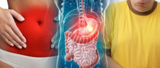
Features of colonoscopy
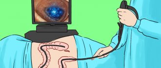
Colonoscopy is an endoscopic technique for examining the colon. The procedure is carried out using a fiberconoloscope - a special optical device, thanks to its flexibility and softness, the intestines are examined.
A large number of people are wary of the procedure, believing unsubstantiated rumors that a colonoscopy can lead to colon rupture or fecal incontinence. However, all these arguments have no scientific confirmation, and the examination itself is safe for the patient.
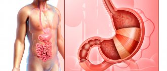
At the end of the fibercoloscope there is a light source and a microcamera, with the help of which a specialist can:
- Assess the condition of the intestinal mucosa
- Detect cancer at the earliest stages
- Study indicators of intestinal motor activity
- Remove foreign bodies from the intestines
- Get rid of bleeding
- Take the necessary pictures
Preparation for colonoscopy begins 2 days in advance and, just like before irrigoscopy, is aimed at cleansing the intestines with the help of a special diet and laxatives prescribed by the attending physician.
The main differences in performing colonoscopy and sigmoidoscopy
The main difference between these two procedures for the patient is that sigmoidoscopy is less painful, takes less time and does not require long and serious preparation. All that is required of the patient before undergoing sigmoidoscopy is to avoid eating waste foods 3 days before the study, and the day before, thoroughly cleanse the intestines with an enema.
Sigmoidoscopy is performed without anesthesia, while during colonoscopy you can use medicated sleep. The proctoscope is inserted into the rectum a short distance, and the colonoscope, through the rectum, is inserted further along the colon, straightening its lumens with air pressure, which creates a certain discomfort and unpleasant sensations in the patient’s abdomen.
There are no absolute contraindications to sigmoidoscopy; there are only relative ones that prevent this diagnostic examination from being carried out only in acute cases. Relative contraindications for the sigmoidoscopy procedure include:
- presence of anal fissure;
- acute inflammation of the tissues surrounding the rectum
- intestinal bleeding;
- the impossibility of its implementation due to pathological narrowing of the lumen of the rectum.
After carrying out therapeutic measures to eliminate these contraindications, or when their symptoms subside, the sigmoidoscopy procedure can be performed.
Stages of a colonoscopy
- Anesthesia. The procedure can be performed under general anesthesia (deep sleep and complete loss of consciousness), sedation (a state similar to sleep, but without blackout) and local anesthesia (numbing a specific area of the body).
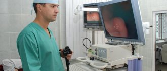
- Direct examination. The doctor inserts a fibroconoscope into the anus, uses air to expand the intestines to get a clearer picture, and takes the necessary pictures. The entire procedure lasts no more than 20 minutes.
- Consultation with a specialized doctor. The specialist conducting the study refers the patient to another doctor to make a diagnosis and, if necessary, prescribe a treatment regimen.
Colonoscopy. Colonoscopy only or is there an alternative?
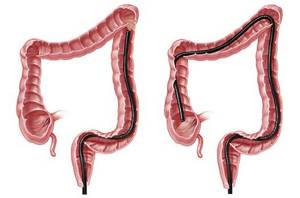
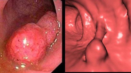
The traditional method of examining the large and small intestines is currently still a colonoscopy. During a colonoscopy, the endoscope is advanced from the rectum to the small intestine. Thus, the entire colon is examined in detail. If necessary, it is also possible to insert a colonoscope into the small intestine. The examination is performed with flexible endoscopes. The light source is an illuminator operating on a halogen or xenon lamp, which eliminates burns to the mucous membrane.
During the study, air is supplied into the intestinal lumen to straighten its wall, this makes it possible to create the so-called. “air cushion”, which minimizes the risk of injury. During the procedure, the endoscopist examines the condition of the colon mucosa and can see almost all possible changes. Knowledge of the features of endoscopic anatomy allows him to navigate the intestinal lumen and determine the location of the apparatus based on characteristic endoscopic signs. Among the currently available methods for detecting colon cancer, colonoscopy is the most reliable. A general examination, radiation diagnostic methods (ultrasound, x-ray, tomography) and laboratory tests can also help in making a diagnosis. However, they are auxiliary, and only colonoscopy allows the doctor to look inside the intestine without surgery and directly see the condition of the intestinal wall.
Currently, the so-called “rotational” method of performing a colonoscope, developed at the State Research Center of Coloproctology, has found widespread use. The principle of the “rotational” method is based on the peculiarities of the anatomical structure of the large intestine. In this case, the colonoscope is inserted not with translational movements, but in a spiral, rotational movements. This eliminates forced insertion of the device and possible trauma to the intestinal wall, and also significantly reduces discomfort for the patient.
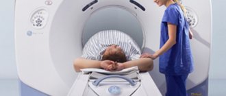
Computed tomography of the colon. This technique is also called virtual colonoscopy, although the principle of the technique is completely different and it has nothing to do with endoscopy. In essence, this is an X-ray examination, which consists of obtaining layer-by-layer (step width 1 cm) sections of the abdominal cavity using a special device - a computed tomograph. In this case, to visualize the surface, a contrast agent (barium enema) is first introduced into the colon; after the examination, a special computer program mounts the resulting sections (hence the word “vertical”), then the results are analyzed by a doctor. The main disadvantages of the method are: the inability to detect formations less than 1 cm (especially if the formation falls between the sections), the inability to take a biopsy (which may subsequently lead to an additional colonoscopy), significant radiation exposure, and the exclusion of the method for patients weighing more than 100 kg.
Irrigoscopy Irrigoscopy is an X-ray examination of the colon with preliminary administration
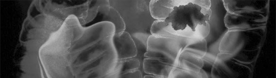
X-ray contrast agent (so-called barium enemas). The main advantage of the method is the ability to accurately determine the location of the pathological focus. In general, the information content of the technique is significantly lower than with colonoscopy, since it is impossible to examine the mucous membrane of the colon in detail, and there is also a danger of obtaining false positive results due to the possibility of mistaking intestinal contents for neoplasms. The technique eliminates the possibility of taking a biopsy (which may subsequently entail an additional examination - a colonoscopy). It must be remembered that the study is quite uncomfortable (the intestine is distended due to the introduction of barium suspension) and there is radiation exposure to the body.
Capsule endoscopy The essence of the capsule endoscopy technique is to pass through all parts of the gastrointestinal tract a special video capsule - a device equipped with a digital micro-video camera and a recording unit. The patient takes the capsule orally, within 6 hours it passes through the gastrointestinal tract and is released naturally. The obtained data recording the condition of the walls of the gastrointestinal tract is subsequently analyzed by a doctor. The main disadvantages of a video capsule come from its advantages:
- Autonomy - does not allow for a targeted and detailed examination of the detected pathological formation. In addition, the capsule may turn its back side towards the pathological focus and “not be noticed”; — The possibility of biopsy and histological confirmation of the diagnosis is excluded; — Often the capsule gets stuck in the small intestine, which requires additional intervention to remove it (even before surgery). From the point of view of complete examination of the colon, the endocapsule is ineffective, since the battery life is not enough for a complete examination of the entire gastrointestinal tract. In this case, additional gastroscopy is often performed with a deeper insertion, pushing the capsule, for example, into the duodenum, that is, the main advantage is that ease of use is eliminated, and in combination with insufficient information content, the method is significantly inferior to colonoscopy for colon pathology. Of course, the method is good for studying the small intestine, but, from my point of view, it is inappropriate as a method of choice for examining the large intestine.
The material was prepared based on data from the State Scientific Center for Coloproctology of Roszdrav and the website colonoscopy.ru. Today, the only scientific and clinical institution in the Russian Federation completely specializing in diseases of the colon and rectum, anus, pelvic organs and perineum.
Features of irrigoscopy
Irrigoscopy is another method of examining the colon using a contrast agent and x-rays, which can detect a large number of diseases of the gastrointestinal tract.
The difference between irrigoscopy and colonoscopy is that during irrigoscopy there is no need for anesthesia or anesthesia. Irrigoscopy is simple to perform and is not accompanied by painful sensations. However, when an examination is carried out using a contrast agent, a person is exposed to x-rays, albeit in normal doses. During a colonoscopy, the camera is inserted directly into the colon, so there is no need to take X-rays - the doctor sees the intestinal space directly on the monitor. In preparation, both of these procedures are practically no different. Both before irrigoscopy and before colonoscopy, it is recommended to follow a diet that cleanses the intestines and drink a lot of water. At the same time, the X-ray method of diagnosing the intestine is very different from the endoscopic method in the technique of the procedure.
Intestinal examination, which method to choose instead of colonoscopy?
Dina
May 27, 2020
Please advise which diagnostic method is best for me to use to examine the intestines and abdominal organs? I am 55 years old, I have been taking hormone replacement therapy (Livial, the main substance is tibolone) for 2 years. Last summer, after switching to proper nutrition (excluding bread, sugar, introducing a water regime of 1.5 liters per day) and cleansing the intestines and lymphatic system with natural preparations, I lost a lot of weight by 14 kg (from 67 kg to 53 kg over 4 months). In December I turned to a preventive medicine physician. Various examinations were prescribed: general blood, blood biochemical analysis, general urine analysis, feces for several types of studies, incl. for dysbacteriosis, ultrasound of the abdominal organs, gastroscopy. After receiving the test results, appropriate treatment was prescribed. I took treatment for 3 months, my condition was good, but my weight did not increase. Currently, I am worried about sensations of being sucked into the upper part of the stomach, a vacuum state (the same symptoms existed before treatment), not related to food intake (they arise suddenly and go away on their own); periodic painless sensations of the presence of something incomprehensible in the solar plexus area, closer to the right side, under the ribs (an ultrasound of the liver in December 2019 revealed a questionable hemangioma), which also suddenly arise and also suddenly go away. The doctor recommended that I have a colonoscopy of my intestines (the results of gastroscopy done in January 2020 were without pathology). I have had many operations on the intestines in the past (at the age of 14 I suffered peritonitis due to appendicitis, 10 days after appendicitis (the intestines were punctured during the operation) - intestinal obstruction, then adhesive disease, infertility 1, in 2006 an endometrioid cyst of the left ovary, tubal plastic surgery (in the hope of getting pregnant). I can’t stand a colonoscopy without anesthesia, because my adhesive process has not gone away. I’m afraid to do a colonoscopy under anesthesia, because there may be a puncture of the intestine again. Currently, there are several alternative methods for examining the intestines and abdominal organs - MRI or CT with and without contrast. Which examination is best for me to do instead of a colonoscopy, taking into account my problems?
Age:
55
Chronic diseases:
Chronic adhesive pelvioperitonitis. Chronic migraines.
The question is closed
intestines
constipation
discomfort in the right hypochondrium
Stages of irrigoscopy
- The patient lies down on a specially equipped table with his arms folded behind his back and his legs bent at the knees.
- A special tube is inserted into the anus, through which barium sulfate enters the intestines.
- The doctor takes x-rays of the colon.
- The patient has a bowel movement from the contrast agent.
- Then the subject lies down on the table again in the same position and their intestines gradually begin to be filled with air. This will help smooth out all the folds of the intestinal mucosa and take new pictures.
Disadvantages of sigmoidoscopy
Why, with all its advantages, has this method not become dominant in diagnostics? Experts say that sigmoidoscopy cannot be a complete alternative to colonoscopy, since it does not provide complete data on the condition of the entire colon.
Sigmoidoscopy, as a method of diagnostic examination, is effective if there are the following indications for its implementation:
- establishing the cause of bleeding from the rectum - it could be anal fissures, polyps, hemorrhoids and even advanced stages of cancer;
- the presence of purulent or mucopurulent discharge from the anus, which may indicate the presence of inflammation of nearby tissues (paraproctitis);
- pain in the rectum during defecation, after it, and at any time, which may indicate not only the presence of internal hemorrhoids or fissures, but also the development of intestinal cancer.
So, sigmoidoscopy is a good method for examining the lower parts of the colon. If it is necessary to examine the entire colon with a detailed study of the mucous membrane, it is necessary to conduct a total colonoscopy, that is, an examination throughout the entire colon. Therefore, in combination with sigmoidoscopy, additional studies of the condition of the large intestine may be prescribed, such as
- virtual colonoscopy, in which the examination is performed using a high-precision computed tomograph;
- Ultrasound of the large intestine, if the clinic has new generation ultrasound equipment;
- radiography using a barium mixture, in which a contrast agent is introduced into the cavity of the colon, allowing one to examine its walls and identify the presence of a defect on it.
Such examinations, together with sigmoidoscopy, are not cheap, and they also take time. Moreover, all of the above studies do not provide such a high percentage of information as colonoscopy and do not resolve the issue of collecting biopsy material from the inner mucous membrane of the large intestine.
Conclusion. In some cases, it is impossible to make an accurate diagnosis to determine effective treatment for colon diseases without a colonoscopy. Therefore, if the patient’s fear and fear are the only contraindications to conducting this highly accurate study, we suggest undergoing the study under anesthesia, or rather in a state of medicated sleep. In this case, you will avoid any possible feelings of discomfort.
Find out the cost of the Videocolonoscopy procedure