Electrical impedance mammography is an innovative technique for diagnosing mammary gland pathologies, based on the difference in electrical conductivity of healthy and altered tissues.
The main tool of this method is an electric current passed through the area under study. Each type of internal tissue has a different ability to conduct electric current and, accordingly, conductivity standards. The principle of the diagnostic method is based on the fact that the electrical conductivity of diseased tissue is strikingly different from normal values. Therefore, when generating results in 3D images, the tumor is clearly visible.
Advantages
The method of multifrequency impedance mammography is quite new in the medical world, but has already gained recognition and respect in various circles. Its undeniable advantages include:
- higher information content of the procedure compared to traditional methods;
- the method is absolutely harmless to the health of women of any age, since ionizing radiation is not used;
- the complete absence of contraindications and restrictions for the procedure, which is incredibly important for the control of rapidly developing diseases;
- the technique allows you to monitor the condition of breast tissue during mastalgia, when other examination methods are not available;
- improved quality of diagnosis of dishormonal pathologies of the mammary glands;
- the technique is capable of not only detecting mastopathy, but also distinguishing its cystic from non-cystic form;
- detects breast cancer at the earliest stages, and detects non-palpable neoplasms with a diameter of up to 1 cm.
results
Each organ of the human body has its own electrical characteristics, which are determined by the electrical conductivity of the tissues that form it, the properties of intra- and extracellular fluid, the number and condition of blood vessels. Electrical impedance mammography [10, 11] allows you to visualize the distribution of electrical conductivity of biological tissues in several cross sections of the patient’s body and detect areas with abnormal electrical conductivity values. During the measurement process, the device in turn, using each of the 256 contact electrodes, injects a weak alternating electric current of 50 kHz (0.5 mA) into the patient’s body and measures the distribution of the corresponding electrical potentials on its surface using the remaining electrodes. The data obtained is then used to reconstruct electrical impedance images of subsurface regions using mathematical algorithms implemented on a personal computer, to which the device is connected via a standard USB port. The result of the reconstruction is electrical impedance images of seven cross sections of the medium under study, parallel to the plane with the electrodes, taken with a step of 0.7 cm in depth.
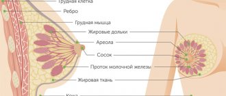
Many years of experience [12] have shown that there is no statistically significant difference in electrical conductivity indicators depending on the position during the study (lying or standing) and the scanning side (right or left mammary gland). The patterns of changes in electrical conductivity indicators are the same at all levels, respectively. Therefore, for maximum clarity, we used average electrical conductivity values from the 2nd scanning level (depth 1.1 cm) without taking into account obstetric history, which were later used for comparison. In addition, taking into account the anatomical structure, it is possible to find all tissues and structures of the mammary gland at this depth (Table 1).
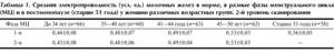
Table 1. Average electrical conductivity (arbitrary units) of the mammary glands is normal, in different phases of the menstrual cycle (MC) and in postmenopause (over 51 years old) in women of different age groups. 2nd scanning level
With age, the average electrical conductivity of the mammary glands increases, reflecting the stages of involutive changes in their structure. A statistically significant difference is observed between all age groups ( p
<0.01), except for the age groups 35-40 years/41-44 years (
p
>0.05) in both phases of the menstrual cycle and in postmenopause.
When comparing the average electrical conductivity indicators between the phases of the menstrual cycle in the corresponding age groups, there is no statistically significant difference ( p
>0.05).
Due to the lack of statistically significant differences in different phases of the menstrual cycle, the study of electrical conductivity characteristics depending on obstetric history was carried out in the first phase of the menstrual cycle. In table 2, 3, 4 show electrical conductivity indicators in women of different age groups depending on the number of births, abortions and the duration of the lactation period in the anamnesis (see Table 2).
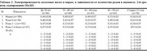
Table 2. Electrical conductivity of the mammary glands is normal, depending on the number of births in the anamnesis. 2nd scanning level (M±SD)
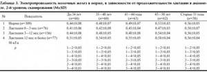
Table 3. Electrical conductivity of the mammary glands is normal, depending on the duration of lactation in history. 2nd scanning level (M±SD)
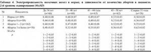
Table 4. Electrical conductivity of the mammary glands is normal, depending on the number of abortions in history. 2nd scanning level (M±SD)
A statistically significant decrease in the electrical conductivity of the mammary glands was determined in all menstruating women in the absence of a history of childbirth ( p
<0.05,
p
<0.01), and an increase in electrical conductivity in the presence of at least one genera (
p
<0.05,
p
<0.01) compared to the average electrical conductivity in normal conditions.
In nonmenstruating women, in the absence of a history of childbirth, electrical conductivity indicators are significantly higher than the average normal values ( p
<0.01), and in the presence of childbirth, they are slightly lower, but the difference is not statistically significant (
p
>0.05).
It should be noted that the number of births in history does not affect the electrical conductivity indicators, since in the compared subgroups (births 1-2/births 3 or more) they are not statistically different ( p
>0.05). In the absence of childbirth, the mammary gland retains its “juvenile” type of structure longer, when glandular tissue predominates in the structure. In addition, according to J. Russo and I. Russo [13], during the growth and development of the mammary gland, 4 types of lobules can form. Type I lobules are the least differentiated and are known as virgin lobules. Type II lobules evolve from type I lobules and present a more complex morphological picture. Type III lobules evolve from type II lobules during pregnancy. Type IV lobules are present in lactating women, but are not found in women who have not had pregnancy. This type of lobule represents maximum differentiation and development of the female mammary gland. After the end of lactation, type IV lobules regress into type III lobules. So, pregnancy promotes true differentiation of the lobules, which never happens in nulliparous women. In nulliparous women, undifferentiated structures are mainly presented - lobules of types I and II, rarely - type III. In women who have given birth, the mammary glands are mainly represented by differentiated lobules of type III and make up 70-90% of their total number. J. Russo et al. [13] concluded that type I lobules in nulliparous women never go through the process of differentiation, while lobules in parous women do. Thus, pregnancy and childbirth leave a lifelong mark on the biological characteristics of the mammary gland. Probably for this reason, there is a difference in the electrical conductivity of the mammary glands in women who have given birth and those who have not given birth in the corresponding age groups. After the first pregnancy, involutive changes begin in the breast tissue, which is reflected in an increase in electrical conductivity compared to the general group.
Menstruating women have statistically significantly lower electrical conductivity compared to average values in the absence or short-term lactation ( p
<0.01,
p
<0.05) in almost all groups and statistically significantly higher with long-term lactation for more than 12 months (
p
<0.01,
p
<0.05) (see Table 3).
Electrical conductivity indicators do not differ from the average in menstruating women with a lactation period of 3 to 12 months and in postmenopausal women, regardless of the duration of the lactation period ( p
>0.05). During pregnancy, the production of prolactin increases, under the influence of which the number of estrogen receptors in the mammary gland increases. Under the combined influence of placental estrogens and gestagens, natural morphofunctional and functional changes occur in the mammary glands, reflecting preparation for lactation. By the end of pregnancy, glandular tissue with a developed network of ducts predominates in the structure of the mammary gland. A drop in the level of placental steroids at the end of pregnancy, through a feedback mechanism, causes a rise in prolactin, which turns on the lactation mechanism [8]. Prolonged lactation leads to the formation of a more pronounced network of milk ducts and their physiological expansion, which persists for a long time. This is accompanied by a slight increase in the electrical conductivity of the mammary glands compared to the average at 50 kHz in women with a history of lactation for more than 12 months. In the absence or short-term lactation, the peak release of prolactin, which turns on the lactation mechanism, leads to an increase in the number of estrogen receptors in the breast tissue, mainly estradiol receptors [14]. Excess prolactin has a direct stimulating effect on proliferative processes in peripheral target organs, realized by increasing estrogens by the ovaries. Possible hyperestrogenism and an increased number of receptors leads to proliferation of the alveolar epithelium and enhances the activity of connective tissue fibroblasts. In addition, in the structure of non-lactating mammary glands there are no highly differentiated lobules of type IV, and mainly there are poorly differentiated structures - lobules of type III and undifferentiated structures - lobules of types I and II [13]. All this is accompanied by a decrease in electrical conductivity compared to average values.
As the data in table shows. 4, a statistically significant difference in electrical conductivity indicators is observed only in women over 41 years of age with a history of 3 or more abortions. This to some extent refutes the conclusion of some authors who claim that the corpus luteum functions for a long time after termination of pregnancy. This is accompanied by the secretion of progesterone, which leads to suppression of the production of luteinizing hormone by the pituitary gland and an increase in the production of follicle-stimulating hormone. As a result, hyperestrogenism and proliferation of target organs, including breast tissue, occurs [4, 15-19]. Pronounced proliferative changes would lead to a decrease in electrical conductivity, which is not observed in our study. On the other hand, changes in the electrical conductivity of the mammary glands in women with a large history of abortions prove that the mammary glands are sensitive to minimal changes in hormonal levels and even short-term pregnancy accelerates involutive changes in the glandular tissue and mammary glands in general.
When is electrical impedance mammography necessary?
Electrical impedance mammography is recommended once a year to prevent the development of neoplasms and for the following indications:
- risk group based on hereditary characteristics;
- after breast surgery;
- for problems during the lactation period;
- obesity or overweight;
- frequent abortions;
- impaired metabolism;
- any pain in the chest area;
- when mastopathy is detected;
- mammary gland injuries.
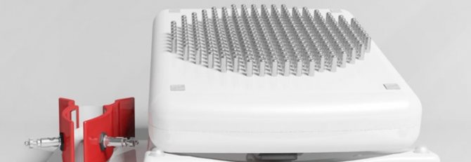
Main parameters and dimensions
- Overall dimensions of the mammograph – 220x190x100 mm
- The weight of the included mammograph is no more than 2.9 kg.
- Equipment:
- A measuring unit consisting of a built-in microprocessor and a matrix (diameter 12 cm) with contact measuring electrodes. The number of gold-plated electrodes is 256 pieces.
- Patient communication cable with double contacts for gel self-adhesive electrodes.
- A stand under the device that ensures that the electrodes do not come into contact with other objects when not in use.
- Software and database on CD
- User's manual on CD
- Atlas of electrical impedance images on CD
- Set of disinfectant wipes
- Disposable gel electrodes
Possible complications or side effects after the procedure
Electrical impedance mammography, as a hardware diagnostic method, is considered the most harmless of all types of mammography. Medicine does not have information about the negative consequences after undergoing it, since the use of X-rays is not practiced.
If the specialist complies with all the conditions of the diagnostic sequence, the likelihood of any complications and negative consequences is excluded.
In isolated episodes, some patients experienced an allergic reaction to the gel or other special liquid used for monitoring. This situation is explained by the individual immunity of the female body to these substances.
Performance characteristics
- The mammograph provides continuous operation for 8 hours in a continuous mode with a constant load.
- The execution time of one scanning cycle is no more than 35 seconds
- The image reconstruction time does not exceed 1 minute (depending on the characteristics of the personal computer).
- Time to complete the examination in screening mode: 3-5 minutes/patient
- The examination time in the inpatient mode is 15-20 minutes/patient
- The warranty period for the electrical impedance computer mammograph "MEIK"® 5th version is 2 years from the date of sale.
- Failure-free operation of electronic components (manufacturers’ warranty) – at least 5 years.