Author
Bordashova Larisa Viktorovna
Leading doctor
Ultrasound diagnostic doctor
until September 30
Appointment with a doctor based on the results of instrumental diagnostics with a 50% discount More details All promotions
Ultrasound of the bladder
is a diagnostic examination of the bladder using ultrasound waves. Different tissues of the body reflect ultrasound differently, which makes it possible to visualize internal organs. Ultrasound diagnostics is non-invasive and safe; ultrasound can be used as often as necessary. It is not recommended to undergo an ultrasound scan only in the very first weeks of pregnancy.
Ultrasound of the bladder can be performed as part of a complete ultrasound examination, as well as as part of an ultrasound of the urinary system. However, quite often an ultrasound of the bladder can be prescribed as an independent study.
How is an abdominal ultrasound of the bladder performed?
In clinics, the transabdominal method of performing ultrasound of the bladder . It is universal, simple and works well for almost everyone, even women during pregnancy and small children. This scan will allow you to:
- assess the general condition of the organ and adjacent structures of the abdominal region and pelvis;
- determine the size and shape, location and structure of the bladder;
- detect pathological changes.
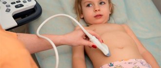
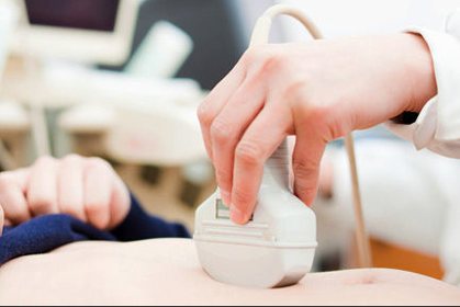
This ultrasound procedure is performed externally. The bladder is scanned with a sensor through the anterior wall of the abdomen. The scanning process itself is quite comfortable. The patient is placed on the couch, then the doctor lubricates the sensor with gel for smoother sliding and consistently moves it in the abdominal area in different directions throughout the entire procedure. An ultrasound of the bladder is performed in children using the same scheme.
General recommendations for preparing for ultrasound
The content of the article
Ultrasound examination is the most accessible, safe and painless method, which has virtually no contraindications. It is used to diagnose pathologies of the abdominal and pelvic organs, brain, mammary glands, heart, kidneys, muscles, etc., as well as to diagnose and monitor the condition of the fetus and woman during pregnancy. Most types of ultrasound require special preparation.

This is due to the peculiarity of ultrasound waves - they do not visualize hollow organs filled with air. With proper preparation, tissue visualization becomes much clearer, and the efficiency of the study increases significantly.
In some cases, it is enough just to have a positive attitude, and sometimes more serious measures are necessary, for example, following a special diet. In emergency situations, an ultrasound is performed at the moment alarming symptoms are detected; no preparation is carried out.
It is not recommended to conduct research after irrigoscopy or x-ray, since these examination methods use a contrast agent - barium. It distorts ultrasound waves, causing ultrasound results to be unreliable. Ultrasound is not recommended after gastro- and colonoscopy. The patient must first notify the doctor if he is taking any medications.
You must take with you the results of your previous study, if any. Many clinics offer to bring a towel or napkins to wipe off the special gel from the body after the examination, as well as a disposable diaper to lay on the couch.
How to do a transvaginal ultrasound of the bladder in women?
Transvaginal ultrasound or TVUS is considered a more informative way to diagnose the bladder in women, since anatomically the bladder is located close to the vagina, and there is no fat layer. This examination is performed only on women for very obvious reasons. An exception will be patients with an intact hymen. For such clients, the doctor will select an alternative method of scanning the bladder. During a transvaginal ultrasound examination, a vaginal sensor is used, onto which a disposable condom is placed, hence a contraindication for this method will be women with allergies to medical rubber. Those patients who have a genital infection will also be exempt from this procedure. Women who are 12 weeks pregnant are likely to be referred for a TRUS scan. The TVUS and TRUS methods are interchangeable, and doctors select one method or another based on contraindications.
| Ultrasound service of pelvic organs | Price, rub | Promotion Price |
| Ultrasound of the bladder with determination of residual urine | 800 rub. | |
| Comprehensive pelvic ultrasound with abdominal and vaginal probe | 1400 rub. | |
| Ultrasound of the prostate and bladder with an abdominal probe | 1900 rub. | |
| Comprehensive ultrasound (ultrasound of the abdominal organs + ultrasound of the kidneys + ultrasound of the thyroid gland + pelvic ultrasound with an abdominal probe + ultrasound of the mammary glands) | 4200 rub. | 2999 rub. |
| Comprehensive ultrasound (ultrasound of the abdominal organs + ultrasound of the kidneys + ultrasound of the thyroid gland + ultrasound of the prostate gland with an abdominal probe) | 3300 rub. | 2499 rub. |
| Comprehensive body diagnostics (MRI of the thoracic spine, MRI of the lumbar spine, ultrasound of the abdominal organs, ultrasound of the kidneys, ultrasound of the bladder, consultation with a neurologist, consultation with a therapist) | 11700 rub. | 7000 rub. |
Contraindications to the procedure
Ultrasound with determination of residual urine is carried out in one of three ways:
- transabdominal - through the anterior abdominal wall;
- transrectally - through the rectum;
- transvaginally - through the vagina.
The first method has no contraindications, with the exception of acute inflammatory diseases of the skin of the abdomen. Transrectal examination is not performed for anal fissures, exacerbation of hemorrhoids, proctitis, paraproctitis. The transvaginal method is not suitable for inflammatory diseases of the genital tract, and is also not used in women who have not begun to be sexually active.
How is transurethral ultrasound of the bladder performed?
Transurethral ultrasound of the bladder, or TUUS, is an invasive type of ultrasound examination. It is carried out exclusively with the introduction of a local anesthetic drug. If the patient is allergic to anesthetics, then this diagnostic method is contraindicated. Transurethral ultrasound of the bladder is also contraindicated when there are any inflammatory diseases of the urinary tract. This scan is carried out using a special sensor inserted into the urethra. This form of diagnosis allows you to examine in more detail pathological lesions of the urethra and assess the degree of damage to the urethra. However, this type of ultrasound service is rarely used in medical centers due to the risk of injuring the urethra.
Symptoms that require an ultrasound
Ultrasound – a specialist makes diagnostics through the anterior abdominal wall. During this study, it is possible to identify diseases and pathologies that have arisen in the organ, as well as monitor the effectiveness of treatment.
A bladder examination is carried out in the following cases:
- suspicion of pathology in any part of the urinary system, or with previously identified diseases of the urinary organs;
- if necessary, find the cause of erythrocyturia if the urine analysis showed a large number of red blood cells;
- when the patient complains of painful urination, which indicates the following diseases: cystitis, prostatic hyperplasia, kidney stones or urolithiasis;
- patient complaints of urinary retention and the appearance of dysuric symptoms;
- for any suspicion of the appearance of neoplasms of any type (malignant or benign tumors), as well as cysts in the urinary duct;
- if there is a suspicion of a cavity formed near the bladder, with accumulated fluid, so-called diverticulosis;
- after injuries and bruises of the bladder, ultrasound will show the possible presence of leaks in the paravesical area;
- after completing a course of treatment to evaluate the results;
- When taking tests, changes were found in the indicators that indicate problems in the genitourinary system.
Preparation
Transabdominal ultrasound is performed with a full bladder. You should drink 1 liter of water 2-3 hours before the procedure. Before the test itself, the person will be given a diuretic to help the bladder fill faster. This is why abdominal ultrasound of the bladder is contraindicated in patients with urinary incontinence. Also, doctors will choose a different method if a person has skin lesions in the lower abdomen of any nature or there are sutures or scars on the walls of the bladder. A necessary condition before transrectal ultrasound of the bladder is bowel cleansing. This can be done at home using a regular enema. To eliminate flatulence, you should give up gas-causing foods 2-3 days in advance. It will not be superfluous to take activated carbon. Transvaginal bladder ultrasound is performed without filling the bladder. For clearer visualization, it is recommended to cleanse the intestines before the study at home and follow a diet for 2-3 days to eliminate the manifestations of flatulence. Preparation for transurethral ultrasound of the bladder is simple. To avoid side effects, you should give up alcohol the day before the scan, and stop smoking 2-3 hours before and limit yourself to breakfast. You should also stop taking medications, with the exception of vital medications. Before starting the procedure, it is important to tell the doctor about the problems of the cardiovascular system (if any), about the medications you take on an ongoing basis, about bad habits and the condition of the body as a whole.
How does the manipulation work?
Ultrasound examination is performed using computer equipment and a special device that interacts with the patient’s skin. The person lies down on the couch, and the doctor treats the area being examined with a special gel-like composition. If we are talking about a pregnant woman, the ultrasound is performed in a standing position.
Features of ultrasound examination:
- During the procedure, it may be necessary to change position if the doctor considers it necessary;
- the patient should be naked to the waist or slightly below;
- The ultrasound examination lasts about twenty minutes.
Manipulation to examine the genitourinary system and kidneys in our clinic is carried out by an experienced specialist who transmits information to a nephrologist. The latter is treating the patient. If necessary, the process can be recorded using a camera.
Contraindications
Unfortunately, abdominal ultrasound is not suitable for all patients. The doctor will not prescribe it to obese people, because the subcutaneous fat layer in such patients is too large; this technique will not be informative for overweight people. Transrectal ultrasound of the bladder also has contraindications. They are connected:
- with diseases of the rectum at the time of exacerbation;
- with the absence of an organ as a result of surgery and its replacement with an artificial one;
- with narrowing or obstruction of the rectum;
- with latex intolerance.
Author: Telegina Natalya Dmitrievna
Therapist with 25 years of experience
Why do you need to fill an organ for an ultrasound, and how to do it?
A filled organ of the excretory system allows you to examine all its walls much better, since they straighten and stretch, there are no additional folds. When this organ is full, it brings the intestinal loops closer to the anterior abdominal wall.
Usually it should be filled naturally, but sometimes it happens that the patient did not know about all the intricacies of the procedure and did not prepare properly. Or the ultrasound diagnostics were urgent and unscheduled. In such cases, rapid filling of the organ is necessary.
This can be done by drinking a liter of liquid about an hour before the test. It could be water, tea, fruit drink or juice. Do not consume milk or carbonated drinks.
If after an hour it is still not full, then you should drink another 500 ml of liquid. When the bladder is full earlier than an hour later, this indicates good kidney function. In this case, you can relieve the overcrowding, but then drink a little more liquid.
If a person is well aware of the work of his excretory system, then he may not drink additional fluid, but simply not empty his bladder for several hours before the procedure. How to prepare for the test
Preparing to conduct research is not too difficult. To do this, you only need a full bladder. There is no need to do any diet or bowel cleansing before the ultrasound.
Preparing for an ultrasound of the bladder
- drinking a liter of liquid an hour before the procedure;
- stopping going to the toilet 4-6 hours before the study;
- if a person suffers from incontinence, then before the procedure, catheterization should be performed, with the help of which the organ is filled with saline solution;
- using diuretics, but this method should not be used by people with kidney or heart disease, as it can cause harm and cause fluid retention or bladder blockage, which will lead to renal colic;
- when conducting research in the morning, do not urinate after sleep;
- when examining the bladder in children under one year of age, they must be fed 10 minutes before the ultrasound.
Patients of different age groups have their own norm of fluid that must be drunk to fill the organ being examined:
- children from one to 2 years old - as long as they can;
- children from 3 to 7 years old - 200 ml;
- from 8 to 11 - 300 ml;
- teenagers from 12 to 18 years old - 400 ml;
- For all people over 18 years of age, it is better to wait for the bladder to fill naturally, and if there is an urgent need, drink up to a liter of liquid.
By performing an ultrasound examination of the excretory organ, you can determine its actual volume and find out why there is a delay in urine output. You can determine the percentage of residual urine volume, which is very important for people after surgery. An organ ultrasound helps to see the exact problem of the excretory system and prescribe the necessary treatment.
In order for the study to obtain the most objective data possible, it is necessary to strictly follow all the recommendations of specialists when preparing for diagnostics. Only then will the doctor be able to obtain the most correct information and prescribe timely and correct treatment that will help cope with the disease.
Indications and contraindications
Ultrasound (ultrasound) examination is prescribed to patients with the following symptoms:
- pain in the lower abdomen;
- frequent urge or painful urination;
- the presence of blood and various impurities in the urine;
- suspicion of urolithiasis;
- inflammation of the prostate and suspicion of adenoma in men;
- the appearance of sharp pain in the pelvic area along with an increase in body temperature.
Ultrasound is indispensable as a tool for preoperative preparation before:
- removal of local tumors;
- cystolithotomy (crushing and removal of stones);
- removal of adenoma by transurethral method;
- other types of surgical manipulations in the area of the ureters, urethra and others.
Using ultrasound, dynamic monitoring of chronic patients is carried out, tumors are identified and the degree of spread of metastases is analyzed, differential diagnosis of pathologies with similar symptoms is performed: inflammation of the prostate gland, female appendages, ureters, kidney diseases, and so on.
This technique allows you to study the pelvic organs in women and at the same time identify deviations in the functioning of the elements of the reproductive system. This diagnosis is recommended for pregnant women to monitor the condition of the fetus and uterus. With its help, patients of childbearing age can promptly diagnose inflammation of the appendages, pathological changes in the uterus and adjacent soft tissues and organs.
Regular ultrasound of the urinary system is also recommended for healthy people for preventive purposes.
Depending on the method of performing ultrasound, the following contraindications are identified:
- with transabdominal – urinary incontinence, excess weight, violation of the integrity of the skin in the area of contact with the sensor, defects of the bladder parenchyma;
- with transrectal – absence, inflammation, narrowing or obstruction of the rectum, latex allergy;
- with transvaginal – acute infectious process, pregnancy more than 12 weeks, virginity, latex intolerance;
- with transurethral - local inflammation, intolerance to anesthesia.
Ultrasound is of great importance in diagnosing bladder diseases, but you need to understand that you need to prepare for this procedure in advance.
Preparing for an ultrasound of the bladder includes several important points, which we will look at later.
Ultrasound examination is needed to study the functioning of the kidneys. In men, it is also performed for prostate adenoma or signs of inflammation.
Read about inflammation of the prostate in men in our article.
Women need a procedure to fully study the functioning of the genitourinary system and a detailed study of the organs.
What will it show?
This process can reveal:
- The shape and size of the organ. Its decrease indicates cystitis, and its increase indicates narrowing of the urethra.
- The presence of neoplasms is established.
- Organ contents. We are talking about urine, blood, pus.
- Foreign bodies.
- Contours.
- Violation of integrity. Diagnostics helps determine the type of damage.
- Increased tone.
- Inflammation.
- Organ prolapse.
- Prostate pathologies.
- Ovarian diseases.
Bladder volume can also be determined using ultrasound. Modern devices automatically calculate this indicator.
Residual urine is an indicator that determines the presence or absence of diseases in the urinary tract.
Normally, the residual urine in the organ cavity should not be more than 10% of the total urine volume. The calculation of this indicator is of great diagnostic importance and helps to establish or exclude the presence of pathology.
To determine this indicator, the study is carried out before and after urination. Having examined the organ twice, in a filled state and without liquid, the specialist can tell about the amount of residual urine. The size of the organ image is assessed. The length of its ultrasound shadow is determined using formulas.
In this situation, an ultrasound scan of a full bladder is performed. This allows you to visualize sediment, contours, and possible changes in the walls of the organ. The study makes it possible to determine not only the presence of flakes and sediment, but also studies in detail its distribution and quantity.
Normal indicators are:
- Shape: if the organ is healthy, the shape is clearly visible. On transverse photographs it is a round organ, and on longitudinal photographs it is ovoid.
- Volume norm: for women 200-500 ml, for men 300-700 ml.
- Structure: normally echo-negative.
- Residual urine: maximum 50 ml.
- The walls of the organ: must be of the same thickness, from 2 to 4 mm.
The process differs depending on the gender and age of the patient.
In women and men
men need to drink 1-1.5 liters of water 2 hours before the procedure. Water must be filled in the bladder; emptying is strictly prohibited.
women need to drink 0.8-1 liters of water 2 hours before the procedure. A woman’s body is different from a man’s, so she only needs a smaller amount of fluid to fill the organ being examined. It is forbidden to have bowel movements before the procedure.
menstruation is not a reason to cancel the procedure. diagnosis can be carried out during menstruation. You should also prepare.

The day before the ultrasound, it is prohibited to drink alcoholic and sweet drinks. they can influence the outcome.
in children
practically no different from the algorithm of adults. children need to drink from 0.5 to 0.7 liters of liquid. A child’s body is smaller than an adult, so this amount of water is quite enough for the procedure. the child should be given liquid 1.5 hours before the test.
If the baby wants to empty his bladder, you need to try to explain to him that this cannot be done. If, nevertheless, the baby could not resist and emptied his bladder, he must be given water again to compensate for the deficiency.
It is better not to give sweet carbonated drinks and juices to your child the day before the procedure.
in pregnant women
Two days before the procedure, you need to exclude spicy, fatty and fried foods from your diet. you need to eat only healthy food.
in the first and second trimester, 2 hours before the procedure you need to drink at least 0.5 liters of liquid. In the third trimester, you do not need to drink water first.
During pregnancy, a woman should know that the procedure is performed on an empty stomach. It is better not to eat in the morning before the examination. this will make it possible to obtain accurate ultrasound results.
There is no special method to quickly fill an organ with urine. It is recommended to drink still water 2 hours before the test. it may be mineral. It’s not only more convenient to drink water from a bottle, it’s easier to calculate the amount of water you drink.
how is it carried out?
There is nothing complicated or dangerous in this procedure. A man enters the office and lies down on the bed. The lower abdomen is lubricated with a special gel. Then, using a special apparatus, the organ is studied. The gel facilitates the sliding of the device, which is passed over the patient’s abdomen. He doesn't feel any pain.
The procedure usually lasts 10 minutes. Then the person gets up, wipes the gel off his stomach, and can go to the toilet to relieve himself.
The results are deciphered immediately or the next day after the procedure. It depends on the doctor's workload. The image shows the absence or presence of organ diseases. Decryption is carried out only by a specialist. He can tell what condition the organ is in.
Ultrasound diagnostic procedure
Diagnosis in men and women is carried out differently. Thus, ultrasound can be used to identify cysts, obstructions and infections in the body. They can also measure blood flow in the arteries to detect blockages, which can be seen on a bladder ultrasound. The kidneys are also included in the study area; they are checked along with other organs of the urinary system. How to prepare for a bladder ultrasound as a woman? Quite often, transvaginal examination is provided for women by inserting a sensor into the vagina. Before starting the procedure, the organ should be emptied to better view the image on the equipment. This ultrasound shows any changes in the organs of the urinary system without problems, which cannot be said about the transabdominal method, which is most often used for men and girls who have not had a sexual partner. Preparation for a bladder examination takes 5 minutes and does not require any intervention from the patient. During most ultrasound exams, the technician may ask you to turn on your side or stomach to get a better image. Once positioned on the couch, the sonographer will apply the gel to the area of the body in question. The gel is applied to the skin so that the air between the sensor and the cover does not interfere with the formation of sound waves. The instrument used during the procedure is called a transducer. It is placed on the body on top of the gel and moved to capture the image. The sensor is capable of emitting high-frequency sound and recording echo signals that determine the size, shape and consistency of soft tissue. This instrument resembles a handheld microphone. The sensor sends sound waves and receives them reflected to create images inside the body that are displayed in real time on the equipment screen. How to prepare for a bladder ultrasound for a man? Urinary ultrasound is used in urology, which specializes in surgical and medical diseases of the urinary tract and reproductive organs in men. What is needed for a bladder ultrasound? There is no preparation as such. You need to take with you everything that the doctor asks (as a rule, the list is limited only to a medical card and shoe covers), and also drink plenty of fluids before the process. Many people wonder how much water to drink before a bladder ultrasound. There is no definite answer - the amount depends on the volume of the organ. For some, 300 ml is enough for the urge to urinate to occur within 30 minutes.