The ideal option for conducting an ultrasound examination is the first half of the cycle before ovulation. At this time, you can obtain the most accurate data on the condition of the organs in this area. But in some cases, ultrasound is performed before ovulation, during or after the ovulatory period. There are several situations in which it is necessary to carry out an ultrasound examination urgently, regardless of the day of the cycle.
Controlling follicle development
One of the reasons for female infertility in the absence of complaints and a regular menstrual cycle is the lack of ovulation. The egg does not mature or leave the ovary in the middle of the cycle. There are many reasons for this phenomenon - hormonal imbalance, premenopausal period, inflammation of the appendages. Even climate change, diet or nervous stress can “knock down” ovulation.
In this case, ultrasound is performed several times during the cycle:
- Immediately after the end of the critical days , at this time two or three follicles with a diameter of 5-9 mm begin to mature in one of the ovaries. The thickness of the endometrium is 2-3 mm, it is smooth, without inclusions.
- The second ultrasound is performed on days 10-11 to determine the dominant follicle, which is ahead of the others in development. As a rule, it reaches 10 mm during this period. The endometrium thickens to 4-5 mm. If the dominant follicle does not appear, the woman is advised to stimulate ovulation.
- The third time an ultrasound is performed on days 13-14 . During this period, the follicle bursts, releasing the egg, and the endometrium increases to 22-23 mm in order to be ready to receive a fertilized embryo.
- The fourth examination is carried out after ovulation - at this time the corpus luteum begins to form in the ovary, and the endometrium becomes three-layered, 13 mm thick.
- The last time a woman is seen is an ultrasound before her expected menstruation . At this time, fertilization and implantation of the embryo into the uterus should occur.
If ovulation does not occur, such a cycle is called anovulatory and requires special treatment. In the absence of fertilization, it is necessary to check the partner’s fertility using a spermogram - a study of seminal fluid.
Preparation for the procedure (ultrasound of the genital organs)
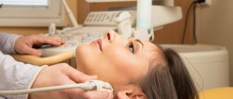
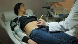
Ultrasound examination of the female reproductive apparatus is carried out in two main ways - transabdominal (through the abdominal wall) and transvaginally (through the vaginal wall). They are often combined.
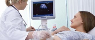
If the doctor examines the reproductive organs through the abdominal wall, the woman will be asked to lie on her back and bend her legs at the knee joints. The lower abdomen is bare. The procedure requires preliminary preparation: the woman must appear in the ultrasound room with a full bladder (to be agreed with the doctor). A conductive gel-like agent is applied to the device’s sensor, the tip is moved over the abdomen and the necessary organs are examined.
The transvaginal method involves inserting a sensor into the vagina and examining the uterus and appendages. There is no need to drink water before the procedure, you cannot douche - hygienic washing is enough. The woman must have a bowel movement. A regular condom is put on the sensor, the gel is applied, and inserted into the vagina. The echo image is displayed on the device monitor.
So, to the very frequently asked question “Is it possible to do an ultrasound during menstruation?”, the answer can be given in the affirmative, if we are talking about the study of non-genital organs, as well as when studying folliculogenesis and in all the emergency situations listed above.
It is advisable to plan a preventive examination of the reproductive system on days 5–10 of the cycle, when the uterus is sufficiently cleared of blood clots and remnants of exfoliated mucosa, and there will be no interference with a quality examination.
You should not interpret the result yourself, or self-medicate if you do not have a medical education. With the received conclusion of the ultrasound examination, you need to contact a specialized specialist - he will give a competent assessment of the data and prescribe therapy if necessary.
Delayed menstruation
In the absence of menstrual periods, women take a pregnancy test and are relieved when it is negative. But the absence of a second strip does not always mean that your period is late and will come later. This is often the first sign of hormonal disorders, which subsequently lead to uterine bleeding.
Under the influence of an increased concentration of estrogen, the internal uterine layer grows, which thickens excessively, but is not rejected. Endometrial hyperplasia occurs.
However, the endometrium cannot grow indefinitely. As a result, its massive rejection occurs with the development of life-threatening bleeding. In this case, the woman has to undergo curettage of the uterus.
In order not to put your life in danger, you need to undergo an ultrasound if your menstruation is delayed. The picture of hormonal imbalance is revealed by thickening of the endometrium and the absence of the corpus luteum in the ovary. In this case, doctors prescribe drugs that induce menstruation.
Sometimes a woman with an irregular menstrual cycle experiences an ectopic pregnancy that she does not expect. The fetus attaches to the fallopian tube, ovary, or even the abdominal cavity and begins to develop. Without an ultrasound scan and timely treatment, the situation can end tragically.
Ultrasound of other organs during menstruation
If there is a need to undergo a diagnostic examination of non-genital organs, the day of the menstrual cycle is not taken into account. You can undergo an examination and get a reliable result at any time, including during monthly bleeding, which will in no way interfere with the diagnosis or affect the result of the study.
Most often, using an ultrasound machine, the following structures of the body are viewed:
- heart, blood vessels;
- The lymph nodes;
- liver, gall bladder;
- kidneys;
- joints;
- thyroid gland.
For a routine examination of the mammary glands, which are considered part of the reproductive system, the doctor will suggest that the woman appear on days 8–12 of the cycle, when the breasts are not swollen and its lobules and ducts can be qualitatively examined. If an urgent reason arises, the date may also be adjusted.
Bladder problems
Although it is located next to the female genital organs, the menstrual phase does not affect its condition. You need to make an appointment for an ultrasound if:
- pain and stinging when urinating , increased urge to go to the toilet;
- , mucus, turbidity in urine
- feelings of incomplete emptying , when after urination it seems that some of the urine remains inside.
In this case, the doctor can make a diagnosis any day. Delay threatens kidney inflammation - pyelonephritis, acute urinary retention caused by blockage of the urethra with a stone, blood clot or mucus. Blood poisoning may occur due to the spread of infection.
Therefore, the opinion that a pelvic ultrasound should be performed only in the first half of the cycle is incorrect. If there are unpleasant symptoms in the lower abdomen, absence of menstruation, problems with the bladder, and also as prescribed by a doctor, this procedure is performed on any day of the cycle.
Therefore, if you have problems with the genitourinary area, you need to sign up for a pelvic ultrasound at any time. When conducting an examination, the doctor always asks the woman about the timing of her last menstruation and evaluates the picture of what she saw from the point of view of the menstrual phase.
On what day of the cycle is an ultrasound of the uterus and appendages performed?
Typically, an ultrasound of the female reproductive system is performed in the absence of menstruation. First of all, because it will be much more difficult for a diagnostician to examine the uterine cavity and assess the condition of the endometrium due to bloody clots. Various neoplasms are difficult to see during menstruation. Accordingly, the diagnosis may be incorrect.
An echography of the uterus and its adnexal structures is prescribed, as a rule, on days 5–10 of the menstrual cycle, when the vast majority of women have already had their periods. A new mucous membrane is formed in the uterus. At the time of the examination, it has not yet hardened, and therefore it can be examined qualitatively, pathological formations can be excluded or confirmed.
Another reason why it is advisable to postpone a planned examination of the organs of the reproductive system until the end of menstruation is psychological discomfort - it is unpleasant for a woman to see a doctor on “special” days.
Not least important is the issue of hygiene. In addition, during a transvaginal examination, the insertion of a special sensor may cause some pain, while on other days the manipulation does not provoke pain.

Ultrasound for fibroids in a paid clinic
The decision on the number and frequency of ultrasound for detected uterine fibroids is made by the doctor and the patient’s task is to strictly follow the recommendations of the prescribed treatment so that it is as effective as possible. But it is much more prudent to take care of your health in advance and after 30 years of age undergo an ultrasound examination annually, without waiting for obvious ailments.
As you know, it is not easy to do ultrasound in public clinics now. Particularly if you need regular, frequent testing. Contacting a paid clinic will save time, get accurate results using modern expert-class equipment and an opinion from doctors of the highest qualification category. You can find all this at Diamed clinics. We invite you to an appointment at our centers. Close proximity to the metro, reception seven days a week and affordable prices will make your stay in our clinics absolutely comfortable.
Features of the breast associated with pregnancy and breastfeeding
With hormonal changes associated with pregnancy and lactation, rapid development of the parenchyma occurs, namely the ducts and acini of the mammary gland. Due to this, the breasts increase in size and often become more sensitive. This is accompanied by regression of the interlobular stroma, the fat lobules of the subcutaneous and retromammary zone are reduced. Only during breastfeeding does a full cycle of development of all mammary gland structures occur, which is of utmost importance in the prevention of breast cancer. But at this time the mammary gland is especially vulnerable to external factors. Therefore, during breastfeeding, especially at the stages of lactation, minimal external influence is very important. Expression should, if possible, be carried out by the nursing mother herself using a breast pump. Spreading with “alien hands” with “crocodile tears” of a nursing woman causes deep trauma to the breast, negates the value of breastfeeding in the prevention of breast cancer, and even vice versa, increases the risk of developing breast cancer in the future.
Benign formations on ultrasound of the mammary glands
Benign breast formations are divided into cystic and solid.
Breast cysts are:
- Simple.
- Complex.
- Combined
Often cysts are a component of fibrocystic mastopathy (dysplasia) of the mammary glands. Fibrocystic mastopathy is the most detected condition during ultrasound of the mammary glands in women of reproductive age. The reason for seeking an ultrasound of the mammary glands is most often pain. In case of mastopathy, a characteristic sign of ultrasound of the mammary glands is compaction of the connective tissue, moderate dilation of the ducts of the mammary glands, and the presence of small cysts. These manifestations are completely individual for each woman.
Solid benign formations on ultrasound of the mammary glands:
- Breast fibroadenoma is a benign tumor of the breast.
- Galactocele is a formation associated with the accumulation of secretions from the mammary gland ducts.
- Breast abscess formation of purulent-inflammatory origin. Etc.
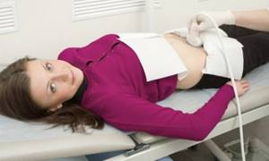
What is pelvic ultrasound
Ultrasound examination is a method of examining a woman’s internal organs using a special apparatus, which consists of a sensor attached to a monitor. The doctor places a transducer that emits ultrasound waves over the area being examined. The so-called echo, reflected from the organ, forms a picture on the monitor, from which the doctor visually observes the state of the body and makes the necessary measurements.
The small pelvis in women is the space in the lower abdominal cavity in which the organs of the excretory and reproductive systems are located. Ultrasound of these organs is an accurate, highly informative way to find out about the presence of disorders of the excretory or reproductive functions, determine their causes, and choose the right tactics for further treatment or observation.
Normal female breast anatomy
The mammary gland consists of stroma and parenchyma.
The parenchymal component of the mammary gland is represented by the ducts and acini of the mammary glands. The stromal component is the support of the mammary gland, i.e. its connective tissue, which connects the parenchyma of the mammary gland itself with adipose tissue. The number and distribution of structural components of the mammary gland are strictly individual, depending on the woman’s age and her hormonal status. The parenchymal component of the mammary gland consists of 15-25 lobes, which have their own ducts (milky ducts) flowing into the common space under the nipple. This space is called the milky sinus. Each milk duct is successively divided into segmental, subsegmental and terminal. The terminal ducts empty into the mammary acinus.
Breast tissue has three levels. The first, most superficial, subcutaneous layer consists of fat, organized into lobules, bounded by fibrous septa by Cooper's ligaments. Cooper's ligaments connect the superficial layer to the middle layer. The middle layer is the mammary gland itself, the mammary layer is represented by the parenchymatous component. The deepest, retromammary layer, a derivative of fat lobules, is bounded in front by the deep mammary fascia and behind by the pectoral fascia.