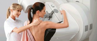
What is mammography?
Mammography is an x-ray of the mammary glands; a research method used to detect (screen) and diagnose breast diseases. Along with regular examinations and consultation with a mammologist, such a study is the main tool for the early detection of breast cancer.
Why get a mammogram?
You should have regular mammograms to make sure you are healthy or to detect a disease early and begin treatment early.
Why is it important?
Breast cancer is a dangerous disease, the leading cancer among women. However, this type of cancer is also the most curable if detected early. Not to scare you, but to give you an honest and objective picture, our center’s mammologist, oncologist of the highest category, Dmitry Karaskov, introduces you to statistical data.
- Malignant neoplasms of the breast rank first among all cancers in women.
- Unfortunately, they also rank first in cancer mortality.
- Every 4-5 malignant tumors in women is breast cancer.
- Every 5th life lost due to cancer is a victim of this type of cancer.
- With age, the problem can overtake an increasing number of women.
- Already at the age of 18-29 years - among women it ranks 3rd in frequency (after thyroid cancer and Hodgkin's lymphoma).
- From the age of 30, it takes 1st place and subsequently “leads” in all age groups up to 75 years.
- The greatest proportion of the disease is observed in 30–54 years of age.
It affects women of the most active and working age.
- So in 2021, 15 thousand women fell ill in Ukraine.
- 6 thousand women died from this disease.
- Every 10th woman did not live more than a year from the moment of diagnosis.
- Less than half of the cases in our country are detected during preventive examinations.
- The woman herself should bear greater responsibility for her health.
- Monthly self-examination of the mammary glands is the key to peace of mind and the earliest possible detection of any problematic changes in the breast.
- In our country, 74% of patients are diagnosed at stages I-II.
- But, accordingly, every 4th case is advanced, which requires much more time, effort and financial resources with a not always good prognosis for recovery.
It is mammography that allows a specialist to see a tumor “in the embryo,” when it is impossible to detect it at the very beginning of development by other means.
High chances of cure if detected early. According to the US Cancer Institute, 98% of women recover when the disease is detected in the early stages and treated correctly.
Methods of performing mammography in St. Petersburg
Mammography at Medpomosch 24 is a painless procedure that takes place in several stages:
- The radiologist or nurse will place your breast on the mammograph stand and gently press it on top with a transparent plastic plate.
- The doctor will take several pictures in different projections, during which you will need to change your body position
- The same steps are repeated for the second breast.
You will need to remain still during the examination in order to obtain the clearest possible images. Upon completion, you will immediately receive results. The whole procedure takes no more than 15 minutes.
Mammography is a preventive examination, during which you can find out whether the neoplasm is malignant or benign and whether it is a single node or several foci at once.
Thus, mammography is the most accurate method for diagnosing breast cancer. Detection of changes can be determined already at the initial stage, when even the woman herself does not feel them. And if you experience discomfort or pain in the mammary glands, then, of course, a visit to a mammologist is simply a must.
Main indications for the procedure
According to international recommendations, women aged 40 years and older should undergo mammography annually. This preventive examination of the mammary glands is called screening. If you have a family history of breast cancer (your mother, grandmother, aunt, or sister had it), your doctor may recommend that you start screening at an earlier age, do it more often, or use other additional methods.
If you come in with any symptoms - for example, you have discovered a lump in the gland, pain or other changes - such a mammogram is called diagnostic.
Digital mammography and computer systems for identifying pathological changes have become a global achievement in medicine, which has made it possible to significantly reduce the radiation dose during the study and increase the accuracy of the results.
In our center, in a diagnostic trailer, you can undergo digital mammography using one of the most modern Siemens Mammomat devices - this is the latest development of the German company Siemens.
Taking care of you!
Medpomoshch 24 is a multidisciplinary medical and clinical diagnostic center. With us you can undergo diagnostics and treatment in 65 areas without queues or fuss. And all this in one building. The combination of highly qualified doctors, modern medical equipment and a caring approach to each patient makes us one of the best in the city. Call right now and our manager will select a convenient time for you to visit. Where to get a mammogram in St. Petersburg? Of course, at Medpomoshch 24!
Rules for preparing for mammography
No special preparation is required. If you menstruate, schedule your exam based on your menstrual cycle. We recommend undergoing examination in the first phase of the cycle. At this time, the density of the gland is less, the procedure will not cause discomfort (many women note that in the second half of the cycle they feel breast tenderness), and visualization will be more accurate.
If you notice a lump in the breast or any other problems, you should immediately consult a doctor - at any time, regardless of your menstrual cycle.
results
What will the doctor look for when reading your mammogram?
If you keep the results from your previous mammograms, your doctor will compare them with the new test results. The doctor will immediately understand whether any new formations have appeared in the breast, whether existing ones have changed, or whether everything is within normal limits. The specialist will definitely pay attention to any deviations, the occurrence of calcifications or tumors. Calcifications are deposits of calcium in the gland tissue: by their size, location and shape, the doctor can determine whether they pose a risk of malignancy and, if necessary, prescribe additional examinations.
Analysis of mammography results is the most important stage of diagnosis. A mistake is unacceptable when it comes to health and life. It all depends on the knowledge and skills of the doctor. Our doctors in Kropyvnytskyi, at TomoClinic, have extensive diagnostic experience. Specialists have the most modern methods - mammography, ultrasound, fine-needle aspiration biopsy, MRI.
FAQ
- At what age can you have a mammogram? Mammography is a test that women aged 40 years and older need to undergo regularly. Your doctor may prescribe a mammogram at an earlier age if you are at risk: close female relatives in your family have had breast or ovarian cancer. It will be very correct if you not only undergo the examination yourself, but spread the information: you can convince your mother, sister, friend, daughter not to skip screening, to undergo a breast examination annually.
- Is it possible to have a mammogram during pregnancy? If you are pregnant, be sure to tell your doctor. Your doctor may recommend another screening method, such as an ultrasound or MRI.
- How long does the procedure take? The time required for mammography varies from one minute to half an hour - this largely depends on the class of equipment. The duration of the examination on a modern digital mammograph Siemens Mammomat, which is equipped with our diagnostic trailer, will not exceed 5 – 6 minutes. At the same time, the Siemens Mammomat mammograph allows you to visualize the smallest microcalcifications ranging in size from 50 microns.
- How is mammography different from ultrasound? Both methods are widely and successfully used to study the condition of the mammary glands, but often patients do not understand what the difference is. There are also significant differences. Mammography is an X-ray method, and ultrasound is an ultrasound method that uses high-frequency sound waves to produce images. Both methods are effective. Mammography is recommended for women over 40 years of age and older, because at this age, for a number of reasons, mammography becomes more informative than ultrasound.
- How long is a mammogram valid? If your mammogram did not show any abnormalities, then you can have your next breast examination in a year. If any pathologies are detected, you will discuss with your doctor when and what additional tests you need to undergo.
- How often can I have a mammogram? We recommend that all women 40 years of age and older undergo a mammogram once a year as screening for early detection of breast disease. Still have questions?
Call the numbers listed on the website. We are always happy to help.
Mammography
Dear women! Why do you need a mammogram?
There are many different diseases in the world that are extremely difficult to identify. Breast diseases are not one of them. Each of you can promptly diagnose breast cancer at an early stage. And, thus, ensure timely treatment and almost complete recovery.
The incidence of breast cancer is very high throughout the world, and ranks first among cancer diseases in women, with a constant trend towards an increase in the number of cases and rejuvenation. But the mammary gland is an organ that is easily accessible for examination. Attentive attitude towards yourself, regular consultations with a mammologist, mammography and ultrasound of the mammary glands are a guarantee of timely detection of mammary gland pathology.
So why do women dislike themselves so much and start getting examined most often when someone close to them gets sick?
Mammography must be performed regularly, starting from the age of 36, once every two years, and from the age of 50 annually.
What is mammography?
Mammography is a method of examining the mammary glands in women using a reduced dose of X-rays. This is the most common, accurate and accessible method of x-ray diagnosis of breast diseases.
How is mammography performed?
Mammography is performed in a special room (X-ray room), using a mammography machine. The breast is placed between two plates, and images are taken with slight compression of the breast. They do this in order to get high-quality photographs. Typically, two images are taken of each gland. In some cases, additional photographs are taken (if there are scars on the chest after surgery).
Information content of mammography
Breast cancer is a potentially curable cancer. Its early diagnosis is the key to a full recovery. In the early stages of development, breast cancer is not detectable by palpation and does not cause symptoms. Thus, mammography is practically the only way to timely detect a small, silent malignant breast tumor. The method has proven itself to be excellent for detecting breast cancer in the early stages. According to WHO, regular digital mammography alone helped reduce breast cancer mortality by 30%.
In addition, the method allows you to detect other changes in the tissues of the gland, benign tumors (fibroadenomas), cysts, calcifications, and inflammatory diseases of the mammary gland.
Preparing for a mammogram
There is no special training. It is recommended to conduct the examination on days 5–12 of the menstrual cycle, when the breasts are least painful. For women going through menopause, mammography is performed at any convenient time.
Be sure to tell your doctor if you are taking hormonal medications, have had any surgeries, or have a family history of breast cancer.
Do not have a mammogram if you are pregnant or suspect you are pregnant. Do not use deodorant or lotion under your arms on the day of the test. They may appear on a mammogram as calcium spots.
Complications with mammography
The study is absolutely harmless since the radiation exposure is insignificant. The method does not cause complications. There are two main methods of performing mammography: film (analog) mammography and digital mammography. In our center, the examination is carried out using the most modern digital mammograph PHILIPS MicroDose L30, which has a minimum radiation dose and maximum resolution. The technique for performing these two techniques is exactly the same. The main advantages of digital mammography are:
- low radiation exposure compared to conventional analog X-ray mammography, so patients can undergo mammography once every six months according to doctor’s instructions without harm to their health.
- the ability to process images to improve its perception (changing brightness and contrast), obtaining a separate fragment with 8 or 12 times amplification, the doctor can use computer functions to examine the image in more detail;
- very high information content, which makes it possible to see such minimal signs of breast cancer as a tumor node measuring 3-5 mm in diameter, an accumulation of microcalcifications measuring 30 microns in size, an area of extensive structural restructuring starting from 5 mm in size, and cancer inside the duct measuring 1-2 mm;
- Images obtained as a result of digital mammography can not only be printed on X-ray film, but also transmitted electronically from one device to another.
We work with private clients (individuals) and corporate clients (legal entities, organizations).
For questions regarding concluding agreements with legal entities, please contact: (863) 223-72-81; e-mail


