Breast cancer is the most common cancer among women in Russia. The tumor occurs in 11% of women during their lifetime, while the incidence continues to grow, increasing annually by 1.2%. About 1 in 2,000 women who are pregnant or breastfeeding will also develop breast cancer. Breast cancer also occurs in men, but it is quite rare - less than 1 in every 180 cases.
This form of cancer ranks third among all causes of death among the female population of Russia after diseases of the circulatory system and accidents in all age groups.
Regular examination by a mammologist will help identify breast diseases in the early stages, provide timely assistance and maintain health.
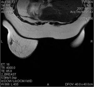
- Breast tumors on ultrasound can be presented as a delimited formation or an area with a violation of the normal echostructure.
- Whether a breast space-occupying lesion belongs to a malignant or benign process is determined based on the following echo signs:
- • the nature of the contours (smooth, uneven, clear, fuzzy, wavy, scalloped, star-shaped).
- • relationship with surrounding tissues;
- • type of internal structure;
- • echogenicity;
- • acoustic effects observed behind the tumor;
- • nature of blood supply.
The nature of the tumor contours is one of the main differential criteria for the type of tumor growth in the breast—expanding or infiltrating.
A benign tumor does not destroy surrounding tissues, but only pushes them apart. Therefore, benign tumors are characterized by clear contours and a delimited (expanding) type of growth.
Malignant breast tumors are mostly represented by cancers. Breast cancer has a heterogeneous pathohistological structure. Most breast cancer (up to 70-80%) are invasive, ductal.
Invasive forms of breast cancer are characterized by an infiltrating type of growth, which is manifested echographically by blurring and unclear contours. With the infiltrating type of tumor growth, it can be difficult to draw a boundary between the edge of the tumor and the surrounding tissues.
Echographic signs of infiltrating growth type are never found in benign tumors. At the same time, ultrasound signs of a limited type of growth can be detected in both benign and malignant tumors.
Both benign and malignant tumors may have their own anatomical capsule, or a pseudocapsule formed by compressed or fibrotic surrounding tissues.
The echogenicity of a tumor may vary, but malignant tumors are more characterized by low echogenicity and heterogeneity of the internal structure.
Acoustic effects in breast tumors are varied: from a slight increase or decrease in ultrasound waves to the appearance of acoustic shadows behind the formation. Acoustic shadow is considered one of the leading ultrasound signs of malignancy. However, statistical data indicate that this echographic sign is detected only behind 30-65% of malignant tumors. It should be noted that in breast cancer, the frequency of occurrence of the dorsal acoustic shadow (ultrasound mammography criterion) approximately corresponds to the frequency of grouped microcalcifications (X-ray mammography criterion).
Biological subtypes of breast cancer
Breast cancer is not a uniform disease. When treating, clinicians take into account whether the tumor belongs to a specific subtype. Genetic testing and immunohistochemical methods can reliably determine the biological subtypes of breast cancer. These subtypes themselves include many risk factors and predictive signs, which allows you to select the most effective therapy for the patient.
The pathogenetic diversity of breast cancer, proven using molecular genetic analysis and immunohistochemical studies, allows for individualized treatment.
Increased survival from breast cancer is associated not only with the widespread implementation of mammography screening, but also with the adequate use of systemic treatments.
Do you need to prepare for an ultrasound of the mammary glands?
It is worth noting that no special preparatory measures are required to conduct an ultrasound of the mammary glands. However, you need to know that a woman’s hormonal background has its own characteristics and manifests itself in monthly cycles. The date of your visit to the diagnostic room depends on this.
So when should you do a breast ultrasound? Doctors recommend carrying out this examination on days 5-10 of the cycle (in the first half). According to scientific data, it is during this period of time that a woman’s hormonal background is most stable. This allows you to reliably and qualitatively determine the condition of tissues and structures in the mammary glands.
Immediately before or after menstruation, the hormonal picture in women is unstable, this can distort the results of the diagnosis.
If a woman uses oral contraceptives or her age corresponds to menopause, then the time of ultrasound diagnostics is not particularly important.
Diagnosis of breast cancer
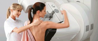
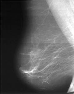
To identify malignant breast tumors, there is a specific diagnostic algorithm, and its primary element is mammographic screening. The sensitivity of this diagnostic method for tumors from 2 mm to 5 mm is about 85%. Mammographic examination is performed in two projections.
Digital mammography for breast cancer
Young women with a dense breast structure need to include ultrasound and MRI in their surveillance program.
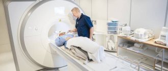
Magnetic resonance imaging
Since traditional mammography screening in women under 40 years of age is ineffective, screening may require an alternative technique - MRI. Modern contrast magnetic resonance imaging is a highly sensitive method for diagnosing breast diseases.
MRI diagnosis of breast cancer
In women with BRCA1 or BRCA2 mutations, MRI can detect breast cancer in its earliest stages.
Ultrasound-guided puncture
For a number of indications, women are prescribed an ultrasound-guided breast biopsy. This study allows you to specifically take biological material for histological examination, determining the level of expression of steroid hormones and Her-2 status.
Mammography or breast ultrasound?
Breast ultrasound and X-ray mammography are the leading methods for early diagnosis of breast diseases.
Ultrasound of the breast, ultrasound mammography is the preferred method for girls and women under 40 years of age.
X-ray mammography is a mandatory preventive standard method of examining women over 40 years of age.
Ultrasound of the mammary glands, ultrasound mammography is advantageous when examining teenage girls, young women who have not breastfed, pregnant and breastfeeding women.
Ultrasound of the mammary glands is preferable when examining the breast immediately after injury, for the purpose of differential diagnosis (difference) between lactostasis (milk stagnation) and mastitis.
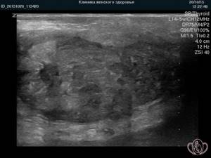
| Ultrasound mammography: Photo of a large lipoma (“fat”) in the left breast of a 16-year-old girl |
|
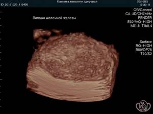
| 3D photo of a breast lipoma. Same case |
|
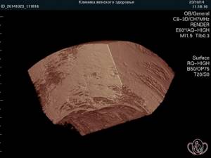
| 3D photo ultrasound of the mammary glands. A fragment of breast tissue from a 48-year-old woman. Predominantly adipose tissue |
|
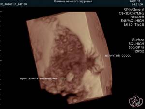
| 3D photo ultrasound of the breast. The nipple is retracted. Ductal papilloma is indicated by an arrow. Pay ATTENTION to the clarity and QUALITY of the picture |
|
The price of an ultrasound scan of the mammary glands with regional lymph nodes is 1,650 rubles.
The price of 3D/4D ultrasound of the mammary glands with regional lymph nodes is 3150 rubles.
The cost of an initial consultation with a specialist to identify breast diseases is 2,300 rubles.
The cost of a repeated consultation with a specialist to identify breast diseases is 1,250 rubles.
| It is IMPORTANT to start moving in the RIGHT direction! EVERYTHING you need to know about the right direction in breast examination, see HERE: |
- Good mammologist in Pyatigorsk
- Cost of mastopathy treatment
- Photo gallery 3D 4D ultrasound of the mammary glands
- Questions and answers about breast screening
Ultrasound of the mammary glands by appointment by multi-channel phone 8 (800) 500-52-74 (toll-free within Russia), or +7.
ONLINE ultrasound doctor can be found at:
REGISTER ONLINE for an ultrasound here.
REGISTER for an ultrasound scan online here.
Sign up for an ultrasound
Reviews of breast ultrasound
H.R., Nalchik
The staff is very attentive to all patients and correct. To Oleg Yurievich - special thanks, the greatest gratitude from the bottom of my heart for your attention, professionalism, attitude towards your work and towards your patients. Also thank you for the good modern equipment, which makes the most accurate diagnosis possible. I wish you further prosperity and expansion. I also wish that you open your own branch in Nalchik, because... It’s hard for us visitors to get to you. All reviews and words of gratitude are only from the good side. May only good luck and success accompany you in everything!
Z.R., Nalchik
First of all, thank you very much for the positive experience that every patient receives. The friendliness and professionalism of the staff is already a guaranteed recovery. Oleg Yuryevich - You are a professional with a capital P, a very subtle psychologist, a real doctor. I wish you further expansion and more health, smiling, happy women around you.
All reviews about breast ultrasound
Expert-class ultrasound devices with specialized mammography sensors used in our Clinic allow you to control the insertion and trajectory of the needle during puncture biopsy, the condition of mammary gland cysts during drug treatment and the completeness of cyst emptying during fine-needle biopsy, and examine the mammary glands in static volumetric image modes (3D Ultrasound of the mammary glands) and dynamic volumetric image (4D ultrasound of the mammary glands), etc.
The resolution of the ultrasound machines of the Women's Health Resort Clinic and the QUALITY of the images (photo quality) rightly deserve admiration!
Each ultrasound doctor has long experience, several specializations and is able to comprehensively assess the situation.
We put our extensive medical experience, all our skills and invest large amounts of money to ensure that the ultrasound examination methods and ultrasound machines of the Women's Health Resort Clinic meet the diagnostic level of the world leaders in ultrasound diagnostics.
At the Women's Health Resort Clinic, ultrasound equipment is upgraded every 3-6 months in accordance with the release of new software versions.
3D ultrasound of the mammary glands allows a more detailed study of the retromamillary area in order to identify dilation of the ducts and intraductal formations (papillomas), visualize the walls of cysts for parietal growths, and examine in detail the blood flow of local formations and the mammary gland as a whole.
Elastography (determining tissue elasticity/stiffness), available with the ultrasound machines of the Women's Health Resort Clinic, is an important additional modern diagnostic option when performing routine breast ultrasound.
Most malignant breast tumors have increased stiffness compared to unchanged tissues.
Ultrasound of the mammary glands and elastography make it possible to identify a tumor with a greater degree of certainty at a very early stage (before palpable changes) and reduce the number of unfounded diagnostic actions.
Elastography allows us to judge the involvement of surrounding tissues in the process (tumor invasion).
Ultrasound of the mammary glands, ultrasound mammography in 3D and 4D modes at the Women's Health Resort Clinic make it possible to identify questionable and unfavorable areas of breast tissue when examined in power Doppler mode based on early changes in blood flow velocity curves.
Conclusion Ultrasound of the mammary glands using an expert-class device from the Women's Health Resort Clinic in some cases is the only basis for revising treatment tactics and choosing the scope of surgical treatment.
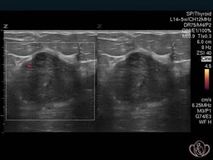
| Photo of breast cancer. The tumor is an irregularly shaped formation 2.8 mm in diameter and has a fuzzy, uneven contour. In power Doppler mode (left side of the image), the feeding vessel is determined |
|
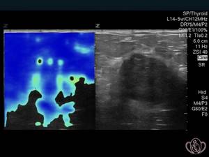
| Photo of breast cancer. The same case. In the elastography mode, moderate elasticity of the formation was revealed and the true boundaries of the spread of the tumor were established, exceeding those visible with conventional ultrasound mammography |
|
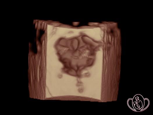
| Photo of breast cancer. The same case. 3D photo of a tumor node. The formation of a coarsely tuberous structure with an indistinct, spicule-shaped (star-shaped) contour is determined |
|
See all photos
Pay attention to the EXCELLENT quality of photographs taken on our expert-class devices.
You can find photos of breast ultrasound taken by our specialists on many Russian and foreign websites.
Ultrasound of the mammary glands and ultrasound mammography do not emit X-rays. Thus,
3D 4D ultrasound of the mammary glands is SAFE for the purpose of dynamic control, including in children, adolescents and pregnant women.
The number of ultrasound examinations in one day is not limited.
It is possible to perform ultrasound of the mammary glands and fluorography of the lungs, X-ray mammography, X-ray examinations of joints, spine, skull, arm bones, legs, etc. on the same day.
You can have an ultrasound of the breast and get advice from a specialist to identify breast diseases at the Resort Women's Health Clinic from 6 to 12 days from the start of menstruation.
You can perform breast ultrasound and ultrasound mammography at the Women's Health Resort Clinic at your own request or as prescribed by a doctor.
You can also do
- Ultrasound of the fetus in 3D/4D modes with extended Doppler study of blood flow
- Ultrasound of the pelvis in 3D/4D and elastography modes
- Ultrasound of the thyroid gland in 3D/4D and elastography modes
- Ultrasound of the abdominal cavity in 3D/4D and elastography modes (ultrasound of the liver, gallbladder, spleen, pancreas and paraortic lymph nodes)
- Ultrasound of the kidneys and bladder in 3D/4D and elastography modes
- Checking the patency of the fallopian tubes WITHOUT PAIN (HSG echo, GHA, hydrosonography)
A well-functioning pre-registration system allows you to conduct an ultrasound examination at a time convenient for you and in the shortest possible time.
Sign up for an ultrasound
Risk factors for breast cancer
There are many factors known to increase the risk of breast cancer. Some of them cannot be influenced:
- family history of breast cancer,
- early menarche (start of menstruation),
- late onset of menopause.
At the same time, there are also modifiable factors, such as:
- excess weight in postmenopause,
- use of hormone replacement therapy,
- alcohol consumption,
- smoking
etc.
Strategic steps aimed at reducing the risk of breast cancer include weight control and the fight against obesity, regular physical activity, and reducing alcohol consumption.
Most risk factors for breast cancer are associated with the effect of hormones on breast tissue (early menarche, late menopause, obesity, use of hormonal drugs). It is believed that it is female sex hormones that stimulate cell growth processes and increase the risk of DNA damage, which can lead to the development of malignant neoplasms.
More details about this in the article Causes and risk factors for developing breast cancer.
When is breast ultrasound prescribed?
There are a number of subjective and objective indicators when it is correct to do an ultrasound of the mammary glands without delay. Any deviation from the norm or development of discomfort should not be ignored. If such phenomena occur, you should urgently seek advice from an appropriate specialist or therapist.
Ultrasound of the mammary glands is prescribed in the following cases:
- the appearance of pain and/or discomfort in any area of the chest;
- the appearance of unusual discharge from the nipple (purulent, putrefactive or bloody);
- with hardening of the nipples;
- asymmetry of the mammary glands;
- detection during palpation of pathological neoplasms or changes in breast shape;
- in inflammatory processes developing in the mammary glands and/or their ducts;
- to monitor the effectiveness of treatment therapy;
- as a preventive measure to detect the development of mammary gland pathologies at an early stage.
This type of study is also used to clarify the diagnosis, for example, when detecting a pathological process using radiography, since ultrasound shows many nuances that are not visible on an x-ray. It is used to monitor the progression of pathology and the effectiveness of the therapeutic measures used. It is worth noting that ultrasound diagnosis of mammary gland tumors is used to select the method and optimal tactics of surgical intervention. Thus, if breast cancer develops at an early stage, it is possible to perform a sectoral mastectomy with minor tissue excision or a radical mastectomy, which involves complete removal of the breast and nearby lymph nodes and ducts (often in later stages).
Hereditary risk factors for breast cancer
Only 5–10% of breast cancer cases are caused by inherited mutant BRCA genes. But at the same time, among mutation carriers, the risk of developing the disease can reach 80%.
The younger the age at which primary breast cancer is diagnosed, the higher the likelihood of developing contralateral cancer, i.e. opposite mammary gland.
A family history of breast cancer in blood relatives, even without association with BRCA mutations, also increases the risk of breast cancer.
Lifestyle
Obesity Various studies have proven the existence of a connection between the incidence of primary breast cancer in postmenopause and obesity. There is evidence of a connection between excess weight and low survival rates for all types of breast cancer.
Alcohol Alcohol is also an established risk factor for primary breast cancer. The evidence for its negative effects on breast cancer survivors is compelling, as it increases the amount of circulating estrogen.
Symptoms of breast cancer
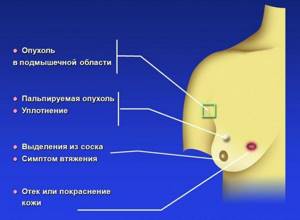
Only a doctor can assess the significance of various symptoms. However, every woman needs to know for what symptoms she should contact a mammologist:
- “Formation”, compaction, node, infiltrate, tumor, “ball” - you found something similar to this. This is not yet a reason to decide that you have breast cancer, but a reason to see a specialist.
- Deformation of the contour of the breast, areola or nipple (skin retraction or, conversely, bulging)
- Nipple retraction; especially if it appeared recently
- Discharge of blood from the nipple
- Swelling of the skin of the entire breast or its local area
- The appearance of irritation, wet “sores”, ulcers, crusts on the nipple or areola
- Ulcerations (long lasting, for no apparent reason) on the skin of the chest
- Discomfort in the armpit and detection of lymph nodes ("balls") in the armpit
- Change in skin color of the breast - redness, increased skin temperature in this area.
All these symptoms can be manifestations of various diseases (there are several dozen of them), perhaps not of a malignant nature. However, this can only be decided by a breast specialist.
Signs and symptoms of breast cancer
Leading ultrasound doctors of the Southern Federal District
Ermolaeva Elvira Kadirovna is a well-known and recognized specialist in the field of ultrasound diagnostics of mammary gland diseases in the North Caucasus. People turn to her to establish an accurate diagnosis and expert ultrasound of the mammary glands.
Ermolaev Oleg Yurievich Candidate of Medical Sciences, gynecologist-endocrinologist, ultrasound diagnostics doctor, specialist in detecting diseases of the mammary glands with 25 years of successful experience in treating advanced forms of mastopathy. Specialized in mammology in 1993 at the Moscow Mammology Dispensary.
Shchepkin Petr Sergeevich Gynecologist, experienced breast ultrasound doctor. Specialist in identifying diseases of the mammary glands.
About the doctors of the Clinic in detail...
| INTERNATIONAL RECOGNITION of the reputation and achievements of the Women's Health Resort Clinic in the development and implementation of effective and safe treatment methods and the quality of medical services provided is the AWARDING of the Women's Health Resort Clinic in Pyatigorsk with the SIQS International QUALITY CERTIFICATE in the field of medicine and healthcare. International Socratic Committee, Oxford, UK and Swiss Institute for Quality Standards, Zurich, SWITZERLAND. |
We work seven days a week and on holidays:
Monday - Friday from 8.00 to 20.00, Saturday, Sunday, holidays from 8.00 to 17.00.
Ultrasound of the mammary glands by appointment by multi-channel phone 8 (800) 500-52-74 (toll-free within Russia), or +7.
The price of an ultrasound scan of the mammary glands with regional lymph nodes is 1,650 rubles.
The price of 3D/4D ultrasound of the mammary glands with regional lymph nodes is 3150 rubles.
The cost of an initial consultation with a specialist to identify breast diseases is 2,300 rubles.
The cost of a repeated consultation with a specialist to identify breast diseases is 1,250 rubles.
| ONLINE ultrasound doctor can be found at: REGISTER ONLINE for an ultrasound here. REGISTER for an ultrasound scan online here. |
Sign up for an ultrasound
The resort clinic for women's health operates both for paid services and in the voluntary health insurance system.
For detailed information about visiting doctors at the Women's Health Resort Clinic, preparing for ultrasound and laboratory tests, living conditions, as well as other questions you are interested in, you can call in Pyatigorsk (toll-free within Russia) or +7 (928) 022- 05-32.
We are at your FULL DISPOSAL if you have any doubts or wishes.
Stages of the disease
Stage 0 This is the stage when the primary tumor is not identified or cannot be assessed, and also in the case of non-invasive breast cancer (which means the tumor does not extend beyond its appearance, so-called in situ cancer).
Stage 1 Cancer cells in this stage invade or grow into neighboring tissues. The tumor node is no more than 2 cm, the lymph nodes are not affected at this stage.
Stage 2 In this stage, the tumor node exceeds 2 cm and can reach up to 5 cm. At this stage, lymph nodes may be affected, but the damage to the lymph nodes is single, they are not fused to each other and are located on the same side as the tumor. In case of lymph node involvement, the tumor size may be less than 2 cm.
Stage 3 Invasive cancer, more than 5 cm or with obvious and significant damage to the lymph nodes. In this case, the lymph nodes can be fused together.
Stage 4 At this stage, the tumor grows into the skin of the chest, chest wall, or internal mammary lymph nodes. It can be of any size.
Stage 4 breast cancer is inflammatory cancer; it occurs in up to 10% of all cases. Symptoms of the inflammatory form of breast cancer are redness of the skin, the gland becomes warm, and there is an enlargement and/or hardening of part or all of the breast. The skin takes on the appearance of an orange peel. This form of cancer must be differentiated from inflammation of the mammary gland - mastitis.
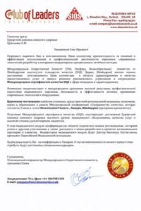
Also, at stage 4, the tumor can spread beyond the chest, into the axillary region, and internal mammary lymph nodes. Metastases to the supraclavicular lymph nodes, as well as to the lungs, liver, bones or brain are possible.
What breast cancer looks like in different stages:
Breast cancer treatment methods
Surgery
Surgery is the main method of treatment for breast tumors, and the outcome of the disease largely depends on the quality of its implementation. According to some Indian states that do not offer surgical treatment for religious reasons, the incidence rates are almost equal to the mortality rates. Typically, the mortality rate from breast cancer is two to four times lower than the incidence rate.
Radical mastectomy
However, surgical treatment is most often followed by radiation. Local treatment without postoperative radiotherapy often leads to local-regional recurrence of the disease. The fact is that after completion of the surgical operation it is impossible to exclude the existence of hidden distant metastases. Even in patients with tumors less than 1 cm in diameter, recurrence of the disease is possible in 10% of cases.
Localization of distant metastases in breast cancer
Organ-preserving operations
Nowadays, there is a worldwide trend towards reducing the volume of surgical intervention without loss of effectiveness. Surgical and radiation treatment of breast cancer is developing and improving in the direction of organ preservation.
Thanks to the introduction of mammography screening, the number of patients with an early stage of the disease, when the lymph nodes are not affected by metastases, has sharply increased. In this case, the “classical” removal of all levels of lymph nodes would be an unnecessary mutilating procedure. The method of biopsy of sentinel lymph nodes came to the aid of surgeons.
Since metastases in the axillary lymph nodes appear sequentially from the first to the second, then to the third level, it is enough to determine the presence of metastases in the first lymph node. It was called “sentinel”: if the sentinel lymph node does not contain metastases, then other lymph nodes are also not metastatic.
Thanks to this organ-preserving method, thousands of patients have avoided unnecessary complete surgical dissection; removal of the cancerous tumor did not lead to breast removal.
The latest results of clinical trials, including with the participation of the Oncology Research Institute named after. N.N. Petrov, confirmed the safety of avoiding complete axillary dissection. Sentinel lymph node biopsy is gradually replacing axillary dissection as the standard procedure for staging breast cancer.
The concept of sentinel node biopsy is becoming increasingly accepted and has been introduced into the surgical treatment standards of the European Organization for Research and Treatment of Cancer (EORTC) for many tumor sites.
Chemotherapy
Chemotherapy, along with surgery, is one of the main methods of cancer treatment. Postoperative chemotherapy improves the results of surgical treatment, as well as the prognosis of the disease.
Previously, the decision to prescribe chemotherapy was based on two factors:
- stage of the disease
- condition of regional lymph nodes.
Thanks to numerous studies by scientists, ideas about the biology of breast cancer are changing, and the choice of chemotherapy regimens is significantly expanding. And today, chemotherapy treatment is prescribed even in the absence of metastases in the lymph nodes, if small tumors have aggressive biological characteristics.
Luminal A cancer In luminal A cancer, chemotherapy is avoided, especially with negative lymph nodes, and endocrine therapy alone is used.
Luminal B cancer Luminal B tumors are characterized by high aggressiveness. In this case, chemotherapy will most often be prescribed, and the choice of treatment is based on an assessment of the risk of relapse.
HER2-positive breast cancer Treatment of HER2-positive breast cancer is based on the use of standard chemotherapy regimens - trastuzumab and chemotherapy based on anthracyclines and taxanes. However, only a small percentage of patients benefit from treatment, but all are susceptible to associated toxicity.
Triple-negative breast cancer Triple-negative breast cancer is usually associated with a poor prognosis. Due to the rare occurrence of specific types of breast cancer, there is insufficient data on the role of adjuvant chemotherapy.
Chemotherapy for breast cancer in very young women Breast cancer at a young age is usually aggressive, and hormone-resistant and HER2-positive tumors with different properties than in older women are common. For such patients under 35 years of age, adjuvant chemotherapy is almost always the necessary treatment.
Chemotherapy for elderly patients In elderly patients (over 65 years of age), when deciding whether to prescribe adjuvant chemotherapy, the general condition of the body and the presence of concomitant chronic diseases must be taken into account.
Ideally, older patients should undergo geriatric evaluation to determine their suitability for adjuvant treatment. The potential effect of treatment must be balanced against the risks to the body that chemotherapy carries. The physician determines the most effective, yet safest, specific regimen based on the tumor subtype and individual patient characteristics.
Hormone therapy Young women with hormone-positive breast cancer remain at risk of disease recurrence for at least 15 years after the initial disease. Oncologists must determine which patients require long-term adjuvant therapy with tamoxifen or aromatase inhibitors.
Neoadjuvant (preoperative) therapy Neoadjuvant therapy plays a leading role in the treatment of women with inoperable breast cancer, and is also important for operable tumors when breast-conserving surgery is performed.
Effect of neoadjuvant treatment BEFORE (left) and AFTER (right)
Questions and answers about breast screening
Question: On what days of the menstrual cycle is breast examination performed? A.S., Cherkessk. Answer: Ultrasound of the mammary glands, X-ray mammography, ultrasound mammography and a mammologist’s appointment are carried out from 6 to 12 days from the start of menstruation.
Women from Cherkessk, Karachaevsk, Ust-Dzheguta, Teberda, Dombay, Zelenchukskaya, Ispravnaya, Kardonikskaya, Storozhevoy, Kyzyl-Oktyabr, Kumysh, Kamennomost, Pravokubansky, Uchkeken, Krasny Kurgan, Pregradnaya, Khabez, Kurdzhinovo and other settlements of Karachaevo come to perform breast ultrasound -Circassian Republic.
Question: My daughter is 13 years old. Her mammary glands are of different sizes. Could this be due to a tumor? R.M., Nalchik. Answer: In order to identify large (nodular) formations in the mammary glands with their asymmetry, ultrasound mammography (3D 4D ultrasound of the mammary glands) is recommended at any age.
Women from Nalchik, Prokhladny, Baksan, Maisky, Tyrnyauz, Terek, Chegem, Khasanya, Adiyukh, Belaya Rechka, Atazhukino, Zhankhoteko, Zayukovo, Islamey, Kuba, Belokamenskoye, Zolsky, Malki, Prirechny, Oktyabrsky, Elbrus, Tyrnyauz, come for breast ultrasound examinations. Kashkhatau, Nartkaly, Anzorey, Zalukokoazhe and other settlements of the Kabardino-Balkarian Republic.
Question: At what age should the breasts be examined regularly? I.N., Kislovodsk. Answer: Examination of the mammary glands in the absence of complaints begins at 14-15 years of age. If there are complaints - if necessary.
Women from Pyatigorsk, Kislovodsk, Zheleznovodsk, Essentuki, Lermontov, Mineralnye Vody, Novoaleksandrovsk, Novopavlovsk, Mikhailovsk, Svetlograd, Nevinnomyssk, Stavropol and other cities of the Stavropol Territory come for breast ultrasound.
| ONLINE ultrasound doctor can be found at: REGISTER ONLINE for an ultrasound here. REGISTER for an ultrasound scan online here. |
Sign up for an ultrasound
Question: My doctor ordered an ultrasound of my breast. You have information on your website that you can do an ultrasound at the Women’s Health Clinic on Saturday, Sunday, weekends and holidays, and you do an ultrasound in the evening?! How to get to you? I live in Pyatigorsk. Answer: We actually perform ultrasounds WITHOUT DAYS OFF. We can
Do an ultrasound of the mammary glands on Saturday, Sunday, evening, New Year, March 8, May and other holidays. Make an appointment by phone in Pyatigorsk 8 (800) 500-52-74 (calls within Russia are free) or +7.
Question: Where is the best place to do breast ultrasound in Pyatigorsk? Answer: Ultrasound of the mammary glands is performed at the Women's Health Clinic of HIGH QUALITY.
Question: During an X-ray mammogram at the clinic, my breasts were severely compressed. Then they were sick for 2 weeks. Is this how it should be? R.Kh., Vladikavkaz. Answer: The mammary glands are flattened (“flattened”) to improve the quality of the x-ray. Modern X-ray mammography units have sensors for the adequacy of compression (squeezing), which makes examination of the mammary glands on modern mammographs less painful.
| ONLINE ultrasound doctor can be found at: REGISTER ONLINE for an ultrasound here. REGISTER for an ultrasound scan online here. |
Sign up for an ultrasound
Question: I didn’t like the ultrasound of the mammary glands in Grozny. I want to do a breast ultrasound in Pyatigorsk at your famous clinic. When should I come to you? Ya.M., Grozny. Answer: An ultrasound of the mammary glands in Pyatigorsk can be done by appointment by phone (calls within Russia are free) or +7 (928) 022-05-32.
Women from Grozny, Argun, Gudermes, Urus-Martan, Shali, Achkhoy-Martan, Bamut, Orekhovo, Dolinsky, Gikalovsky, Komsomolsky, Krasnostepnovsky, Pervomaisky, Raduzhny, Sadovoy, Vinogradny, Bragun, Novogroznensky, Chkalovo, Krasnaya Gorka, come for breast ultrasound. Yuzhny, Bratsk, Kalaus, Znamensky, Levoberezhny, Obilny, Savelyevskaya, Guli, Lyazhgi, Olgeti, Dzhugurty, Shatoya, Shelkovskaya and other settlements of the Chechen Republic.
Question: At the age of 45, my breast size has increased, although my weight has not changed. How can this be explained? Z.K., Makhachkala. Answer: The reasons for an increase in the volume (size) of the mammary glands are numerous. An increase in breast size occurs due to the use of contraceptives (COCs), medications and foods that cause fluid retention in the body, excessive water consumption for a particular organism, due to wearing a tight bra or top that interferes with blood circulation and a number of other reasons. In order to exclude oncological causes, consultation with a mammologist and ultrasound of the mammary glands are necessary.
Women from Makhachkala, Derbent, Dagestanskiye Ogni, Buinaksk, Kizlyar, Izberbash, Karata, Khasavyurt, Kizilyurt, Yuzhno-Sukhumsk, Kaspiysk, Leninkent, Semender, Novy Khushet, Bavtugay, Dubkov, Novy Sulak, Komsomolsky, Tserkhimakhov, Lutkun, come for breast ultrasound. Hamamatyurt, Botlikh, Verkhniy Kazanishche, Karamakhov, Gergebil, Maali, Mekhelta, Kuli and other settlements of the Republic of Dagestan.
Question: Where can I get an ultrasound of the mammary glands on the weekend? O.P., Pyatigorsk. Answer: You can get an ultrasound of the mammary glands on Saturday and Sunday in Pyatigorsk at the Women's Health Clinic, phone 8 (toll-free within Russia) or.
| ONLINE ultrasound doctor can be found at: REGISTER ONLINE for an ultrasound here. REGISTER for an ultrasound scan online here. |
Sign up for an ultrasound
Question: “The resort clinic for women’s health operates both for paid services and in the voluntary health insurance system.” In the voluntary health insurance system, is it covered by medical policies? Can you explain please! Answer: We accept Insureds at SOGAZ, AlfaStrakhovanie, Alliance, Ingosstrakh under policies issued by these insurance companies. You can find out detailed information about which medical institutions your insurance company works with by calling your insurance company’s hotline number. If the Women's Health Resort Clinic is not on its list, you can leave a request and, perhaps, your insurance company will meet you, enter into an agreement with us, and we will be happy to accept you. Sincerely, Chief Accountant of the Women's Health Resort Clinic.
Question: How to do an ultrasound of the mammary glands in the POLIS Clinic? What is needed for this: a referral from a doctor, or is just a desire and an insurance policy enough? Answer: You should contact your insurance company to obtain a cover letter. If you have a cover letter, you will be able to conduct an ultrasound of the mammary glands in our Clinic. At the appointment you must have your passport and insurance policy with you. Sincerely, Chief Accountant of the Women's Health Resort Clinic.
Question: Do you provide documents to obtain a tax deduction for breast ultrasound? Answer: The resort women's health clinic provides documents to obtain a tax deduction for breast ultrasound (13% tax refund).
We work seven days a week and on holidays:
Monday - Friday from 8.00 to 20.00, Saturday, Sunday, holidays from 8.00 to 17.00.
Ultrasound of the mammary glands, ultrasound mammography by appointment by multi-channel telephone (calls within Russia are free), or +7 (928) 022-05-32.
| ONLINE ultrasound doctor can be found at: REGISTER ONLINE for an ultrasound here. REGISTER for an ultrasound scan online here. |
Sign up for an ultrasound
Radiation therapy
Radiation treatment after surgery plays an important role in the treatment of breast cancer and the prognosis of the disease. At the National Medical Research Center of Oncology named after. N.N. Petrov conducted a study of the role of radiation therapy after organ-conserving operations (sectoral resection with axillary lymphadenectomy) in patients with minimal breast cancer.
An analysis of ten-year relapse-free survival proved a higher effectiveness of treatment in the group of patients where postoperative radiation therapy was used.
Breast cancer prevention strategies
- Chemoprophylaxis
- Preventive surgical interventions
- Lifestyle correction
Using medications to reduce the risk of disease is called chemoprophylaxis. The currently approved drugs for the prevention of breast cancer are tamoxifen and raloxifene.
Tamoxifen can be used by both premenopausal and postmenopausal women. Taking tamoxifen leads to a 38% reduction in the risk of breast cancer over a period of more than 10 years. The most common adverse events while taking the drug are hot flashes.
Clinical trials are currently investigating the role of another class of drugs, aromatase inhibitors, to evaluate the risk-reducing effects of breast cancer, which are currently used only for the treatment of breast cancer. Preliminary results are promising. Aromatase inhibitors only work in women with nonfunctioning ovaries.
Preventive surgery to remove the mammary glands is carried out only in one case - if a woman is a carrier of mutations in the BRCA1 and BRCA2 genes, known as “Angelina Jolie syndrome”. World practice has proven that when tissue from both mammary glands is removed, the risks of breast cancer are reduced by more than 90%. Such operations are performed in clinics in the USA and Israel. In Europe, the approach to this issue is more conservative.
At the National Medical Research Center of Oncology named after. N.N. Petrov, during surgical treatment, women with BRCA1 mutations are offered prophylactic removal and reconstruction of the mammary gland.
An active lifestyle is beneficial and should be encouraged among breast cancer survivors. Experts from such world communities as the American Cancer Society and the American College of Sports Medicine came to this consensus in their research. In addition to regular physical activity, experts recommend that women maintain a consistent weight and limit their alcohol intake to reduce their risk of breast cancer.
How is the research conducted?
Ultrasound of the mammary glands
carried out in a specially equipped ultrasound diagnostic room. The patient is asked to strip naked to the waist and lie down on the couch. After this, it is necessary to ensure a position in which the diagnosis will be convenient for both the patient and the doctor. To do this, you need to place your hand behind your head on the side being examined.
After this, the doctor applies a special hypoallergenic hydrogel to the skin, which ensures close contact with the skin.
Then, in real time, through the action of ultrasonic waves, tissue is visualized on the monitor. The specialist takes the necessary measurements using a specific program, after which he analyzes the data obtained.