Hysteroscopy with separate diagnostic curettage - RDV - one of the effective methods of instrumental diagnostics in gynecology. The procedure consists of alternately scraping the epithelium of the cervical canal and the surface layer of the uterus. The purpose of the examination is to accurately determine the location of foci of pathology and take material for further research. If the purpose of the intervention is to remove the pathologically altered mucosa, then we are talking about therapeutic curettage.
The information content of hysteroscopy with RDV of the uterine cavity and endocervix, according to statistics, exceeds 90%. Despite the fact that the method is considered minimally invasive, it refers to surgical interventions and is performed in a small operating room.
Find out the cost by phone: +7
Duration of the examination: 20-30 minutes
Time in hospital: 1-2 days
General concepts
Hysteroscopy is a technique for examining the internal cavity of the uterus using endoscopic equipment and simultaneously taking a biopsy. This is not a primary intervention - it is not carried out without clear indications, it is prescribed only at the end of the diagnostic stage, after ultrasound, colposcopy and other tests.
To choose the optimal treatment method and make the correct diagnosis, the visual image provided by ultrasound is not enough; you need to know exactly which cells form the pathological focus in the endometrium, the mucous membrane lining the internal cavity.
The procedure is not considered invasive because it does not involve penetration through tissue cutting. Unlike the standard colposcopy performed during the initial appointment—examination of the mucous membrane of the uterine cervix through a microscope—the hysteroscope invades the sterile uterine space. In essence, this is the same endoscopy as gastroscopy, only the uterine cavity, unlike the stomach, normally does not contain microflora and its appearance there is not at all beneficial for the woman.
Endoscopy of the uterine cavity allows targeted diagnostics - a detailed examination and taking the most informative piece of tissue - a biopsy, simultaneously solving treatment problems - removing polypous growths, an internal fibroid nodule, or separating mucosal adhesions that interfere with normal menstruation, formed during the inflammatory process.
What is curettage?
Scraping the endometrium is similar to scraping; the skin of young potatoes is also scraped off, only evenly and using a special technique. The inner mucosa is completely shed during menstruation, so scraping it off is quite easy.
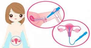
The term “separate diagnostic curettage” involves the removal of the surface layer of the endometrium and separately the cervical mucosa, since different pathological processes that require a different therapeutic approach can simultaneously appear and coexist in different parts of the organ - the body and cervix.
Lecture “How to avoid becoming a victim of a gynecologist”
Most women in their lives are faced with a situation where the gynecologist, after an examination, prescribes curettage. “cleaning” among themselves
Not all patients are told in an accessible form what this operation is, and this ignorance gives rise to unfounded worries.
Let's figure it out
.
- What is scraped out (a little anatomy)?
- Explanation of names
- Why is curettage performed?
- What preparation for curettage
- How does scraping happen?
- Complications of curettage
- What's next?
What is scraped out (a little anatomy)?
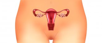
The uterus is a muscular organ shaped like a “pear”, in which there is a cavity that communicates with the external environment through the cervix, which is located in the vagina. The uterine cavity is the place where the fetus develops during pregnancy. The uterine cavity is lined with mucous membrane (endometrium). The endometrium differs from other mucous membranes (for example, in the oral cavity or in the stomach) in that it is capable of attaching a fertilized egg to itself and giving rise to the development of pregnancy.
During the entire menstrual cycle, the lining of the uterus (endometrium) thickens, various changes occur in it, and if pregnancy does not occur, it is rejected in the form of menstruation and begins to grow again in the next cycle.
During curettage, it is the mucous membrane of the uterus - the endometrium - that is removed, but not the entire mucous membrane is removed, but only the superficial (functional layer). After curettage, a germinal layer of the endometrium remains in the uterine cavity, from which a new mucous membrane will grow.
For example, every autumn a rose bush is cut at the root and in the spring a new rose bush grows from this root. In fact, curettage is similar to regular menstruation, only performed with an instrument. Why this is done - read below.
During this operation, the cervical canal (the place where the entrance to the uterus is located) is also scraped. This is where the curettage procedure usually begins - the mucous membrane that lines this canal also down to the germ layer is scraped off. The resulting scraping is sent for examination separately.
Explanation of names
Scraping
- this is the main action during manipulation, but the manipulation itself can have different names.
Russian Far East
– separate diagnostic (sometimes an addition: therapeutic and diagnostic) curettage of the uterine cavity. The essence of this name: will be fulfilled
- separate
(first curettage of the cervical canal, then the uterine cavity) - treated-diagnostic
- the resulting scraping will be sent for histological examination, which will allow an accurate diagnosis to be made, “treated” - since in the process of curettage the formation (polyp, hyperplasia) for which it was prescribed is usually removed. - curettage
- description of the process.
RDV+ GS
– separate diagnostic curettage under hysteroscopy control is a modern modification of curettage. Conventional curettage is performed virtually blindly. When using hysteroscopy (“hystero” - uterus; scopia - “look”), the doctor inserts a device into the uterine cavity with which he examines all the walls of the uterine cavity, detects the presence of pathological formations, then performs curettage and finally checks his work. Hysteroscopy allows you to evaluate how well the curettage was performed and whether there are any pathological formations left.
Why is curettage performed?
Curettage is carried out for two purposes: to obtain material
(scraping of the mucous membrane) for histological examination - this allows for a final diagnosis; the second goal is to remove the pathological formation in the uterine cavity or cervical canal.
Diagnostic purpose of curettage
- If a woman’s ultrasound shows changes in the mucous membrane, ultrasound does not always allow an accurate diagnosis; most often we see signs indicating the presence of a pathological process. Sometimes ultrasound is performed several times (before and after menstruation). This is necessary in order to be sure that the pathological formation actually exists and is not just a variant of the structure of the mucous membrane only in this cycle (an artifact). If the formation that was found remains after menstruation (that is, rejection of the mucous membrane), then it is a true pathological formation, it has not been rejected along with the endometrium, curettage should be performed.
- If a woman has heavy, prolonged menstruation with clots, intermenstrual bleeding, pregnancy and other, rarer conditions do not occur for a long time, and according to ultrasound and other research methods it is not possible to establish the cause
- If there are suspicious changes on the cervix, a diagnostic curettage of the cervical canal is performed
- Before a planned gynecological operation
or procedure for uterine fibroids, in which the uterus will be preserved.
Therapeutic purpose of curettage
- Mucosal polyps (polyp-like growths of the uterine mucosa) - there is no other type of treatment, they do not disappear with medication or on their own (there will be a separate article on the site)
- The hyperplastic process of the endometrium (hyperplasia) - excessive thickening of the uterine mucosa - is treated and diagnosed only by curettage, followed by drug therapy or instrumental methods (there will be a separate article on the site)
- Uterine bleeding – the cause may not be known. Curettage is performed to stop bleeding.
- Endometritis is an inflammation of the uterine mucosa. For complete treatment, the mucous membrane is first scraped off.
- Remains of membranes and embryonic tissues - treatment of complications after abortion
- Synechia - fusion of the walls of the uterine cavity - is performed using a hysteroscope and special manipulators. Under visual control, adhesions are dissected
How to prepare for curettage?
If curettage is not performed for emergency reasons (as, for example, during uterine bleeding), but as planned, the operation is performed before menstruation, a few days before its onset. This is necessary so that the curettage process itself practically coincides in terms of the physiological period of rejection of the uterine mucosa (endometrium). If you plan to undergo hysteroscopy with removal of a polyp, the operation, on the contrary, is performed immediately after menstruation so that the endometrium is thin and the location of the polyp can be accurately seen.
If curettage is carried out in the middle of the cycle or at the beginning, this can lead to prolonged bleeding in the postoperative period. This is due to the fact that the mucous membrane of the uterus grows synchronously with the growth of follicles in the ovaries - if the mucous membrane of the uterine cavity is removed significantly before the onset of menstruation, the hormonal background created by the ovaries will “come into conflict” with the absence of the mucous membrane and will not allow it to fully grow . This condition is normalized only after synchronization between the ovaries and the mucous membrane occurs again.
It would be logical to propose curettage during menstruation, so that the natural rejection of the mucous membrane coincides with the instrumental one. However, they do not do this, because the resulting scraping will not be informative, since the rejected mucous membrane has undergone necrotic changes.
Tests before curettage (basic set):
- General blood analysis
- Coagulogram (assessment of the blood coagulation system)
- ECG
- Tests for hepatitis B and C, RW (syphilis) and HIV
- Vaginal smear (there should be no signs of inflammation)
On the day of curettage, you need to come on an empty stomach, the hair in the perineum should be removed. You bring a robe, long T-shirt, socks, slippers and pads.
How does curettage occur?
You are invited to a small operating room, where you sit on a table with legs, like a gynecological chair. The anesthesiologist will ask you about your previous illnesses and any allergic reactions to medications (prepare for these questions in advance).
The operation takes place under intravenous anesthesia - this is a type of general anesthesia, but only it is short-term, on average 15-25 minutes.
After the drug is injected into a vein, you immediately fall asleep and wake up in the ward, that is, you sleep throughout the operation and do not experience any unpleasant sensations, but on the contrary, you may have sweet dreams. Previously, heavy drugs were used for anesthesia, which caused very unpleasant hallucinations - now they are no longer used, although the skill of the anesthesiologist in administering anesthesia is of great importance.
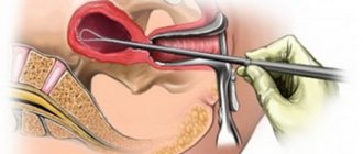
The operation itself is performed as follows. The doctor inserts a speculum into the vagina to expose the cervix. Using special forceps (“bullet pins” there is a tooth at the ends of this instrument) it catches the cervix and fixes it. This is necessary to ensure that the uterus remains motionless during the procedure - without fixation, it easily moves, as it is suspended by ligaments.
Using a special probe (iron rod), the doctor enters the cervical canal and penetrates the uterine cavity, measuring the length of the cavity. After this, the stage of cervical dilatation begins. Extenders are a set of iron sticks of varying thickness (in ascending order from the thinnest to the thickest). These sticks are alternately inserted into the canal of the cervix, which leads to a gradual expansion of the canal to a size that freely passes the curette, the instrument used to perform curettage.
When the cervical canal is dilated, the mucous membrane of the cervical canal is scraped. This is done with the smallest curette. A curette is an instrument similar to a spoon with a long handle, one edge of which is sharpened. A sharp edge is used to scrape. The scraping obtained from the cervical canal is placed in a separate jar.
If curettage is accompanied by hysteroscopy, then after dilation of the cervical canal, a hysteroscope (a thin tube with a camera at the end) is inserted into the uterine cavity. The uterine cavity and all walls are examined. After this, the lining of the uterus is scraped. If a woman had polyps
– they are removed with a curette during the curettage process. After the curettage is completed, the hysteroscope is reinserted and the result is checked. If something remains, reinsert the curette and scrape it out until the result is achieved.
Some formations in the uterine cavity cannot be removed with a curette (some polyps, synechiae, small myomatous nodes growing into the uterine cavity
), then special instruments are inserted into the uterine cavity through
a hysteroscope
and, under visual control, these formations are removed.
After the scraping
Forceps are removed from the cervix, the cervix and vagina are treated with an antiseptic solution, ice is placed on the abdomen so that under the influence of cold the uterus contracts and the small blood vessels of the uterine cavity stop bleeding. The patient is transferred to the ward, where she wakes up.
The patient spends several hours in the ward (usually sleeping, with ice on her stomach) and then gets up, gets dressed and can go home (if this is not a day hospital, but a hospital, discharge is carried out the next day).
Thus, curettage occurs without any painful or unpleasant sensations for the woman
, takes about 15-20 minutes, the woman can go home the same day.
Complications of curettage
In general, curettage in the careful hands of a doctor is a fairly safe operation and is rarely accompanied by complications, although they do occur.
Complications of curettage:
- Perforation of the uterus
- the uterus can be perforated using any of the instruments used, but most often it is perforated with a probe or dilators.
Two reasons: the cervix is very difficult to dilate, and excess pressure on the dilator or tube causes it to pierce the uterus; Another reason is that the uterus itself can be greatly changed, which makes its walls very loose - because of this, sometimes the slightest pressure on the wall is enough to pierce it. Treatment:
small perforations heal on their own (observation and a set of therapeutic measures are carried out), other perforations are sutured and surgery is performed. - Cervical tear
– The cervix is most often torn when the bullet forceps fall off.
Some cervixes are very “flabby” and bullet forceps do not hold well on them - at the moment of tension, the forceps fly off and tear the cervix. Treatment:
small tears heal on their own; if the tear is large, stitches are applied. - Inflammation of the uterus
- this occurs if curettage was performed against the background of inflammation, the requirements of septic and antiseptics were violated, and a prophylactic course of antibiotics was not prescribed.
Treatment:
antibacterial therapy. - Hematometra
is an accumulation of blood in the uterine cavity.
If, after curettage, a spasm of the cervix occurs, blood, which normally should flow from the uterine cavity for several days, accumulates in it and can become infected and cause pain. Treatment
: drug therapy, bougienage of the cervical canal (spasm relief) - Damage to the mucous membrane
(excessive curettage) – if you scrape very hard and aggressively, you can damage the germinal layer of the mucous membrane, which will lead to the fact that the new mucous membrane will no longer grow.
A very bad complication - practically untreatable.
In general, complications can be avoided if this operation is performed carefully and correctly.
.
Complications of curettage include situations when, after this operation, the entire pathological formation (polyp, for example) or part of it remains in place. More often this happens when curettage is not accompanied by hysteroscopy
, that is, it is impossible to evaluate the result at the end of the operation. In this case, curettage is repeated, since it is impossible to leave the pathological formation in the uterine cavity.
What's next?
After curettage, you may have spotting and spotting for several days (from 3 to 10). If the bleeding immediately stops and abdominal pain appears, this is not very good, since there is a high probability that a spasm of the cervical canal has occurred and a hematometra
.
You must immediately contact your doctor
and inform him about this. He will invite you for an ultrasound and if the spasm is confirmed, they will quickly help you.
To prevent hematomas in the first days after curettage, you can take No-Spa 1 tablet 2-3 times a day.
In the postoperative period, you should be prescribed a short course of antibiotics
– this is necessary to prevent inflammatory complications.
The results of histological examination are usually ready 10 days after surgery, do not forget to pick them up and discuss them with your doctor.
In conclusion, I would like to note that curettage is one of the most frequent and most necessary minor operations in gynecology
. It is indispensable in the treatment and diagnosis of some gynecological diseases. Now this operation is very comfortable and can probably be called one of the most comfortable interventions available in gynecology, since you do not experience pain or discomfort. Of course, if you get to a careful gynecologist and anesthesiologist.
Indications and contraindications
The indication for hysteroscopy with RDV is any pathology of the endometrium, which often manifests itself with bleeding outside the menstrual cycle - acyclic or after menopause. Ultrasound allows you to preliminarily determine the nature of pathological changes and their localization; an accurate diagnosis can only be obtained by studying a piece of tissue under a microscope. Inspection of the cavity through the eyepiece of a hysteroscope helps to assess the extent of the lesion and the volume of pathological tissue for curettage.
The manipulation will provide information about the causes of infertility and menstrual irregularities, and at the same time as diagnosis, destroy adhesions in the fallopian tubes that interfere with the movement of sperm.
There is a need for hysteroscopy with RDV when removing synechiae - pathological adhesions of the mucous membrane that disrupt endometrial rejection and prolong menstrual bleeding. Under visual control, the exact location and optimal depth of the treatment effect is determined.
Taking a biopsy after long-term hormonal treatment will demonstrate the achieved result and allow you to decide on a further strategy.
Contraindications to hysteroscopy with RDV:
- inflammatory processes in the genital organs in the active phase;
- infections or severe pathology of other organs with significant dysfunction;
- blood clotting disorders, including with excessive formation of blood clots;
- pregnancy;
- extensive cancerous lesion of the cervix with fusion of the cervical canal.
Methodology
Hysteroscopy with RDV is performed under short-term intravenous anesthesia in a small operating room. The woman is placed on a gynecological chair, the external genitalia and the perineal area are treated with a disinfectant solution. Spoon-shaped speculums are inserted into the vagina. The neck is secured by the anterior lip with bullet forceps. The upper mirror is removed, the lower one is held by the operating nurse. Using a small sharp curette No. 1, the cervical canal is scraped out without inserting the instrument into the internal os. The scraping is placed in a test tube with formaldehyde, and the tube is sent to the histology laboratory. A smear is applied to a glass slide for cytological examination.
To determine the length and position of the uterus, probing of the uterine cavity is performed. A metal probe is inserted through the cervical canal to the fundus of the uterus. To carry out hysteroscopy with RDV, it is necessary to dilate the cervical canal, which is carried out with Hegar dilators up to No. 10-10.5. The dilators are advanced beyond the internal os, starting with small sizes. A hysteroscope tube is inserted into the uterine cavity. In practice, liquid hysteroscopy with a single-channel fluid supply and its independent outflow through the dilated cervix is more often used. After the expansion of the uterine cavity with fluid, the gynecologist sequentially examines the walls, bottom and area of the mouths of the fallopian tubes. Polyps, hyperplasia, cancer, foreign bodies, fibroids and other diseases of the uterus have characteristic hysteroscopic signs that are easily detected during examination.
After determining the localization and extent of the pathological process of the endometrium, the hysteroscope is removed. Next, the uterine cavity mucosa is scraped, removing the entire functional layer. Using sharp curettes No. 6.4, scrape out all the walls in the direction from the bottom to the internal pharynx. Control curettage of the uterine angles, where polyps are most often located, is carried out with a small curette No. 2. If the scraping is abundant with blood clots, it is washed and placed in a separate tube with formaldehyde. To control the completeness of curettage, the hysteroscope is reinserted and the entire uterine cavity is examined. The instruments are removed and the cervix is treated with iodine. The duration of hysteroscopy with RDV is on average 20-30 minutes.
Preparation
Preparation for hysteroscopy with RDV is standard - as for minor gynecological operations, these are detailed tests:
- blood and urine
- on the microflora of the genital tract,
- for common infections (HIV, hepatitis, syphilis).
This implies a complete gynecological examination and for anesthesia, an ECG and fluoroscopy of the lungs, consultation with a therapist and other specialists are required, if the state of health requires it.
On the eve of the manipulation, the colon is cleansed; before the intervention, you must refuse to eat.
Hysteroscopy with RDV requires a short hospital stay for optimal preparation and a calm attitude. The best time is three to four days before the expected menstruation, when, under the influence of hormones, the cervical canal has expanded as much as possible, and the endometrium is almost ready for rejection.
On what day of the menstrual cycle is it done?
RDV in gynecology is a procedure that is performed on a certain day of the menstrual cycle. Only in this case will the data obtained be as informative as possible. However, the exact day or period for performing RDV depends on the risk of complications and pathology that the doctor suspects. Symptoms are also taken into account.
For example, if a patient has uterine bleeding, then diagnostic and therapeutic measures begin immediately, regardless of the day of the menstrual cycle. If there is a suspicion of menstrual dysfunction, then a study of this type is recommended to be carried out in the period from 5 to 10 days of the cycle.
Also, the menstrual cycle can occur without ovulation. In this case, RDV is carried out 2-3 days before the start of menstruation itself.
If there is a suspicion of tumor development, then, as a rule, curettage is performed as quickly as possible. Doctors prefer not to wait for a specific day of the cycle, but to begin treatment as soon as possible.
How is hysteroscopy with RDS performed?
The patient is taken to a small operating room, the manipulation is performed on a gynecological chair.
The woman is put under brief anesthesia - half an hour is enough for the manipulation.
It begins, like any gynecological examination, with the installation of gynecological speculum and treatment of the genital tract with antiseptics. The cervix is fixed with special forceps, its canal is expanded to the required diameter, then the hysteroscope is inserted. For a high-quality inspection, gas is pumped inside, straightening the walls of the cavity. In a certain order, all the walls are examined and the image is recorded on a disk; a biopsy is taken from places suspicious for pathology.
The hysteroscope is removed and RDV is started, collecting the mucous membrane of the cervix and cavity in different jars.
Recovery period
A woman can leave the hospital in the evening if there are no contraindications from other organs. For prophylaxis, antibiotics and pain relief with nonsteroidal anti-inflammatory drugs (NSAIDs) are prescribed.

For several days, there is discharge from the genital tract, initially blood-colored, becoming lighter every day. Their volume is individual, but not comparable to menstruation. If a smell appears, you should immediately consult a doctor.
A woman needs to give up sex for at least a week, avoid douching, and limit physical activity.
How to prepare for the procedure?
After undergoing a gynecological examination, the patient receives a referral from the doctor for the procedure. If a few days before the RDV the patient begins to experience vaginal discharge, this may indicate thrush. You must inform your doctor about this fact.
There are several important rules for preparing for the procedure:
- 6 hours before the RDV, provided that intravenous anesthesia will be administered, you must completely eliminate the intake of milk and any fermented milk products. You should not drink juices, especially those with pulp.
- 4 hours before the procedure, you should refuse any drink, including water.
- 10-12 hours before a diagnostic and therapeutic study, you should not eat food. If this rule is violated, mechanical asphyxia may be caused during the procedure.
- You should stop taking medications for 2 weeks. If there are medications that need to be taken in a course, or a woman cannot refuse medications for health reasons, she must inform the gynecologist.
- Before the Russian Far East, you cannot dye your hair, get your nails extended, or do any cosmetic procedures using chemicals. It is also recommended to avoid using decorative cosmetics.
- In the morning, on the day of the operation, you need to remove hair from the pubic area, perform a cleansing enema and carry out standard hygiene measures.
You cannot come to the procedure while driving a vehicle yourself. During the RDV process, drugs are introduced into the patient’s body that will greatly inhibit the reaction. Therefore, you cannot drive for the next 24 hours.
Some anesthesiologists ask to purchase special compression stockings for the procedure. Therefore, it is worth checking this issue with your doctor first.
The first few hours after curettage, the woman should rest in a hospital setting, so it’s worth taking a book with you and charging your mobile phone. It is recommended to lay a special absorbent film on the bed. Often after the procedure, a woman experiences slight bleeding.
Possible complications
An unpleasant consequence is that the lifting forceps coming off the “flaccid” neck can injure it. The tears are small and do not hurt, but later the neck may become deformed with scars.
The introduction of gas into the uterine cavity can cause short-term arrhythmia due to its absorption and changes in the gas balance of the blood.
Excessive scraping of the mucosa, which removes the endometrial growth zone, as well as bleeding from a damaged vessel, can subsequently lead to scars and synechiae.
The most dangerous thing is a breakthrough through the uterine wall into the abdominal cavity and damage to the nearby pelvic organs.
Preparatory procedures for the Russian Far East
Diagnostic curettage of the uterine cavity should be performed only in a hospital setting. Carrying out RDV can lead to various complications, and then the patient will immediately need help.
RDV of the uterus is curettage of the uterine cavity, which is carried out according to the rules of asepsis and antiseptics in a gynecology hospital.
Before scraping, you need:
- blood test for hepatitis, HIV infection, syphilis;
- clotting test, which will help avoid bleeding after medical intervention;
- smear of the vaginal mucosa;
- fluorography;
- electrocardiogram.
Surgery is performed before eating, since the intestines must be free. It is advisable to empty the bladder as well. This preparation will help the patient feel more comfortable during RDV.
Alternative methods
Today there is nothing to replace diagnostic hysteroscopy with RDV, but treatment without RDV is possible and has advantages for some pathological conditions of the uterine mucosa.
Modern hysteroscopes allow the use of laser and electric current to remove large amounts of tissue; the disadvantage is that nothing remains for analyzing the cell structure under a microscope, because the entire pathological substrate is “burned out”.
With electrical destruction, the likelihood of bleeding is higher, with laser ablation it is lower than with conventional RDV. Laser ablation virtually eliminates perforation of the uterine wall.
In terms of the stages of manipulation, hysteroscopy using modern alternative technologies is no different from the standard RDV.
Our clinic conducts a full range of gynecological examinations; we will choose the most optimal and informative for you, making your stay within the walls of our clinic not only healthy, but also enjoyable.
Book a consultation around the clock +7+7+78
Anesthesia
The RDV procedure is quite painful, so general anesthesia is used during the procedure. Local anesthesia is allowed extremely rarely, if it is impossible to use standard anesthesia. With local anesthesia, it is impossible to completely eliminate pain.
Sometimes spinal anesthesia is used. In this case, during the RDV process, the patient remains conscious, but her lower body is devoid of sensation. However, this method of using anesthesia is associated with certain risks, so if possible, doctors do not recommend using this method of immobilization and dulling of sensitivity.
Before administering anesthesia, the patient must sign a consent form.