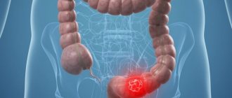
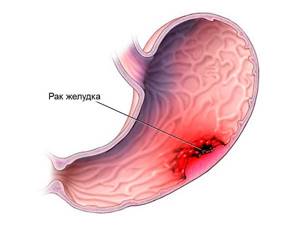
Stomach cancer kills up to 800,000 people worldwide every year. Stomach cancer ranks fourth among malignant tumors in the world. This disease has a high mortality rate, which makes it second in the structure of cancer mortality after lung cancer.
Israeli oncology clinics undertake the treatment of patients with stomach cancer at almost any stage of the disease. Of course, at the very last stage, hopes for a complete recovery tend to zero. Therefore, it is especially important to detect stomach cancer at the earliest possible stages. To do this, you need to undergo an examination - both in case of certain symptoms, and in general for people over fifty years of age.
Causes
Gastric cancer is one of the most common cancers in the world, characterized by a long asymptomatic course and high mortality. The exact causes of the tumor are not known, but scientists have identified the prerequisites for its development:
- hereditary predisposition;
- unhealthy diet (violation of its regime; eating large amounts of animal fat, carcinogenic foods; low content of fresh vegetables and fruits in the diet);
- occupational hazards;
- bad habits (smoking doubles the risk of developing gastric dysplasia);
- Helicobacter pylory infection;
- the presence of background precancerous processes (chronic gastritis, especially with atrophy of the mucous membrane, polyps, Ménétrier’s disease, pernicious anemia);
- operations on the stomach.
Help Stomach cancer does not occur against a background of complete health. Its development is preceded by many years of exposure to unfavorable external and internal factors.
Stages
According to the degree of malignancy and prevalence of the process, the stages of the disease are distinguished:
- 0 – single atypical cells are found only in the upper layer of the gastric mucosa;
- I – tumor cells affect the mucous and submucosal layers of the gastric wall.
Stage I is divided into substages:
- IA – only the submucosal layer is affected;
- IB – the muscular layer of the stomach is also affected, metastases were detected in 1-2 nearby lymph nodes.
Stage II is divided into substages:
- IIA – the tumor spreads: to the submucosal or muscular layers of the gastric wall, metastases were detected in 1-2 nearby lymph nodes;
- submucosal layer with metastases in 3-6 nearby lymph nodes.
- IIB – the tumor spreads: to the outer layer of the gastric wall;
- subserous layer of tissue under the outer membrane with metastases in 1-2 nearby lymph nodes;
- muscular layer of the gastric wall with metastases in 3-6 nearby lymph nodes;
- submucosal layer of the gastric wall with metastases in 7 or more nearby lymph nodes.
Stage III is divided into substages:
- IIIA – the tumor spreads: to the outer layer of the gastric wall with metastases in 1-2 nearby lymph nodes;
- serous layer of tissue under the outer membrane with metastases in 3-6 nearby lymph nodes;
- the muscular layer of the gastric wall with metastases in 7 or more nearby lymph nodes.
- to nearby organs - spleen, liver, diaphragm, kidney, adrenal gland, pancreas, transverse colon or small intestine with metastases to 1-2 nearby lymph nodes;
- to nearby organs - spleen, liver, diaphragm, kidney, adrenal gland, pancreas, transverse colon or small intestine with metastases to 3 or more lymph nodes;
Signs

The initial phase of the tumor process is often hidden and asymptomatic . Moreover, patients often do not pay attention to their condition (attributing poor health to fatigue, gastritis, etc.) and do not seek medical help for a long time. The first signs of the disease include:
- general weakness, fatigue, decreased performance (for several months or even years);
- vague dyspeptic disorders (nausea, belching, lack of satisfaction after eating);
- poor appetite;
- selectivity in food and aversion to certain types of food (for example, meat);
- discomfort in the stomach (feeling of heaviness, fullness after eating), less often - pain;
- weight loss for no apparent reason;
- persistent decrease in hemoglobin level in the blood;
- pale skin;
- apathy, indifference to what is happening around.
In some cases, these symptoms are absent, and the disease manifests itself with bloody vomiting or sudden dysfunction of the digestive tract.
In the later stages, patient complaints become more pronounced:
- dull pain in the stomach area after eating or at night, not related to the nature of the food;
- nausea, vomiting of gastric contents, such as “coffee grounds” (due to the presence of blood);
- belching, heartburn;
- increasing weakness;
- lack of appetite;
- weight loss.
Stomach cancer
Gastritis
3604 02 September
IMPORTANT!
The information in this section cannot be used for self-diagnosis and self-treatment.
In case of pain or other exacerbation of the disease, diagnostic tests should be prescribed only by the attending physician. To make a diagnosis and properly prescribe treatment, you should contact your doctor. Stomach cancer: causes, symptoms, diagnosis and treatment methods.
Malignant neoplasms of the stomach occupy one of the leading places in terms of prevalence among cancer diseases. Atypical cells develop from the epithelium of the gastric mucosa, subsequently forming a malignant tumor. Nonspecific (characteristic of other diseases) symptoms of stomach cancer lead to late diagnosis. Therefore, the presence of dyspeptic disorders (nausea, vomiting, etc.), discomfort in the stomach, decreased appetite, anemia and unintentional weight loss is a reason to visit a gastroenterologist. Very often, stomach cancer develops against the background of atrophic gastritis and gastric ulcer.
The bacteria Helicobacter pylori play a significant role in its occurrence.
Causes of stomach cancer
Impaired division of epithelial cells in the gastric mucosa leads to the appearance of atypical cells that contribute to the development of a tumor. Factors whose influence leads to the development of malignant tumors are called carcinogenic.
External carcinogenic factors include nitro compounds, which are formed from nitrites and nitrates found in food products. Even in small quantities, these substances provoke a tumor process. Vitamin C prevents the formation of nitro compounds in the body. Also, the formation of nitro compounds from nitrates slows down with increased stomach acidity. In contrast, low acidity is accompanied by an increase in the number of nitrite-producing bacteria and an increase in the level of carcinogenic nitro compounds.
A decrease in the acidity of gastric juice and atrophy of mucosal cells is caused by the bacterium Helicobacter pylori, the presence of which leads to the development of a malignant process in the stomach.
External provoking factors also include the composition of the diet. Frequent consumption of too hot food, carbonated drinks, salty, smoked and canned foods, as well as foods containing dietary nitrates and nitrose-containing components, increases the risk of developing stomach cancer.
Patients with diagnoses such as chronic atrophic gastritis, gastric ulcer, Ménétrier's disease (changes in the gastric mucosa with subsequent development of cysts and adenomas) should be regularly observed by a gastroenterologist and carefully examined for the appearance of tumors in the stomach.
Classification of stomach cancer
Depending on the clinical objectives, several classifications of gastric cancer are used. First of all, the tumor is classified according to its location - in the fundus, body of the stomach, in the vestibule of the pylorus or in the pylorus itself, on the lesser or greater curvature of the stomach. In addition, the histological type of the tumor is specified, which must be taken into account when prescribing therapy, since each type has biological and structural features. Some types of tumors are considered more aggressive and difficult to treat. Thus, to determine treatment tactics, it is necessary to establish the type of tumor, which is possible after a biopsy.
Depending on the biological activity and the primary affected cells, intestinal, diffuse and mixed types of gastric cancer are distinguished. The intestinal type is similar to the structure of an intestinal tumor. The specificity of the diffuse type is its rapid growth and the ability to affect nearby organs. This tumor is aggressive and has a high tendency to metastasize. The mixed type combines the characteristics of the previous two.
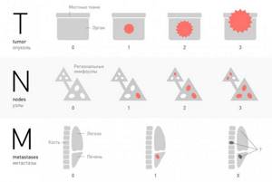
Cancer is also classified according to the TNM system (from the English tumor, nodus and metastasis - stages of tumor development), where the size of the tumor, damage to the lymph nodes and the presence of metastases are assessed.
Symptoms of stomach cancer
In the early stages, malignant tumors do not have characteristic symptoms.
However, doctors identify minor oncological signs of stomach cancer, and if they appear, it is recommended to consult a doctor.
These include decreased appetite and the appearance of dyspeptic disorders (feelings of heaviness, fullness in the stomach, belching, nausea). The progression of the disease leads to asthenia (severe weakness), anemia, which causes pallor, shortness of breath, and weight loss. If the tumor is localized in the cardiac (upper) part of the stomach, dysphagia (impaired swallowing) may occur. Cancer of the pyloric part of the stomach is accompanied by a violation of the movement of food into the duodenum and can cause vomiting of undigested contents (or food eaten the day before). If blood vessels are involved in the cancer process, the vomit and feces will be black. This type of vomiting is called “coffee grounds,” and loose, tarry stools are called melena. Pain and the inability to digest food lead to sudden weight loss and weakness of the patient.
Pain in the stomach appears already in the later stages of the disease. In this case, girdle pain (from the back) may indicate metastases to the pancreas, and pain in the heart area may indicate growth into the diaphragm. If metastases have spread to the intestines, symptoms may include bloating, rumbling and constipation. The disintegration of the tumor is accompanied by vomiting “coffee grounds” or blood.
Diagnosis of stomach cancer
When dyspeptic disorders or discomfort in the stomach appear, patients are referred primarily to gastroscopy, which allows one to see areas of altered gastric mucosa and perform a biopsy (taking biomaterial for histological examination).
Classification
Before proceeding with the classification of stomach cancer, it should be noted that it can grow into the lumen of the organ (exophytic growth) or spread deep into its wall (endophytic growth). These features determine the clinical picture of the disease and the possibility of its detection by various diagnostic methods. For example, with endophytic growth, a tumor may not cause any symptoms for a long time and is more difficult to detect during endoscopy. In such cases, X-ray diagnostics is especially important.
Currently, there are several classifications of stomach cancer. Let us dwell on those of them that are important in the process of primary diagnosis.
Table 1. Main types of cancer according to the classification of A. S. Holdin.
| Tumor types | a brief description of | X-ray changes |
| Fungiform and polypoid | Tumor nodes on a narrow stalk or wide base. | Round filling defects with clear, even contours. They are difficult to distinguish from each other and from a simple polyp. |
| Cabbage-shaped | Lumpy neoplasms resembling cauliflower in appearance. | They are manifested by a filling defect with bumpy contours, which is clearly demarcated from the intact mucous membrane and protrudes deeply into the lumen of the organ. |
| Cup or saucer shaped | Tumors with raised edges and a disintegrating central part. They have a tumor-like shaft that separates the neoplasm from the healthy mucous membrane. | Filling defects are oval or round in shape with smooth and clear contours; in later stages, the tumor may ulcerate in the center, which creates a depot of barium suspension with uneven outlines. |
| Ulcerative-infiltrative | Partial infiltrative germination of all layers of the stomach with ulceration. | Uneven contours of the organ; weakening of peristalsis; malignant relief of the mucous membrane - shapeless accumulations of contrast, alternating with structureless zones |
| Diffuse | They affect the wall of the stomach over a large area and disrupt its motor activity. | Thickening and rigidity of the stomach wall; its deformation and change in size; narrowing of the lumen of the organ. |
Of these, the first 3 options relate to limited growing forms of cancer (exophytic), and the last 2 – to infiltratively growing tumors (endophytic). There are also mixed forms that combine features characteristic of its various types.
international classification TNM
To assess the extent of the process, the international TNM classification is used, where T- characterizes the condition of the tumor itself:
- The neoplasm affects the wall of the stomach to the submucosal layer (T₁).
- The tumor grows to the subserosal layer (T₂).
- This formation grows into the serosa, but does not spread to the surrounding structures (T₃).
- The pathological process involves nearby organs (spleen, liver, pancreas, intestines, adrenal gland) or the walls of the abdominal cavity (T₄).
- In the event that the primary focus is not determined, the stage of the process corresponds to T₀.
- There is also a concept called TIS, which means “carcinoma in situ” or “cancer in situ” (occurs inside the epithelium and does not spread, can only be detected by biopsy).
Please note: If cancer is detected at stage Tіs, then with adequate treatment, patient survival approaches 100%.
N – damage to regional (where lymph flows from the stomach) lymph nodes:
- are not involved in the pathological process (N₀);
- affected in quantities of 1-2 (N₁);
- metastases in 3-6 lymph nodes (N₂);
- pathological change in 7 or more lymph nodes (N₃).
M – presence of distant metastases (accordingly, if there are none - M₀, if there are - M₁).
4 stages of stomach cancer
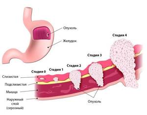
Taking into account all these characteristics, 4 stages of the disease are distinguished. Let us briefly describe them from a clinical point of view:
- Stage 0 : corresponds to Tіs; the disease does not manifest itself clinically and is not detected by conventional research methods.
- Stage 1 : the extent of the tumor corresponds to T₁ or T₂ without involvement of the lymph nodes (N₀), as well as T₁ with a single lesion (N₁). During this period, with the help of modern treatment methods, good results can be achieved.
- Stage 2 : occurs when the tumor grows into the submucosal layer of the stomach (T₁) with multiple lesions of the lymph nodes (N₃, N₂) or when the subserous layer (T₂) and several lymph nodes (N₃, N₂) are affected or when the serous membrane (T₃) and single metastases to regional lymph nodes (N₁). It has a typical clinical picture and a more serious prognosis.
- Stage 3 : characterized by the growth of tumor tissue to different depths (from T₂ to T₄) and various types of damage to the lymph nodes, but the absence of metastases. In this case, it either spreads to the surrounding tissues, or its screenings are found in a large number of lymph nodes. These patients have a poor prognosis despite treatment. Only some of them have a chance of recovery.
- Stage 4 : associated with the presence of distant metastases of any size of the primary tumor. The prognosis is severe, most patients do not survive the 5-year mark.
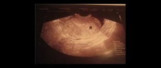
Norm
An X-ray examination of the abdominal cavity shows the stomach in the upper left region . It is partially covered by 8-10 ribs. Depending on the development of the ligamentous apparatus and the constitution of the body, its location may differ significantly in healthy patients.
The lower border of the stomach is located approximately 2-5 cm above the upper edge of the left iliac crest. The shape of the body of the stomach in asthenics is more elongated and resembles a hook . In patients with a wide body (hypersthenics), the stomach on an x-ray has the shape of a horn . The minor curvature is located on the inside and has an even contour. Large - runs from the outside, closer to the wall of the abdomen and can be uneven and jagged due to folds.
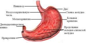
Structure of the stomach
With conventional radiography, the mucous membrane is difficult to determine. Therefore, if it is necessary to visualize it, a Barium-based contrast agent . It should not have filling defects, erosions or protrusion into the stomach cavity. The standard sizes of the stomach are collected in the following table:
| Index | Normal, cm |
| Length | 20-27 |
| Transverse size | 10-14 |
| Anteroposterior size | 8-10 |
Important In a healthy patient in an upright body position, there should be no air under the dome of the diaphragm.
Gas bubble
The gas bubble of the stomach is of important diagnostic importance - this is the area of the organ in which air accumulates . When radiography is performed, it gives a pronounced clearing in the image when compared with the surrounding structures.
If the study was carried out with the patient in an upright position, then it is located in the left hypochondrium in the area of the fundus of the stomach. Normally, it is located 1-2 cm below the edge of the diaphragm. Depending on the filling of the stomach, the gas bubble may be spherical or pear-shaped . Also noteworthy is the clear lower contour, which is located at the border of air and liquid food masses.
If radiography was carried out in a horizontal position, then the phenomenon of “pneumorelief” occurs - when gas accumulates in the highest areas of the body and the bottom of the organ. If Barium contrast is used at the same time, it clearly visualizes the gastric mucosa in this part of the stomach.
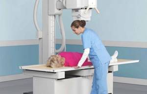
Possibility of the technique
X-rays are used to detect stomach cancer along with other diagnostic methods. It allows you to detect a tumor in the early stages, thanks to its good resolution and spatial perception. To increase its information content use:
- double contrast;
- studying the state of the organ during tight filling;
- special positions and projections.
During the procedure, the doctor is able to assess:
- location of the stomach;
- its shape and contours;
- organ size;
- displacement and motor activity;
- the condition of its inner surface;
- wall thickness.
However, X-rays alone cannot accurately diagnose cancer . His conclusion only suggests the presence of the disease in a person. In addition, there is always the possibility of missing this process, so a negative result of one test cannot exclude the diagnosis .
What does the study show?
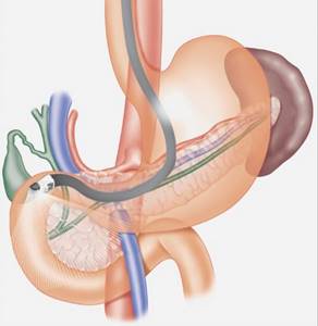
X-ray of the stomach is used not only to visualize this organ, but also neighboring anatomical structures:

lower third of the esophagus;- diaphragms;
- duodenum;
- transverse colon;
- upper abdominal cavity;
- pancreas;
- right lobe of the liver;
- lymph nodes of the upper abdominal cavity;
- spleen.
When performing a targeted X-ray of the stomach, the doctor should pay attention to the following characteristics of the organ:
- location in the abdominal cavity;
- organ shape and presence of developmental anomalies;
- surface of the mucous membrane;
- wall thickness and its structure;
- contours of the greater and lesser curvature of the stomach;
- volume of the gas bubble of the stomach;
- presence/absence of air under the diaphragm dome;
- activity of gastric peristalsis (during fluoroscopy).
Help The use of contrasts based on Barium preparations significantly increases the information content of diagnostics. However, in this case, it is necessary to prepare the patient.
Preparation

To obtain reliable information during the study, proper preparation is needed, aimed at reducing the accumulation of gases and preventing congestion in the stomach cavity. Its most important part is limiting food and water intake, since the procedure is always carried out on an empty stomach. More information about this and other features of preparing for an x-ray examination can be found in the article of the same name.
In what cases is it prescribed?
![]()
The study is prescribed by a physician of a therapeutic or surgical specialty after a patient contacts him with the following symptoms:
- a feeling of heaviness in the upper abdomen , which intensifies after a small snack;
- pain of varying intensity of a spasmodic, cutting or aching nature in the stomach area;
- the appearance of white plaque on the mucous membranes of the oral cavity;
- sensation behind the sternum or heartburn;
- a sharp decrease in appetite;
- loss of body weight;
- nausea and/or vomiting (possibly with blood or bile);
- intolerance to meat products;
- tendency to constipation or diarrhea;
- a feeling of bitter or sour taste in the mouth;
- with symptoms characteristic of a violation of the integrity of the gastric cavity (peptic ulcer or injury).
In real life, X-ray examination of the stomach is used if it is not possible to quickly carry out ultrasound diagnostics, FGDS, or MRI. Often this need arises when a patient enters the clinic on weekends or at night.
Important During pregnancy, X-rays of the stomach are strictly prohibited. For these patients, it is necessary to use alternative diagnostic methods (ultrasound, FGDS).
Decoding the results
A specialist can suspect the presence of a cancerous tumor in a person based on a combination of radiological signs:
- change in the relief of the mucous membrane in the tumor area (lack of folds, thickening, presence of various outgrowths, erosions or ulcers);
- gastric filling defect;
- the rigidity of its walls;
- the presence of a zone devoid of peristalsis;
- organ deformation;
- the undermining of its contours;
- extension of the lesser curvature;
- narrowing of the lumen and obstruction of patency.
This article will help you analyze the doctor’s conclusion in more detail.
Additional Methods
The final diagnosis of stomach cancer can be established during a comprehensive examination of the patient, which, in addition to radiography, includes other diagnostic procedures:
- endoscopic examination;
- Ultrasound of the stomach;
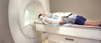
- computed tomography;
- magnetic resonance imaging;
- laparoscopy.
These diagnostic methods provide more accurate results in determining tumor lesions of the stomach and surrounding structures. However, they are not always available and are sometimes contraindicated. In such cases, x-ray examination is indispensable.
The scope of additional examination is determined by the doctor in each specific case. The most reliable way to confirm or refute the diagnosis is by EGD with taking a biopsy from suspicious areas of the inner surface of the stomach.
Additional Research
X-ray examination has limited information content , and in many situations does not allow making a final diagnosis . In such situations, the doctor prescribes additional tests:
- Ultrasound diagnostics of the abdominal organs. Allows you to detect developmental abnormalities, tumors and inflammatory processes in the stomach. There are no contraindications for use.
- Computed tomography (possibly with contrast). X-ray diagnostic method with high information content. It visualizes benign and malignant tumors and developmental anomalies well.
- Magnetic resonance imaging. Allows you to visualize the stomach and other anatomical structures of the abdominal cavity with high image quality.
- Fibrogastroduodenoscopy is an endoscopic diagnostic method that allows you to visualize the mucous membrane of the upper abdomen. Additionally, you can measure the acidity in the lumen of the stomach or take a tissue sample for cytological examination.
- Endoultrasound. A method that combines the advantages of FGDS and ultrasound.
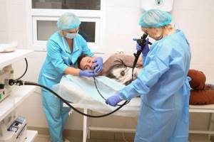
FGDS procedure
What to do if cancer is discovered?
The reaction of different people who are diagnosed with cancer may be different. Some of them become depressed, others begin to panic, and others refuse to believe doctors. But they all must understand that not a minute of precious time should be lost . After all, the earlier treatment is started, the higher its effectiveness . What should a person do if, based on the conclusion of an X-ray examination, he is supposed to have such a diagnosis?
- Firstly, do not despair , because during the diagnostic process, assumptions may not be confirmed . And if there really is cancer, then with proper treatment you can achieve success.
- Secondly, he needs to undergo a full examination using various diagnostic methods.
- Thirdly, follow all doctor's recommendations .
After a final diagnosis is made, patients with stomach cancer are prescribed treatment with the possible use of:
- surgical methods (radical intervention - removal of part of the stomach or the entire organ with regional lymph nodes; palliative operations aimed at alleviating the patient’s condition, for example, carried out to restore gastric patency);
- chemotherapy (prescribing a combination of drugs from the group of cytostatics);
- irradiation (as an adjunct to other treatment methods);
- targeted therapy (the use of drugs that have molecular properties and can selectively affect cancer cells).
The choice of treatment method is made by the doctor, taking into account the type of tumor , the stage of the pathological process and the general condition of the patient. In this case, surgical intervention is considered the most effective, which can be supplemented with chemotherapy or intraoperative radiation.
Help Inoperable patients in the later stages of the disease are prescribed chemotherapy or radiation therapy to reduce the size of the tumor, slow its growth and alleviate symptoms.