Pathological discharge from the respiratory tract is called sputum
. This is a secretory product that is secreted by epithelial cells and accumulates on the walls of the respiratory organs. Normally, there is no sputum, and the secretion produced by the respiratory system is swallowed. During illness, it becomes too much and he coughs up.
Sputum examination is necessary in the presence of a pathological process in the lungs and bronchi. The analysis will determine the causes of the pathology, the stage of the process and the nature of the disease. This study is prescribed over time, which makes it possible to assess the adequacy of therapy and adjust it, if necessary.
The attending physician may prescribe a sputum test if there is a prolonged cough, a chronic pathological process in the organs of the respiratory system, or if the diagnostic picture is unclear.
Types of sputum examination
The following types of sputum examination are distinguished:
- Macroscopic
- Microscopic
- Microbiological
- Chemical.
Go to analyzes
Macroscopic analysis allows you to evaluate the general properties and nature of sputum. The amount of sputum, its consistency, color, and smell are assessed. Also, impurities, their nature and quantity, and various fibers are studied. You can identify pus, mucous particles, serous fluid, elements of putrefaction or tissue decay, blood, fibrin fibers. These elements may be absent, found alone or in combination with each other.
Microscopic analysis gives a more accurate idea of the composition of sputum. Magnification allows you to determine the presence of cells and tissue elements. These may be traces of epithelium, leukocytes, eosinophils, erythrocytes.
Bacteriological testing helps confirm or exclude the presence of microorganisms in sputum. It could be bacteria, fungus, parasites. An analysis is also carried out for sensitivity to antibacterial therapy, which makes treatment more effective, reduces its duration and eliminates incorrect patient management tactics.
Chemical analysis of sputum is less informative. A reaction to hemosiderin is carried out, which suggests an admixture of blood. It also evaluates the acidity of the biomaterial.
Sources
- Calderwood CJ., Wilson JP., Fielding KL., Harris RC., Karat AS., Mansukhani R., Falconer J., Bergstrom M., Johnson SM., McCreesh N., Monk EJM., Odayar J., Scott PJ ., Stokes SA., Theodorou H., Moore DAJ. Dynamics of sputum conversion during effective tuberculosis treatment: A systematic review and meta-analysis. // PLoS Med - 2021 - Vol18 - N4 - p.e1003566; PMID:33901173
- Gannon AD., Darch SE. Tools for the Real-Time Assessment of a Pseudomonas aeruginosa Infection Model. // J Vis Exp - 2021 - Vol - N170 - p.; PMID:33900293
- Liu Y., Zhang L., Li HL., Liang BM., Wang J., Zhang X., Chen ZH., Zhang HP., Xie M., Wang L., Wang G., Oliver BG. Small Airway Dysfunction in Asthma Is Associated with Perceived Respiratory Symptoms, Non-Type 2 Airway Inflammation, and Poor Responses to Therapy. // Respiration - 2021 - Vol - NNULL - p.1-13; PMID:33895739
- Badi YE., Pavel AB., Pavlidis S., Riley JH., Bates S., Kermani NZ., Knowles R., Kolmert J., Wheelock CE., Worsley S., Uddin M., Alving K., Bakke PS ., Behndig A., Caruso M., Chanez P., Fleming LJ., Fowler SJ., Frey U., Howarth P., Horváth I., Krug N., Maitland-van der Zee AH., Montuschi P., Roberts G., Sanak M., Shaw DE., Singer F., Sterk PJ., Djukanovic R., Dahlen SE., Guo YK., Chung KF., Guttman-Yassky E., Adcock IM. Mapping atopic dermatitis and anti-IL-22 response signatures to Type 2-low severe neutrophilic asthma. // J Allergy Clin Immunol - 2021 - Vol - NNULL - p.; PMID:33891981
- Xu C., Zhang W., Wang X., Li Z., Guan G., Wang S., Fan N., Guo Y., Rao W., Wang J., Zheng T., Zang Y. Correlation analysis of pleural effusion and lung infection after liver transplantation. // Minerva Surg - 2021 - Vol - NNULL - p.; PMID:33890441
- Zhang X., Pang L., Lv X., Zhang H. Risk factors for bronchiectasis in patients with chronic obstructive pulmonary disease: a systematic review and meta-analysis. // Clinics (Sao Paulo) - 2021 - Vol76 - NNULL - p.e2420; PMID:33886788
- Velen K., Podewils LJ., Shah NS., Lewis JJ., Dinake T., Churchyard GJ., Reichler M., Charalambous S. Performance of GeneXpert MTB/RIF for Diagnosing Tuberculosis Among Symptomatic Household Contacts of Index Patients in South Africa . // Open Forum Infect Dis - 2021 - Vol8 - N4 - p.ofab025; PMID:33884274
- Feng JN., Gao L., Sun YX., Yang JC., Deng SW., Sun F., Zhan SY. . // Beijing Da Xue Xue Bao Yi Xue Ban - 2021 - Vol53 - N2 - p.320-326; PMID:33879905
- Hu SY., Long F., Long L., Huang WT., Fu P., Dong HB., Gan JF., Huang ZH. . // Zhonghua Yi Xue Za Zhi - 2021 - Vol101 - N15 - p.1071-1076; PMID:33878834
- McEvoy NL., Clarke JL., Mc Elvaney OJ., Mc Elvaney OF., Boland F., Hyland D., Geoghegan P., Donnelly K., Friel O., Cullen A., Collins AM., Fraughen D., Martin-Loeches I., Hennessy M., Laffey JG., Mc Elvaney NG., Curley GF. A randomised, double-blind, placebo-controlled, pilot trial of intravenous plasma purified alpha-1 antitrypsin for SARS-CoV-2-induced Acute Respiratory Distress Syndrome: a structured summary of a study protocol for a randomised, controlled trial. // Trials - 2021 - Vol22 - N1 - p.288; PMID:33874981
Types of sputum
Sputum can be classified according to several criteria. The main parameter is its character. There are the following types of sputum:
- mucous membrane
It is observed in asthma and inflammatory processes. It has a viscous consistency, transparent color, and glassy character.
- purulent
Accompanies the breakthrough of an abscess or empyema into the lumen of the bronchus. It is white in color with a tinge of yellow or green, opaque, thick, and has a characteristic odor.
- Mucopurulent sputum is produced during inflammatory processes with a bacterial pathogen. It is a viscous mass, cloudy, heterogeneous, interspersed with pus and mucus.
- bloody
May contain streaks of blood or formed clots. This occurs during the oncological process, tuberculosis. The blood may be scarlet or crimson in color, depending on the type and duration of bleeding.
- serous
Sputum of a liquid consistency is the result of sweating water from the capillaries into the lungs; it is observed when blood is retained in the pulmonary circulation, pulmonary edema. May have a pinkish color.
How is the material for analysis collected?
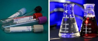
The material for research is collected in sterile containers. After coughing up the material, the container is sealed and sent to the laboratory. It is important that the sputum is fresh, otherwise the acidity changes and some of the liquid dries out. Individual spittoons with a tight-fitting lid are provided. Before transfer to the laboratory, the container with the collected material is stored in the refrigerator.
How are the analysis indicators interpreted?
Evaluation of the results is always carried out in conjunction with an analysis of the clinical picture, symptoms and other studies of the patient. For example, a small amount of sputum is submitted for analysis, but it is important to know the amount of substance secreted per day.
The presence of mucus is observed in acute bronchitis and asthma. Mucus and pus are released during bronchiectasis, pneumonia, abscess formation, and various bronchitis. Blood in the sputum is observed in severe cases of the disease, in pulmonary infarction, malignant growth, and tuberculosis. Dark color and unpleasant odor, the presence of decaying tissues indicate gangrene of the lung.
Sputum microscopy can exclude or confirm the presence of parasites. It could be roundworms, echinococcus. Pus plugs form in sputum during decay processes. Areas of tumor or lung tissue may be observed, indicating organ collapse. If a large amount of squamous epithelium appears in the sputum, most likely the material is mixed with saliva and a repeat analysis is required. Cylindrical epithelial cells in sputum accumulate during an acute inflammatory process in the respiratory tract, asthma or cancer. Leukocytes are part of purulent and mucous sputum and indicate inflammation of the corresponding nature. The presence of eosinophils is characteristic of asthma or a corresponding type of pneumonia. The cells are examined for atypicality - if any are found, tumor growth may be present. Different fibers indicate tissue breakdown. In asthma, characteristic spirals and crystals are also found. These are casts of the respiratory tract that form mucus elements.
The presence of fungus, mycelium, and bacteria indicates the presence of a corresponding infection.
Leukocytes in bile:
Bile is a substance that, in practice, is not very often taken for analysis. Previously, such a study as duodenal intubation was common, in which bile was obtained from patients for study. But now there are highly informative studies (ultrasound, MRI) that provide information about the condition of the biliary tract without the need to study their contents.
However, sometimes bile analysis is still performed. The procedure for “extracting” it is quite complex, but if it is carried out correctly, the result is three different portions of bile. They have slightly different indicators, and the norms of leukocytes, accordingly, are also different. But, in any case, the presence of leukocytes is allowed provided that there are very few of them. The norm of leukocytes in children and adults can differ significantly. However, this study is practically not carried out on children, so it is not necessary to know their norms.
Studying the indicator of leukocytes in bile is not easy: this process requires not only highly qualified specialists, but also efficiency. The fact is that within a few minutes after the bile has been received, the white blood cells in it are destroyed.
Bile
If biliary leukocytes are elevated, the reasons for this may not only be inflammation of the gallbladder or its ducts. Leukocytes are able to enter the sample from the duodenum, stomach, even from the oral cavity. So, in general, the results of bile analysis cannot be called very indicative. The study provides some information mainly if it is carried out together with other types of diagnostics.
What diseases are diagnosed using sputum analysis?
Sputum analysis is a key step in the diagnosis of a number of respiratory pathologies. Let's take a closer look at these diseases.
Acute bronchitis
Sputum begins to be produced in the first stages of the disease. At first it is mucous and viscous, but gradually acquires a mucopurulent character. The amount of separated material gradually increases. Under a microscope, leukocytes, many epithelial cells, and single red blood cells can be detected.
Chronical bronchitis
Patients with chronic bronchitis note regular expectoration of large amounts of mucopurulent sputum. Occasionally there are streaks of blood, especially after an intense cough. Alveolar macrophages, fibrinous casts of the respiratory tract, as well as representatives of the flora appear in the sputum.
Asthma
Sputum in asthma is mucous and viscous, has a glassy character. Kurshman spiral elements and Charcot-Leyden crystal fragments, eosinophils are observed.
Bronchiectasis
This pathology is characterized by a large amount of sputum, which can reach 1 liter. The discharge has a dirty, gray-green tint. If you leave sputum in a bowl for a while, it will separate into several types: mucus, pus and serous fluid. Dietrich's plugs, a significant number of leukocytes, and biochemical impurities are observed.
Pneumonia
Characteristic sputum is produced during lobar pneumonia. It has a viscous consistency, rusty color, and is released in small quantities. As the disease progresses, its quantity increases and it acquires a mucopurulent character. Among the impurities, fibrin and altered red blood cells are observed. Gradually, there are fewer red blood cells, and the number of leukocytes increases.
Lung abscess
The sputum is two-layered, contains a large amount of pus and mucus. Microscopic examination can detect leukocytes, tissue fibers, fatty acid elements, hematoidin and cholesterol. Bacteriological analysis allows you to assess the nature of the flora.
Tuberculosis
Sputum is produced in the cavernous form of the disease. This is accompanied by purulent discharge, mixed with blood and mucus. Microscopy allows you to determine the presence of okha lenses, fibers, and acid crystals. If calcified areas are observed, this indicates the disintegration of the old tuberculosis focus.
Malignant tumor
The appearance of sputum is observed during decay. It contains areas of tissue, fibers, blood, and atypical cells. Character: bloody, slimy.
As you can see, many diseases have common sputum indicators. This once again reminds us of the need for a holistic assessment of the clinical picture, in conjunction with symptoms and the results of other studies.
Leukocytes in the cerebrospinal fluid:
Liquor is a clear liquid that fills the ventricular system of the brain and the space under the membranes surrounding the brain and spinal cord. Through a puncture at a certain point on the back, this fluid can be obtained for analysis.
In the cerebrospinal fluid, the norm of leukocytes in children and adults is the same: these cells should not be in it. They are found only in diseases. What kind of diseases could these be?
For example, hemorrhage under the membranes of the brain. In this case, the cerebrospinal fluid contains a lot of red blood cells and leukocytes, the ratio of these cells is approximately 700:1. Neutrophils are found there in large numbers during blood poisoning, bacterial, fungal infections of the central nervous system (meningitis, arachnoiditis), and toxic meningitis. Lymphocytes in the cerebrospinal fluid are found in cases of parasitic lesions (toxoplasmosis), brucellosis, viral infections, tumors, and multiple sclerosis.