Magnetic resonance imaging is a diagnostic method based on obtaining layer-by-layer “slices” of the area under study. This technique allows you to visualize different organs and parts of the body with high quality. MRI is based on the phenomenon of nuclear magnetic resonance. If we describe the principle of operation in simple words, we can say that an MRI machine is a large magnet that affects the entire human body, as a result of which the response of atomic nuclei, namely the nuclei of hydrogen atoms, changes. These changes are recorded by the device and converted into image format by special algorithms. Simply put, the method is based on the level of “saturation” of various tissues and organs with hydrogen.
- Safety
- What does an MRI show?
- Indications and contraindications
- Preparation and execution
Safety
Initially, the method was called nuclear magnetic resonance tomography. However, the mention of “nuclear” origin caused a certain fear in people after the accident at the Chernobyl nuclear power plant. Therefore, they decided to abandon this term in favor of magnetic resonance imaging. In fact, MRI, unlike X-rays or CT scans, does not expose the body to radiation. The magnetic field is quite safe. It surrounds us everywhere. Mobile phones, microwave ovens, televisions and other household appliances are a source of magnetic field, but it has less “power”.
The influence of MRI on human health has been studied since the introduction of the method into the medical field. For more than 40 years, scientists have not been able to identify negative aspects that would allow the study to be assigned a certain hazard class. This is why MRI is considered a fairly safe diagnostic method, but with some caveats. Due to the relatively short period of time, it is impossible to understand how the research affects subsequent generations. Therefore, MRI is not recommended to be performed unsupervised. Like any other type of research, it should be prescribed when indicated, and not at the request of the patient.
Indications for the study
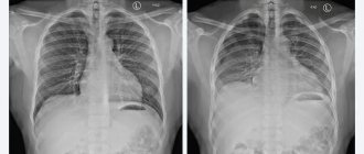
| CT | It is more often performed when examining the spine, traumatic injuries and acute conditions (for example, strokes), examining the ENT organs, chest organs and kidneys with the adrenal glands, as well as to identify metal foreign bodies. |
| MRI | It is preferable to study the brain and spinal cord, mediastinal organs (esophagus, aorta, vena cava, etc.) and pelvic organs (bladder, uterus and appendages, prostate and seminal vesicles), as well as foreign bodies of a non-metallic nature. |
What does an MRI show?
In addition to safety, magnetic resonance imaging has another important advantage - high information content. The study allows us to obtain high-quality images and study the smallest structures in detail. It is actively used in identifying diseases of the thyroid gland, kidneys, liver, spinal cord and spine, joints, blood vessels, pancreas, etc. MRI is indispensable in diagnosing:
- Neoplasms of benign and malignant nature, including metastases.
- Brain diseases.
- Diseases of the musculoskeletal system.
- Pathologies of the biliary tract, etc.
The information content of the study can be increased if it is performed using contrast agents. To do this, before or directly during the procedure, the patient is given intravenous injections of special drugs that allow them to determine the characteristics of the blood supply to the tumor, study the pituitary gland, identify demyelinating diseases, determine the exact boundaries of the tumor, etc.

Tissue sensitivity
| CT | Here the sensitivity of tissues and their ability to be reflected on the information carrier is of great importance. In this regard, it is believed that CT, which is an x-ray method, is best suited for studying bone structures and relatively contrasting soft tissues (tumors of the chest and abdominal cavity, fresh hemorrhages, etc.), as well as hollow organs that can be contrasted using special contrast agents (for example, angiography of cerebral vessels, excretory urography, intestinal contrast, etc.). |
| For MRI | characteristic very subtle and clear differentiation of soft tissues, therefore, here additional contrast is not a prerequisite for studying a number of organs (diseases, traumatic injuries and volumetric processes of the brain, spine, pelvic organs, retroperitoneal space, etc.). At the same time, it is possible to conduct magnetic resonance spectroscopy to study tissue metabolism. |
Indications and contraindications
The list of indications for MRI is huge. For each group of diseases, a characteristic list of diseases and conditions can be identified for which it is preferable to perform this study. For example, indications for MRI of head and neck vessels are:
- Suspicion of an aneurysm or malformation.
- Assessment of the degree of vascular stenosis.
- Hypertonic disease.
- Diagnosis of congenital anomalies.
- Migraine, etc.
However, it cannot be said that MRI is a unique or irreplaceable diagnostic method. There are many other studies that can help the doctor in making a diagnosis, assessing the effectiveness of treatment, or early detection of a particular pathology. The decision to include MRI in a comprehensive diagnostic plan is always made individually and only if there is such a need.
Like any other research method, MRI has a number of contraindications. They can be divided into several groups. The first group is related to the limitations of the tomograph itself. Such restrictions are usually related to the patient's weight and abdominal circumference. Most tomograph models are designed for weights up to 130-150 kg, but modern models allow examination of patients weighing 180 and even 200 kg. Moreover, in almost all cases there is a width limitation of up to 80 cm maximum. The only option for such patients is an open-type MRI, when the examination is carried out not in a “tunnel”, but on a table (like an x-ray).
The next group of indications includes conditions in which MRI cannot be performed in principle. This group includes:
- Implanted electronic devices (pacemaker, insulin pump, middle ear implants, etc.).
- The presence of hemostatic clips, which are made of ferromagnetic materials.
- The presence in the patient’s body of fragments or other metal structures made of ferromagnetic materials.
Finally, there are contraindications in which MRI can be performed, but only after certain preparation or if there is a vital need. Such contraindications include:
- Claustrophobia.
- Decompensated heart failure.
- Early pregnancy.
- Excited state of the patient.
- The need for continuous monitoring of the patient’s condition, etc.
For example, if the patient has a fear of closed spaces or if he is in an agitated state and cannot remain still in one position, then sedation is performed before the MRI.
Physical basis of the method
A water molecule consists of an oxygen atom and two hydrogen atoms. The hydrogen nucleus has its own mechanical torque. Due to the fact that it is a charged particle that has a mechanical moment, it can produce an electromagnetic field and has a magnetic moment.
The hydrogen nucleus can be likened to the needle of a compass: when hydrogen molecules are placed in a magnetic field, they will begin to orient themselves along the direction of the magnetic field. If in a calm state hydrogen molecules are randomly oriented in space, then when they are placed in a magnetic field, they line up in one direction, according to the field strength lines.
When electromagnetic pulses begin to influence particles oriented in space, hydrogen atoms absorb the energy of the pulses and move to a higher energy level.
When the external influence stops, the hydrogen atoms return to their normal state, emitting energy. High-frequency radiation that comes from hydrogen atoms is recorded by tomograph sensors and converted into an image using special software.
Preparation and execution
In most cases, MRI does not require special preparation. The exception is those cases when examination of the abdominal organs is carried out. In such situations, it is necessary to refrain from eating foods that contribute to gas formation the day before, and on the day of the MRI, you should come to the clinic on an empty stomach. In all cases, you must remove any metal objects from yourself before the examination.
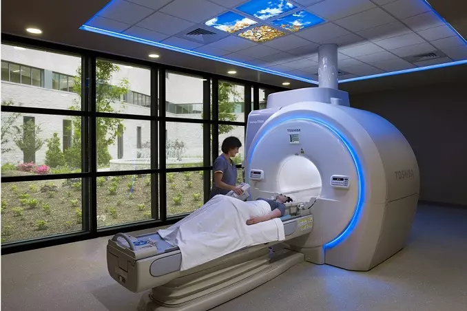
MRI is performed in the supine position. The duration of the study largely depends on the size of the scanning area. For example, an MRI of the brain lasts about 15 minutes, and an MRI of the abdominal organs takes 40-45 minutes. You must lie as still as possible during the scan. During an MRI, the patient does not feel pain or any other discomfort. Discomfort can only be associated with the relatively high noise level that is present during operation of the device. This problem can be solved using special headphones.
After completing the study, the doctor interprets the obtained images, makes a diagnosis, prescribes treatment or further examination. If necessary, the data can be recorded on removable media and taken with you.
Book a consultation 24 hours a day
+7+7+78
FAQ
Which implants can be used for MRI?
With metal-ceramic dentures, crowns. They are not considered implants. Also, the results are not affected by the intrauterine device. Any metal or ferromagnetic implants are questionable. This also applies to cochlear prostheses of the inner ear. In these cases, it is necessary to provide the doctor with an implant passport.
Is it possible to do an MRI during pregnancy?
Among the contraindications for undergoing MRI are the first 3 months of pregnancy, when the formation of fetal organs occurs. During this period, the procedure can be carried out only in case of urgent need and only with the direction of a doctor. The device does not use x-rays, as with computed tomography or x-rays. The magnetic field with which MRI works does not in any way affect the development of the fetus, and over 30 years of using the technology, no negative cases have been identified. Otherwise, MRI is the safest procedure and is used to diagnose the health of both the mother and the fetus. The device is capable of identifying a wide variety of malformations, diseases and collecting all the information about the development of the fetus.
Does MRI affect the body during breastfeeding?
The electromagnetic waves and magnetic field generated during the study do not in any way affect the body of the nursing mother and do not cause any changes in the composition of the milk. After the procedure, you can immediately begin feeding.
What is an MRI procedure?
An assistant helps the patient position himself on the machine table. One of the conditions is fixation of the examined body area to ensure immobility. For this purpose, soft belts and rollers are used. The patient does not experience discomfort; he can blink, swallow and talk with the doctor. The part of the body being examined is located in the coil of the device, which emits and receives reflected magnetic waves.
Examination of one organ or part of the body takes 30-40 minutes, but if we are talking about a complex case, it will take more time. The process itself consists of several series of images that last several minutes each. While the device is operating, you cannot move; slight movement is allowed in between, but the part of the body being examined must remain motionless. Otherwise, the next series of pictures will not coincide with the previous one. To take a series of images, the doctor will ask you to hold your breath for 10-15 seconds.
How is an MRI with contrast performed?
In some cases, solely on the direction of a doctor, the procedure involves the use of a contrast agent. It is first injected into a vein in the patient's arm through a catheter. This is an absolutely safe solution, which for some time makes the vessels of the organ being studied (for example, the brain) visible to the MRI machine. It does not cause allergic or adverse reactions. Within 24 hours, the substance is completely eliminated from the body naturally.
What does an MRI feel like?
The procedure is completely painless. The only thing that can cause a feeling of discomfort is the need to be alone in the office during the study. But the doctor will be in the control room and talk to the patient via speakerphone. Patients with fear of closed spaces also have nothing to fear. Our device leaves enough open space. In addition, the Siemens tomograph has an extremely low noise level during operation. If the study is carried out with a contrast agent, then when it is administered, the patient may feel coolness and a slight rush of blood associated with the entry of the substance into the vessels. The sensations last no more than a couple of minutes.
Do you need to prepare for an MRI?
No, no special preparation is required. But the doctor must know about your past illnesses, recent injuries, surgeries, etc. If you wear metal jewelry, you will have to remove it. If the doctor does not limit the intake of medications or food before the MRI, no changes in the usual daily routine occur.
Why do patients prefer to have MRI done at the Moscow clinic?
- Fast. A few minutes after the diagnosis, you will be able to receive the results of the examination.
- Affordable price. With the proper quality of services provided, highly qualified specialists and the availability of high-tech equipment, we try to set prices at a level accessible to our patients. The price of MRI in our clinic depends on the area being diagnosed. When compared with CT, we can say for sure that MRI is very inexpensive.
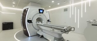
- Accurate diagnosis. MRI is performed using an American modern 1.5 Tesla tomograph, which allows you to obtain detailed information.
- Doctors are high-class specialists.
How is the examination carried out?
MRI is prescribed by the attending physician if there are appropriate indications. The patient lies down on the tomograph platform, which is pushed inside the device. During the entire procedure, the person must remain completely still. The doctor who will perform the procedure is in another room, separated by a transparent window. If you need to report discomfort or pain, you should use audio communication.
The research process is accompanied by noise and knocking. This is how any magnetic resonance imaging scanner works, so you shouldn’t be scared. The duration of the procedure is determined by the area being examined and can range from 15 minutes or more. After completing the examination, its results are recorded on electronic media and given to the patient in the form of images along with a doctor’s report.
What types of MRI scanners are there?
Based on the magnetic field strength, three types of devices are distinguished. Devices are classified into:
- Low-field (up to 1.0 Tesla). Diagnostics on such units is not always informative. Due to low power, the quality of images deteriorates. The advantage of the devices is the lower price of the procedure.
- High-field (1.5-3.0 T). Scanning is informative in 97% of cases due to the high clarity of the images. Studying the anatomical area takes less time (compared to low-field devices).
- Ultra-high-field (from 3.5 Tesla). High power provides highly detailed images and reduces scanning time.
The effectiveness of the study depends on the type of MRI device. Scanning on a low-field device sometimes requires repeating the procedure on more powerful equipment.
Depending on the design, open and tunnel tomographs are distinguished. Each variety has advantages and disadvantages
Open MRI
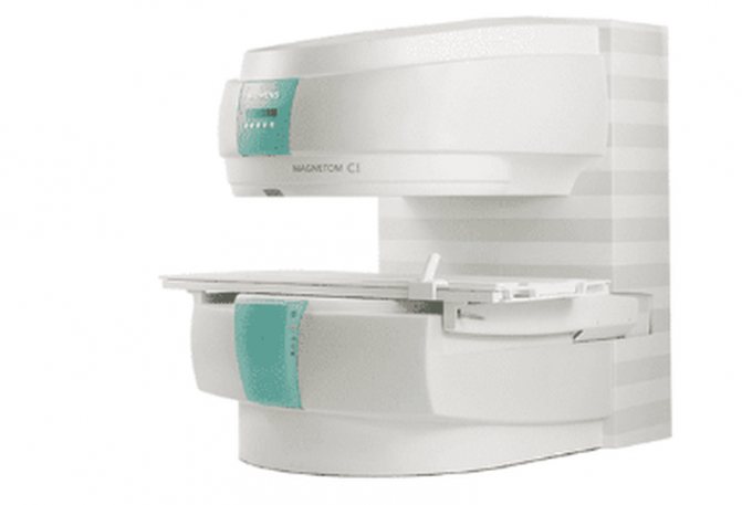
Open-type MRI scanner
The devices are distinguished by a planar arrangement of magnets. A person is placed on the platform of the apparatus, the sources of the induction field are above and below the area under consideration. Advantages of open tomographs:
- suitable for people with claustrophobia and small children;
- can be used for examining obese people (the design can withstand a weight of 200 kg);
- low noise level;
- the procedure is cheaper.
Disadvantages: less detailed and contrasty sections, the study takes longer, and may require repeated scanning on a more powerful machine.