Pro trace in urine - what is it?
All laboratory tests have terminology encrypted for the ignorant person. Everything that is called protein by the common man is designated by the doctor as pro. As a rule, this indicator is absent in tests for a healthy person, but there may be a trace value that indicates the presence of protein. Literally in the period the phrase Pro trace means traces of protein in the urine.
A general urine test is performed to diagnose the body. And a large number of meanings are subject to study. There are more than 10 of them on the form, and each is responsible for a specific pathology.
When examining urine, pay attention to protein, as it is an indicator of many diseases. Proteinuria is the name given to protein in the urine. If we look at protein molecules, we can see that these are the materials from which the entire body consists.
For reference. Protein is found everywhere - in muscles, bones, hair. He himself is involved in many processes occurring in the body. Blood, circulating throughout the body, reaches the kidney, where it begins its filtration.
And it turns out that physiologically all unnecessary substances passing through the kidneys, as if through a filter, are excreted in the urine, and the necessary ones are absorbed back into the blood. And this process lasts indefinitely as long as the renal corpuscles are intact. Once destruction or inflammation of the kidney tissue begins to occur, traces may appear in the urine, otherwise called a pro trace.
Protein gets into the urine more often than others, but this does not immediately mean pathology. As a rule, doctors force you to take tests multiple times. This allows you to study the patient's body in more detail.
Very often, traces of protein can be detected if the patient did not perform proper personal hygiene, or used an unsterile container for analysis.
References
- Diapeutics in urology / ed. A.V. Morozova. - M.: IPO "Polygran", 1993.
- Urology: textbook / ed. P.V. Glybochko, Yu.G. Alyaeva. - Rostov n/d: Phoenix, 2013. - 528 p.
- Urology. From symptoms to diagnosis and treatment. Illustrated manual: textbook / ed. P.V. Glybochko, Yu.G. Alyaeva, N.A. Grigorieva. - M.: GEOTAR-Media, 2014. - 148 p.
- Guide to urology in 3 volumes / ed. ON THE. Lopatkina. - M., 1998.
- Philip M. Hanno, S. Bruce Malkovich, Alan J. Wein. Guide to clinical urology. Clinical Manual of Urology. - M.: MIA, 2006.
- Donat MD, Herr HW Transitional cell carcinoma of the renal pelvis and ureter: diagnosis, staging, management and prognosis. In: Urologic Oncology / Eds.JE Osterling, JP Richie. - Philadelphia: WB Saunders Harcourt Brace Co, 1997. - P. 215.
- Foley SJ, Soloman LZ, Wedderburn AW et al. A prospective study of the natural history of hematuria associated with benign prostatic hyperplasia and the e?ect of ?nasteride // J. Urol. - 2000. - Vol. 163. - P. 496.
Decoding the analysis - normal values
Knowing the terminology of doctors, you can easily decipher your tests . Of course, at the first stage you should not make any diagnosis for yourself, since its confirmation requires additional research. Let's give examples of values in the analyzes and see what is considered normal:
- Red blood cells (BLd): should be absent. If found, the person is immediately sent for further examination. Acceptable BLd values are no more than 1-2 in the field of view.
- Bilirubin (Bil): It should not be detected in a healthy person. If pathological changes in the liver begin, it can be detected.
- Ketones (KET): Normally absent, if detected, it may be caused by diabetes.
- Protein (PRO): Normal is none. Detection may be caused by pathological changes in the kidney, inflammatory processes (cystitis, prostatitis).
- Bacteriuria (NIT): absent, or can be detected in small quantities. A healthy person has sterile urine. But when the process of urination occurs, microbes enter it; their number should not exceed 10,000 per milliliter of urine.
- Glucose (GLU): also should not be detected at normal levels. If it was detected, this means diabetes is possible.
- Urine acidity (pH): the norm for the human body is 5.0 – 6.0. Above these values may indicate a deviation of the thyroid gland and kidney failure.
- Density (SG): the norm for the body is 1030, more means diabetes, less than the norm causes kidney failure.
- Leukocytes (LEU): diseases associated with the kidneys and urinary tract show an increase in the content of LEU in urine.
- Urobilinogen (UBG): The urine of a healthy person may contain traces of this substance. Indicators above the norm will indicate the disease jaundice, toxic and inflammatory processes in the liver and intestines.
For reference. In addition to these values, the analysis determines the color, smell, and weight of urine, which should also be normal. A healthy person is unlikely to come to the clinic for examination, so those who undergo these tests already have inflammation in the body.
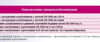
Microalbuminuria is a marker of glomerular damage
Microalbuminuria is a pathology in which the kidneys secrete the protein albumin in quantities exceeding the norm, but not reaching the level of proteinuria.
In recent years, the number of cases of end-stage renal failure (ESRD) has been continuously growing in the world. The most common causes of ESRD are diabetes and hypertension.
Microalbumin in urine is an important indicator of renal dysfunction and the possible development of nephropathy.
The main reason for the appearance of microalbumin in the urine is an increase in the permeability of the renal filter. This condition occurs as a complication of diabetes mellitus, arterial hypertension, chronic heart failure, and inflammatory kidney diseases.
Normally, a small amount of albumin is present in the urine, which is not determined by conventional methods. But if the renal glomeruli are damaged (even slightly), the content of albumin in the urine rapidly increases. As a rule, small albumins (microalbumin) are excreted first. But as the disease progresses, larger fractions of albumin are found in the urine. In this case, the analysis shows the presence of protein in the urine. Further development of the disease threatens to disrupt the general blood circulation in the kidneys, reduce their function and develop chronic renal failure.
Timely detection of microalbumin in urine makes it possible to diagnose damage to the glomeruli of the kidneys at an early stage. If treatment is started promptly, this will prevent the development of nephrotic syndrome and avoid chronic renal failure.
Patients with certain diseases and pathologies should undergo a urine test for microalbuminuria. Among them:
- Diabetes;
- Hypertonic disease;
- Heart failure;
- Lower back pain, weakness, swelling;
- Systemic lupus erythematosus;
- Hereditary forms of dyslipidemia;
- Impaired sensitivity to glucose.
The laboratory of our medical center offers a semi-quantitative method for testing urine for microalbumiuria.
Rules for collecting urine for microalbuminuria
The daily or morning average portion of urine is examined. Collecting morning urine is easier and faster.
Before collecting urine, it is important to toilet the external genitalia. Next, you need to collect the morning average portion of urine in a clean container (it is better to use special plastic containers that are sold in pharmacies). Urine should be delivered to the laboratory within 1.5-2 hours after collection.
Repeated urine testing for microalbumin over a period of 3-6 months is of diagnostic value.
Analysis reception time: from 8.00 to 14.00 (including Saturdays and holidays). Technical break 12.00-12.30. Execution time: 1 day.
Reference values
| Name | Morning average urine sample | Daily urine |
| Norm | Less than 20 mg/l | Less than 30 mg/day |
| Microalbuminuria | 20-200 mg/l | 30-300 mg/day |
| Proteinuria | More than 200 mg/l | More than 300 mg/day |
An increased content of microalbumin in urine is observed with:
- Diabetic nephropathy;
- Nephropathy associated with arterial hypertension, congestive heart failure;
- Inflammatory kidney diseases (glomerulonephritis, pyelonephritis, cystitis, urethritis);
- Amyloidosis of the kidneys;
- Polycystic kidney disease;
- Systemic lupus erythematosus;
- Sarcoidosis;
- Physical activity;
- Pregnancy;
- Hyperthermia;
- Hypothermia.
Detailed description of the study
Proteinuria is a condition characterized by increased protein excretion in the urine.
The range of proteins that are found in urine is quite diverse. These include blood plasma proteins (especially albumin), the renal epithelium's own protein (Tamma-Horsfall), and proteins of the genitourinary tract epithelium. Blood plasma is filtered through the nephron glomeruli, the structural unit of the kidneys. Further in the glomerular tubules, most of the proteins return back to the blood. Normally, proteins are retained by the kidney filter and only about 0.08 g per day enters the urine.
Proteinuria can be physiological or pathological. Physiological occurs when disorders in the body are not associated with pathology of the kidneys and urinary tract. It can occur during physical exertion, fever, hypothermia, prolonged orthostatic stress and stress. In this case, protein losses are usually minimal.
Pathological proteinuria is divided into several types.
Prerenal, or stress, occurs with increased protein synthesis in the blood or increased breakdown of proteins in tissues. The filtration barrier in the kidneys cannot cope with the reabsorption of large quantities. Prerenal proteinuria is not associated with renal pathology and occurs in malignant diseases such as myeloma or lymphoma, as well as in hemolysis.
Glomerular is associated with impaired permeability of the renal barrier. Normally, low molecular weight plasma proteins—albumin and microglobulins—are filtered through the glomeruli. Glomerular proteinuria can occur directly with damage to the glomeruli of the kidneys (glomerulonephritis) or as a complication of other diseases (diabetes mellitus).
With pronounced changes in the nephron, larger proteins enter the urine. Massive loss of protein, more than 3 g per day, occurs with nephrotic syndrome. It is also accompanied by a decrease in the amount of albumin in the blood and an increase in lipid levels. Clinically manifested by edema, high blood pressure.
Tubular - low molecular weight proteins that are filtered in the glomeruli are reabsorbed in large quantities in the renal tubules. When these renal structures are damaged, moderate proteinuria is generally observed. Occurs in hereditary (renal tubular acidosis, Fanconi syndrome), as well as acquired kidney diseases (taking nephrotoxic antibiotics, heavy metal poisoning, toxic effects of vitamin D).
Postrenal is associated with inflammation, infection or tumor of the urinary tract: ureters, bladder, urethra.
Determination of protein in a single portion of urine is a screening method. If the indicator increases, to clarify the nature of proteinuria, you should take daily urine for protein content and other tests, according to the recommendations of the attending physician.
What does it mean
Pro trace does not necessarily indicate the presence of a disease. Protein penetrates into the urine due to an incorrect procedure for collecting biomaterial:
- Contaminated container;
- Failure to comply with intimate hygiene rules before donating urine;
- Excessive protein intake from food;
- Recent infectious disease;
- Active physical exercise.
Factors contribute to the detection of protein particles in urine.
Pro trace is often found in urine test results in adolescents. This is facilitated by hormonal changes in young people. At the end of this age period, the substances disappear from the urine.
To accurately determine whether a person is healthy or not, the laboratory test is repeated. If the pro trace appears again in the conclusion, this indicates an active pathological process.
Protein above normal may indicate diseases of the internal organs. First of all, diseases of the urinary system should be suspected.
Prolonged detection of protein in urine is called proteinuria.
What to do
If the pro trace value is higher than the permissible value, additional blood biochemistry must be prescribed. This method allows you to determine the active inflammatory response in the body. The urine test should be repeated in a week.
If there are no protein particles in the biological fluid, it means that mistakes were made in collecting urine the previous time. In this case, there is nothing to worry about, the body is normal.
Detection of protein in the analysis above normal values indicates a pathological process. Further diagnosis with tests or imaging techniques is necessary. After identifying the disease, the doctor determines treatment tactics.
Total protein in urine
This is a clinical and laboratory sign of kidney damage, used to diagnose kidney diseases and monitor treatment.
English synonyms
Urine total protein, urine protein, 24-Hour Urine Protein.
Research method
Colorimetric photometric method.
Units
G/l (grams per liter), g/day. (grams per day).
What biomaterial can be used for research?
The average portion of morning urine, daily urine.
How to properly prepare for research?
- Do not drink alcohol for 24 hours before the test.
- Avoid taking diuretics for 48 hours before donating urine (in consultation with your doctor).
General information about the study
Total protein in urine is an early and sensitive sign of primary kidney diseases and secondary nephropathies in systemic diseases. Normally, only a small amount of protein is lost in the urine due to the filtration mechanism of the renal glomerulus - a filter that prevents the penetration of large charged proteins into the primary filtrate. While low molecular weight proteins (less than 20,000 daltons) freely pass through the glomerular filter, the supply of high molecular weight albumin (65,000 daltons) is limited. Most of the protein is reabsorbed into the bloodstream in the proximal tubules of the kidney, with the result that only a small amount is ultimately excreted in the urine. About 20% of the protein secreted normally is low molecular weight immunoglobulins, and 40% each is albumin and mucoproteins secreted in the distal renal tubules. Normal protein loss is 40-80 mg per day, the release of more than 150 mg per day is called proteinuria. In this case, the main amount of protein is albumin.
It should be noted that in most cases, proteinuria is not a pathological sign. Protein in the urine is detected in 17% of the population and only 2% of them cause serious illness. In other cases, proteinuria is considered functional (or benign); it is observed in many conditions, such as fever, increased physical activity, stress, acute infectious disease, and dehydration. Such proteinuria is not associated with kidney disease, and protein loss is insignificant (less than 2 g/day). One of the types of functional proteinuria is orthostatic (postural) proteinuria, when protein in the urine is detected only after prolonged standing or walking and is absent in a horizontal position. Therefore, with orthostatic proteinuria, an analysis of total protein in the morning urine will be negative, and an analysis of 24-hour urine will reveal the presence of protein. Orthostatic proteinuria occurs in 3-5% of people under 30 years of age.
Protein in the urine also appears as a result of its excess production in the body and increased filtration in the kidneys. In this case, the amount of protein entering the filtrate exceeds the possibilities of reabsorption in the renal tubules and is ultimately excreted in the urine. This “overflow” proteinuria is also not associated with kidney disease. It can accompany hemoglobinuria with intravascular hemolysis, myoglobinuria with muscle tissue damage, multiple myeloma and other plasma cell diseases. With this type of proteinuria, it is not albumin that is present in the urine, but some specific protein (hemoglobin in hemolysis, Bence Jones protein in myeloma). In order to identify specific proteins in urine, a 24-hour urine test is used.
For many kidney diseases, proteinuria is a characteristic and constant symptom. According to the mechanism of occurrence, renal proteinuria is divided into glomerular and tubular. Proteinuria, in which protein in the urine appears as a result of damage to the basement membrane, is called glomerular. The glomerular basement membrane is the main anatomical and functional barrier to large and charged molecules; therefore, when it is damaged, proteins freely enter the primary filtrate and are excreted in the urine. Damage to the basement membrane can occur primarily (in idiopathic membranous glomerulonephritis) or secondary, as a complication of a disease (in diabetic nephropathy due to diabetes mellitus). The most common is glomerular proteinuria. Diseases accompanied by damage to the basement membrane and glomerular proteinuria include lipoid nephrosis, idiopathic membranous glomerulonephritis, focal segmental glomerular sclerosis and other primary glomerulopathies, as well as diabetes mellitus, connective tissue diseases, post-streptococcal glomerulonephritis and other secondary glomerulopathies. Glomerular proteinuria is also characteristic of kidney damage associated with certain medications (non-steroidal anti-inflammatory drugs, penicillamine, lithium, opiates). The most common cause of glomerular proteinuria is diabetes mellitus and its complication – diabetic nephropathy. The early stage of diabetic nephropathy is characterized by the secretion of a small amount of protein (30-300 mg/day), the so-called microalbuminuria. As diabetic nephropathy progresses, protein loss increases (macroalbuminemia). The degree of glomerular proteinuria varies, often exceeding 2 g per day and can reach more than 5 g of protein per day.
When protein reabsorption function in the renal tubules is impaired, tubular proteinuria occurs. As a rule, protein loss with this option does not reach such high values as with glomerular proteinuria, and amounts to up to 2 g per day. Impaired protein reabsorption and tubular proteinuria are accompanied by hypertensive nephroangiosclerosis, urate nephropathy, intoxication with lead and mercury salts, Fanconi syndrome, as well as drug-induced nephropathy when using non-steroidal anti-inflammatory drugs and some antibiotics. The most common cause of tubular proteinuria is hypertension and its complication – hypertensive nephroangiosclerosis.
An increase in protein in the urine is observed in infectious diseases of the urinary system (cystitis, urethritis), as well as in renal cell carcinoma and bladder cancer.
The loss of a significant amount of protein in the urine (more than 3-3.5 g/l) leads to hypoalbuminemia, a decrease in blood oncotic pressure and both external and internal edema (edema of the lower extremities, ascites). Significant proteinuria provides an unfavorable prognosis for chronic renal failure. Persistent loss of small amounts of albumin does not cause any symptoms. The danger of microalbuminuria is the increased risk of coronary heart disease (especially myocardial infarction).
Quite often, as a result of a variety of reasons, the analysis of morning urine for total protein is false positive. Therefore, proteinuria is diagnosed only after repeated testing. If two or more tests of the morning urine sample are positive for total protein, proteinuria is considered persistent, and the examination is supplemented by an analysis of 24-hour urine for total protein.
Testing morning urine for total protein is a screening method for detecting proteinuria. It does not allow assessment of the degree of proteinuria. In addition, the method is sensitive to albumin, but does not detect low molecular weight proteins (for example, Bence Jones protein in myeloma). In order to determine the degree of proteinuria in a patient with a positive morning urine sample for total protein, 24-hour urine is also tested for total protein. If multiple myeloma is suspected, 24-hour urine is also analyzed, and it is necessary to conduct additional research for specific proteins - electrophoresis. It should be noted that analysis of 24-hour urine for total protein does not differentiate the variants of proteinuria and does not reveal the exact cause of the disease, so it must be supplemented with some other laboratory and instrumental methods.
What is analysis used for?
- For the diagnosis of lipoid nephrosis, idiopathic membranous glomerulonephritis, focal segmental glomerular sclerosis and other primary glomerulopathies.
- For the diagnosis of kidney damage in diabetes mellitus, systemic connective tissue diseases (systemic lupus erythematosus), amyloidosis and other multiorgan diseases with possible kidney involvement.
- For the diagnosis of kidney damage in patients at increased risk of chronic renal failure.
- To assess the risk of developing chronic renal failure and coronary heart disease in patients with kidney disease.
- To assess renal function during treatment with nephrotoxic drugs: aminoglycosides (gentamicin), amphotericin B, cisplatin, cyclosporine, non-steroidal anti-inflammatory drugs (aspirin, diclofenac), ACE inhibitors (enalapril, ramipril), sulfonamides, penicillin, thiazide, furosemide and some others.
When is the test scheduled?
- For symptoms of nephropathy: edema of the lower extremities and periorbital region, ascites, weight gain, arterial hypertension, micro- and gross hematuria, oliguria, increased fatigue.
- For diabetes mellitus, systemic connective tissue diseases, amyloidosis and other multi-organ diseases with possible kidney involvement.
- With existing risk factors for chronic renal failure: arterial hypertension, smoking, heredity, age over 50 years, obesity.
- When assessing the risk of developing chronic renal failure and coronary heart disease in patients with kidney disease.
- When prescribing nephrotoxic drugs: aminoglycosides, amphotericin B, cisplatin, cyclosporine, non-steroidal anti-inflammatory drugs, ACE inhibitors, sulfonamides, penicillins, thiazide diuretics, furosemide and some others.
What do the results mean?
Reference values
Concentration: 0 - 0.14 g/l.
Excretion: 0.15 g/day.
Reasons for increased levels of total protein in urine:
1. Kidney diseases:
- primary kidney diseases: lipoid nephrosis, idiopathic membranous glomerulonephritis, focal segmental glomerular sclerosis, IgA glomerulonephritis, membranoproliferative glomerulonephritis, pyelonephritis, Fanconi syndrome, acute tubulointerstitial nephritis;
- kidney damage in systemic diseases: diabetes mellitus, arterial hypertension, systemic connective tissue diseases, amyloidosis, post-streptococcal glomerulonephritis, preeclampsia, urate nephropathy, malignant neoplasms (lungs, gastrointestinal tract, blood), sickle cell anemia, etc.;
- kidney damage during treatment with nephrotoxic drugs: aminoglycosides, amphotericin B, cisplatin, cyclosporine, non-steroidal anti-inflammatory drugs, ACE inhibitors, sulfonamides, penicillins, thiazides, furosemide and some others;
- kidney damage due to poisoning with lead and mercury salts;
- renal cell carcinoma.
2. Increased protein formation and filtration in the body (overflow proteinuria):
- multiple myeloma, Waldenström's macroglobulinemia;
- hemoglobinuria with intravascular hemolysis;
- myoglobinuria due to damage to muscle tissue.
3. Transient (benign) proteinuria:
- dehydration, stress, high protein diet, significant physical activity, fever;
- orthostatic proteinuria.
4. Other reasons:
- congestive heart failure, subacute infective endocarditis;
- hyperthyroidism;
- diseases of the central nervous system;
- bladder cancer;
- intestinal obstruction;
- trauma and others.
A decrease in the level of total protein in the urine is not diagnostically significant.
What can influence the result?
A false positive indicator can be obtained when:
- use of medications (aspirin, chlorpromazine, penicillin, radiocontrast agents, sodium bicarbonate, sulfonamides, acetazolamide);
- with macrohematuria, leukocyturia;
- when the sample is contaminated with copious urethral and vaginal discharge (including purulent), sperm, feces.
A false negative result is caused by:
- low relative density of urine (less than 1.015), alkaline reaction of urine (pH more than 7.5), urease-positive microflora (Proteus mirabilis, Proteus vulgaris);
- the presence of specific proteins (Bence Jones protein, myoglobin).
Important Notes
- The urine total protein test is very sensitive to albumin but is not designed to detect Bence Jones protein, globulins, or mucoproteins.
Also recommended
- General urine analysis with sediment microscopy
- Complete blood count (without leukocyte formula and ESR)
- Antibodies to nuclear antigens (ANA), screening
- Antistreptolysin O
- Complement component C3
- Complement component C4
- Erythrocyte sedimentation rate (ESR)
- Plasma glucose
- Serum albumin
- Total cholesterol
- Potassium, sodium, chlorine in serum
- Ionized calcium
- Serum phosphorus
- Serum uric acid
Who orders the study?
General practitioner, nephrologist, endocrinologist, cardiologist.
Literature
- Naderi AS, Reilly RF. Primary care approach to proteinuria. J Am Board Fam Med. 2008 Nov-Dec;21(6):569-74.
- Johnson DW. Global proteinuria guidelines: are we nearly there yet? Clin Biochem Rev. 2011 May;32(2):89-95.
- Chernecky CC Laboratory Tests and Diagnostic Procedures / S.S. Chernecky, V.J. Berger; 5th ed. — Saunder Elsevier, 2008.
- Kashif W, Siddiqi N, Dincer AP, Dincer HE, Hirsch S. Proteinuria: how to evaluate an important finding. Cleve Clin J Med. 2003 Jun;70(6):535-7, 541-4, 546-7.
- Carroll MF, Temte JL. Proteinuria in adults: a diagnostic approach. Am Fam Physician. 2000 Sep 15;62(6):1333-40.