The effectiveness of modern medicine is determined by the correct diagnosis. In some cases, it is impossible for clinicians to prescribe treatment without professional hardware diagnostics of the patient, which is carried out by a radiologist. The description of the profession explains its demand: he is a specialist in radiation studies of the body, his interpretation of diagnostic results becomes the basis for a doctor’s diagnosis.
Tell us how and why you became a doctor. Is this a childhood dream? Are you from a family of doctors?
I was born on September 16, 1993 in Novorossiysk into an ordinary family that had nothing to do with medicine. I was interested in studying English, music and computer technology (like many of my peers - fans of technological progress). I always liked music, I was even a DJ at school discos, I learned to play the guitar, but I quickly got tired of it and quit.
After graduating from school in 2010, he entered the Kuban State Medical University in Krasnodar at the Faculty of General Medicine. After graduating in 2021, he entered radiology residency. Having received a narrow specialty, from 2021 to this day I have been working as a radiologist.
The guidance of my parents, in particular my mother, who constantly said that being a doctor was a good thing, played a very important role in my choice. This means helping people and always being in demand.
While still at school, I managed to try my hand at medicine - I got to perform a real autopsy, and only then, after some time, I managed to get a job as an orderly in the operating unit of the First City Hospital of Novorossiysk. This is where my journey in medicine began.
Pros and cons of the profession
As with all professions, radiology has its positive and negative sides. Positive aspects of the work of radiologists:
- work in an office, at a clinic, hospital or sanatorium, does not require visiting patients at home;
- shortened working day (no more than 6 hours);
- additional leave is provided;
- an increased level of wages for work with radiation harmful to health compared to other medical specialties.
According to statistics, for specialists in this profile the salary range is from 20 to 100 thousand rubles, the average salary in the country is 36,400 rubles.
The disadvantages include the following:
- Not all radiology departments in Russia are equipped with modern equipment;
- hazard factor due to contact with radiation, especially when working with old equipment;
- the need to spend a long time at the computer, which negatively affects vision.
But these negative phenomena are gradually being corrected. Clinics, although slowly but surely, are being converted to safer models of diagnostic devices. And computer re-equipment, in turn, does not lag behind this process.
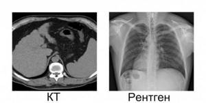
Does CT have an advantage over classical X-ray? What is it?
Clear advantages of computed tomography do exist. This, at a minimum, means that on a classic X-ray we see a two-dimensional picture, and with computed tomography it is possible to create a 3D reconstruction of the organ under study and examine the area of interest to us in three planes.
But each study has its own indications and contraindications. For example, in case of primary injuries and suspected fractures, it is enough to undergo classical radiography, but for examining the gastrointestinal tract, fluoroscopy with a contrast agent (barium sulfate BaSO4) is more effective.
As for brain studies, this is only CT or MRI. Recently, due to the coronavirus pandemic, computed tomography of the chest organs has become very popular, since the resolution of CT allows one to see minimal changes in the parenchyma of the lung tissue, in contrast to a classic radiograph. Here is another advantage of computed tomography over classical x-rays.
Reviews by specialty
Previously, working in this field could cause you to lose your health. But in our times, thanks to the development of technology, the risks are minimized.
Sergey
You can become a radiologist after two years of residency training after medical school. In general, the work is not difficult and the salary is not bad.
Victoria
As in all fields, in radiology there is no place without experience.
Fortunately, before becoming a doctor, you can work as an x-ray technician to understand the profession. Professional websites are very helpful in improving your skills. Larisa
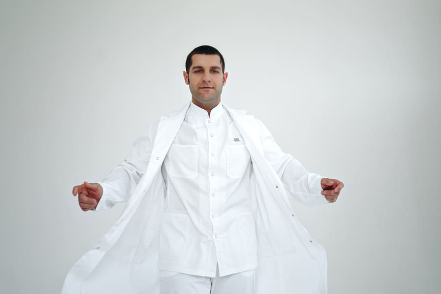
Is it true that the color of a doctor’s uniform somehow affects the patient’s condition?
It is unlikely to affect the state of health, but, according to one study, white color has a beneficial psychological effect on patients, instilling trust and goodwill towards the doctor. Although a good doctor even in colorful or even black overalls will remain a competent specialist, and, accordingly, vice versa. Therefore, it is not the color of the uniform that is important, but the quality of knowledge and attitude towards patients that you carry in everyday practice.
Although, I admit, I wear exclusively white suits because I like the color white, and it is the color that is associated with the image of a doctor.
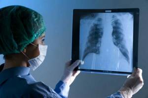
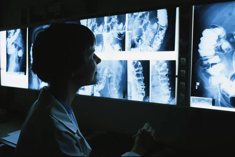
What does a radiologist do?
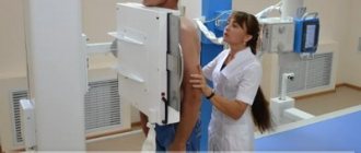
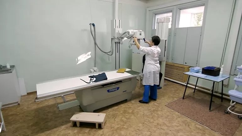
The radiologist examines the patient using special equipment - an X-ray machine . At the same time, the doctor is not required to adjust the equipment - this is the responsibility of the technicians. Routine maintenance of the X-ray is performed by an X-ray technician.
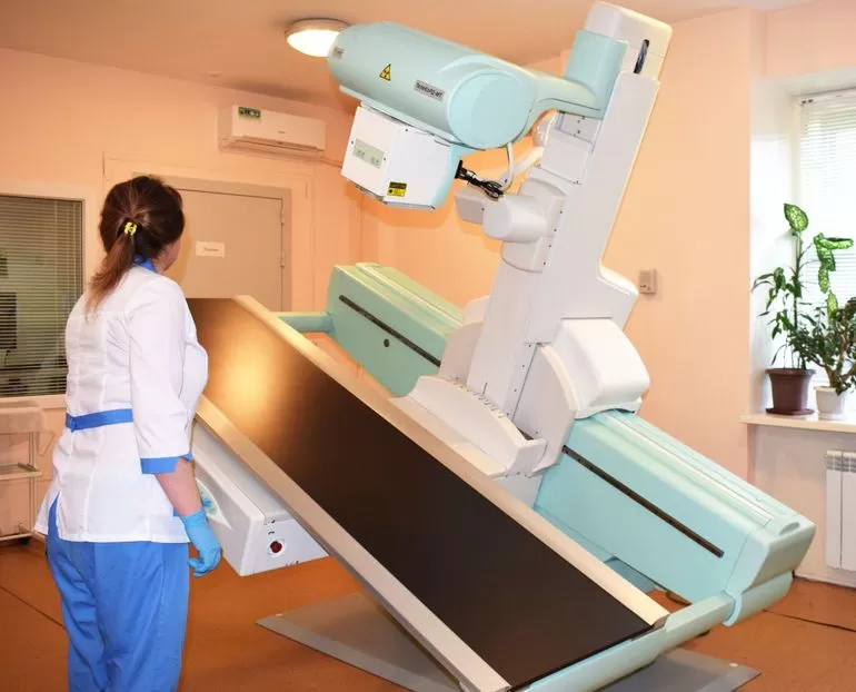
Since working with complex equipment requires a laboratory technician to have a higher education, his functions are often performed by a young radiologist gaining experience. He is responsible for the condition of the X-ray equipment, for the quality of the images, maintains all primary medical documentation, and carries out work to prepare the patient for the study.
A radiologist supervises the activities of the laboratory assistant. An x-ray technician can take pictures independently only on the instructions of a doctor or under his supervision. The main skill of a radiologist is to interpret x-rays and help therapists and specialized specialists in determining the cause and consequences of diseases, pathologies, and injuries.
How do you spend your free time and vacation? Do you have any hobbies or hobbies?

Even in my free time, I try to improve my skills, expand my knowledge base, and constantly learn. I am actively involved in maintaining a medical blog on Instagram. I share interesting cases not only with colleagues and students. I think it is very important to convey information to patients about the need for diagnostics, so that they do not put off taking care of their health until later. We have only one. And timely diagnosis is the key to health.
I work in four places, I don’t have much free time, but I try to spend it usefully and as actively as possible. I go to the gym at any time of the year, it’s already a kind of hobby. And in winter, I try to go to the mountains to enjoy beautiful views, clean air and go snowboarding. I really like visiting the Elbrus region. From time to time I get on my bike to go for a walk along the city streets or over rough terrain with trips outside the city.
Responsibilities of a Radiologist
A specialist should always be aware of all the new developments in the field of diagnostic methods . His responsibilities include:
- receiving patients;
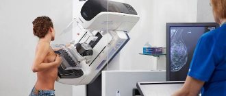
- carrying out fluorography, mammography (as part of a medical examination, medical examination or for therapeutic purposes);
- knowledge of various diagnostic methods - radiography, fluoroscopy, computer, magnetic resonance and positron emission tomography;
- compliance with research safety requirements for both patients and personnel;
- monitoring the work of the laboratory assistant and X-ray room technician;
- deciphering and recording research results;
- maintaining medical records.
Thus, he is engaged not only in his direct responsibilities, but also in administrative activities. Which, in fact, in modern times is inherent in almost all senior medical workers.
IV. Responsibility
The radiologist is responsible for:
1. For improper performance or failure to fulfill one’s job duties provided for in this job description - within the limits determined by the current labor legislation of the Russian Federation.
2. For offenses committed in the course of carrying out their activities - within the limits determined by the current administrative, criminal and civil legislation of the Russian Federation.
3. For causing material damage - within the limits determined by the current labor and civil legislation of the Russian Federation.
4. ____________________________________________________________________
This job description has been developed in accordance with the provisions (requirements) of the Labor Code of the Russian Federation dated December 30, 2001 No. 197 FZ (Labor Code of the Russian Federation) (with amendments and additions), the professional standard “Radiologist” approved by order of the Ministry of Labor and Social Protection of the Russian Federation Federation dated March 19, 2021 No. 160n and other legal acts regulating labor relations.
Also see:
- Occupational safety instructions for X-ray department personnel
Does X-ray show coronavirus?
Can you see Covid in pictures?
Due to the unpredictability and aggressiveness of the virus, scientists do not stop searching for new methods for diagnosing a dangerous disease. And, if at the beginning of the pandemic it was believed that x-rays could not show infection with coronavirus, today the opinion of experts has radically changed.
Will an X-ray show coronavirus in the lungs? It was found that the images show the affected tissue in 9 out of 10 patients. The essence of radiation methods, which include x-rays, is the ability of rays to change intensity when passing through body tissues of different densities. Reflecting on a light-sensitive film, they transfer images of organs onto it.
Dark areas represent seals, and light areas represent cavities filled with air or liquid. To better see the condition of the respiratory tract, an x-ray is taken in two or more projections.
Research effectiveness
It is no secret that X-ray radiation is harmful to humans, but during a 1-2 second examination he receives a negligibly small dose. This means that the benefit clearly outweighs the risk. For adults, X-rays may be taken up to several times a week if needed to diagnose and monitor the effectiveness of treatment for pneumonia.
The information value of radiography in relation to coronavirus lung damage was proven at the beginning of the pandemic by Chinese scientists. It allows you to detect changes characteristic of a new infection. This is a simple and affordable method with which you can examine almost all patients with appropriate indications. X-ray machines are available in every clinic, among them there are mobile units that are convenient to use even in a hospital ward.
- Pathological changes on X-ray films of the lungs will be visible no earlier than 10 days after infection.
It is worth noting that the most informative instrumental study that can detect coronavirus even in the early stages is CT - computed tomography. This is also a radiation method, but the image is called a tomogram and is produced in 3D format.
However, CT is a more expensive and complex method.
Not everyone can afford to be examined for a fee, and there is a catastrophic shortage of free installations in the country. CT scans also have disadvantages - computed tomographs use stronger radiation. What do the lungs look like in the pictures?
Studying the pictures of now numerous Covid patients made it possible to identify the most characteristic signs of lung damage due to coronavirus:
- against the background of the inflammatory process, the structure of the pulmonary alveoli is disrupted, the organ is affected on both sides;
- compactions occur due to the accumulation of inflammatory exudate - liquid mixed with mucus and blood. As a rule, they are noted in both lungs, over the entire surface;
- fluid accumulates between the lungs and chest, forming a so-called “pleural effusion”;
- pulmonary pattern intensifies;
- the affected areas are localized mainly on the periphery - further from the heart, closer to the chest;
- a symptom of frosted glass appears - due to a decrease in density, the lungs seem to be covered with a cloudy film.
The last symptom - ground glass - is shown most often by x-rays, in almost half of all cases.
It is best determined by tomography, but it is also clearly visible on x-rays. The ground glass symptom appears as a large-scale darkening as the surface of the lungs loses definition and appears blurred. This phenomenon is due to the fact that there is less air in the alveoli, their ventilation becomes more difficult, and the walls thicken.
X-ray is a fairly informative diagnostic method, but its use is only advisable at the stage when the coronavirus affects the lungs.
Early diagnosis is possible only by polymerase chain reaction - PCR analysis. Lungs after coronavirus
After an illness, dense areas may appear in the lungs as a result of the replacement of healthy tissue with fibrous tissue. At the same time, the function of the organ deteriorates, and physical activity, which was easy before the illness, now causes shortness of breath.
The inflammatory process in the lungs gradually goes away, which takes about 2 weeks on average. From the time of recovery, when viral particles are no longer released into the blood and PCR tests show a negative result, the lungs work worse by about a third.
The decrease in organ activity is explained by the fact that the tough fibrous tissue cannot open completely. This is how pulmonary fibrosis manifests itself, the danger and consequences of which have not yet been established.
It is unknown whether the lungs will recover, and if so, to what extent. While doctors cannot completely rule out the irreversibility of the fibrotic process, patients are recommended to be observed by a pulmonologist for at least 6 months.
Risk factors for fibrotic degeneration
Fibrosis of the pulmonary structures does not occur in all patients. Factors that increase the risk of scar tissue growth include:
- a significant area of damage - half of the entire lung or more;
- severe form of pneumonia with the development of acute respiratory distress syndrome (ARDS);
- connection to a ventilator;
- addition of a secondary bacterial infection.
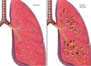
With fibrosis, scars form in the lungs, significantly impairing their ability to enrich the blood with oxygen.
If Covid-19 had a mild or moderate form, and the treatment was carried out on time and correctly, then fibrosis after recovery, as a rule, is not observed. This has been established precisely because recovered patients undergo X-rays, fluorography or computed tomography.
The first two studies are performed in the absence of signs of respiratory failure, if there is no shortness of breath and palpitations with moderate physical activity. X-rays or fluorography are also recommended for patients who have no residual effects in the lungs during a follow-up examination, and blood tests confirm the absence of inflammation.
Control photographs are taken 3 and 6 months after the patient is discharged from the hospital. If compactions are detected, he should be observed at a local clinic by a pulmonologist. The doctor prescribes a course of rehabilitation therapy, and to monitor its effectiveness - CT or X-ray.
Advantages of radiography in diagnosing coronavirus
| Study | Accuracy, % | Period of execution | Availability |
| X-ray | >80 | Max several hours | Equipment is everywhere |
| PCR test | From 60, there is a possibility of false positive and false negative results | From 2 days | Not everyone has enough test kits, you need a referral from a doctor |
Tips on when it is better to take an x-ray
Tip No. 1
In the early stages, there is no point in taking an x-ray. It will not be useful not only during the incubation period, which lasts for 2-14 days after infection, but even when the first symptoms appear - fever, body aches and cough.
It is advisable to take an X-ray when and if the intensity of symptoms increases. In this case, it will be extremely useful and informative.
Tip #2
If you have had contact with a coronavirus patient, and there is a hypothetical possibility of infection, then you need to go to the doctor. If there are characteristic symptoms, it is better to call him at home.
- Attention! A high temperature of more than 38.5°, prolonged coughing attacks, and difficulty breathing are reasons to call an ambulance.
PCR tests are carried out 2 times for contact persons and if coronavirus is suspected. If typical symptoms are present, even if the test result is negative, the test is done twice more. Frequently asked questions being developed that will make it possible to quickly and accurately make the correct diagnosis?
After all, even the results of the PCR test are far from 100%. Leading companies like Googl and IBM have long been working on creating a specific form of artificial intelligence (AI) capable of analyzing X-rays. This will not only make the work of doctors easier, but will also reduce the percentage of medical errors.
- Tech giant Alibaba has already announced an AI system that can detect Covid-19 in 20 seconds.
In addition, the neural network will help eliminate the so-called “search satisfaction,” when a doctor sees a problem in an image and immediately makes a diagnosis without checking other assumptions. As a result, an incorrect diagnosis is made, the consequences of which can be very serious for the patient, since the detected problem is not always a symptom of the underlying pathology.
The results of recent experiments conducted by Canadian scientists have shown the high sensitivity and accuracy of AI in detecting coronavirus. The reliability of the new study was 83%, while the neural network is able to find the symptom of ground glass and the localization of inflammation.
How is coronavirus pneumonia different from regular pneumonia?
With pneumonia caused by bacteria, the organ is partially affected, while the coronavirus infection affects it completely. After encountering Sars-CoV-2, the lungs fill with inflammatory exudate (fluid) and stop receiving oxygen.
In addition, ordinary pneumonia can be cured with antibiotics, but there is no medicine that kills the Covid pathogen yet.
Source: https://apkhleb.ru/rentgen/pokazyvaet-legkih-koronavirus