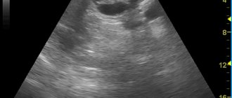
- Ultrasound machine
Ultrasound machine
SAMSUNG SONOACE R7
Class:
Expert (installation year 2019)
What is included in the cost of ultrasound:
Ultrasound diagnostics, interpretation of images, written diagnostic report
Ultrasound of the pancreas
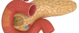
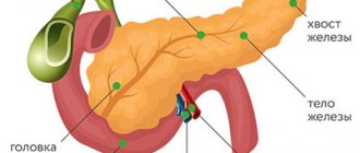
The pancreas is located behind the stomach and is quite tightly adjacent to the duodenum. It consists of a head, body and tail. This organ of the digestive system promotes the release of enzymes for the breakdown of proteins, fats and carbohydrates. The pancreas also produces insulin, which is necessary to regulate blood sugar. Ultrasound is a diagnostic method
, based on ultrasonic waves, allows you to identify various abnormalities and pathologies of internal organs, including the pancreas.
Key points in preparing for the study
Talking about how to prepare for an ultrasound of the pancreas, it should be noted that the preparation itself is divided into 3 main stages. The first is carried out 2-3 days before the planned date of the examination, the second is carried out a day before it, the third - on the day of the ultrasound.
2-3 days before ultrasound
To eliminate the possibility of obtaining unreliable data during the study, pancreatic rest should be ensured several days before the procedure. And since its main task is the production of digestive enzymes, which are activated when food enters the stomach, you will need to follow a special diet.

During the examination, no acute processes should occur in the pancreas, as this will lead to incorrect data. Therefore, it is necessary to exclude the influence of all factors that may cause activation of pathological processes in the tissues of the gland.
You will need to eliminate foods from your diet whose consumption causes increased gas formation in the gastrointestinal tract. These are:
- legumes;
- fresh, boiled and steamed vegetables;
- greenery;
- grape;
- melon;
- milk and dairy products;
- carbonated drinks, including mineral waters;
- black bread;
- spices;
- fatty meats;
- fried foods;
- alcoholic drinks.
Also, while preparing for the procedure, you will need to stop eating foods that contain a lot of protein, as they also contribute to increased gas formation in the gastrointestinal tract (fish, cottage cheese, cheese, etc.).
When performing an ultrasound examination, the pancreas should be functioning at rest. These dietary restrictions contribute to this. But what then can you eat before an ultrasound? During this period you are allowed to consume:
- porridges made from cereals and oatmeal in water;
- lean meats - chicken, turkey, rabbit, etc. (the skin cannot be eaten);
- boiled eggs or steam omelettes (no more than 1 egg per day);
- water and herbal teas.

Herbal teas help improve the functioning of the pancreas and prevent exacerbation of pathological processes in it
In this case, it is necessary to eat food correctly:
- food should be warm (hot and cold dishes and drinks have a negative effect on the functioning of the pancreas);
- Food should be consumed in small quantities, but not less than 5 times a day;
- You should not eat 2-3 hours before bedtime.
Remember that lack of preparation for the examination reduces the accuracy of the examination by 60%! These are serious indicators. Therefore, in order to avoid an incorrect diagnosis and incorrect course of treatment, it is necessary to prepare for an ultrasound in advance.
One day before the ultrasound
The second stage of preparation for an ultrasound examination, starting a day before the procedure, is the most important. During this period, it is recommended to take enterosorbents, especially if errors in nutrition were made during the previous stage of preparation. These drugs help reduce gas formation in the digestive tract and provide more accurate information during the examination.
Such drugs include:
- activated carbon (dosage is calculated individually depending on the patient’s weight - 1 tablet per 10 kg);
- Espumisan;
- Enteros-gel, etc.

Enterosorbents help reduce gas formation in the gastrointestinal tract
The last meal should be 12–14 hours before the ultrasound. Bowel movement is also mandatory. If bowel movements do not occur within 24 hours before the procedure, this may also cause unreliable results and an incorrect diagnosis, since fermentation processes may be activated in the gastrointestinal tract. If defecation does not occur, the situation can be corrected with the help of special rectal preparations (suppositories, microenemas, etc.) or a cleansing enema.
Preparation rules
In case of improper preparation, the information content of the study can be reduced by up to forty percent. Therefore, the preparation stage must be approached extremely responsibly and strictly follow all the doctor’s instructions.
On the eve of the study, dinner should be as light as possible, since the procedure is carried out strictly on an empty stomach.
Due to the accumulation of gases in the intestines, the pancreas may be difficult to see. In this regard, three days before the examination, it is necessary to switch to a gentle diet, namely, exclude gas-forming foods from the diet: milk, yeast bread, raw vegetables and fruits.
Directly on the day of the procedure, you should not smoke, drink alcohol or take medications. The doctor will warn you in advance about the need to take laxatives or anti-flatulence medications.
Ultrasound of the gallbladder with determination of function
When pancreatitis is detected, the doctor usually checks the condition of the gallbladder - these two organs are not only “connected” by a common duct, but are also functionally dependent on each other.
Often chronic pancreatitis is provoked by disturbances in the normal motility of the gallbladder; sometimes, on the contrary, inflammation of the gland can lead to the development of acute cholecystitis.
In the morning, on an empty stomach, the bladder is clearly visualized, being filled with bile. This is a hollow organ, measuring from 3x6 to 5x10 cm. Normally, it should not have kinks or structural anomalies. Wall thickness – up to 4 mm. The development of cholecystitis is indicated by an increase in the size of the bladder and thickening of the walls due to edema.
In the cavity of the gallbladder, ultrasound observation can detect stones, both single and multiple. Formations called polyps may be observed on the walls of the organ.
To assess the functional state of the gallbladder, ultrasound observation is carried out in 4 stages:
- on an empty stomach;
- 10 minutes after the test meal;
- two control observations with an interval of 15 minutes.
If normal motility is maintained, the gallbladder should shrink by 60–70% within 45 minutes of examination. The discrepancy between the indicators and the norm is a sign of a violation of the contractile function of the organ.
The pancreas plays an important role in the digestion process. It produces enzymes for the breakdown of organic substances, regulates carbohydrate, fat and protein metabolism. The pancreas produces insulin, a hormone needed to lower glucose (sugar) levels in the blood. Various diseases can lead to disruptions in the functioning of this organ. Therefore, it is important to promptly identify signs of possible deviations from the norm. Their diagnosis has its own characteristics, because the pancreas is located deep in the abdominal cavity.
Simple examination methods (for example, palpation) do not provide an idea of the condition of the organ. One of the most informative diagnostic methods is ultrasound of the pancreas. During the procedure, the organ is clearly visualized. This makes it possible to assess its shape and size, identify possible neoplasms and signs of inflammation.
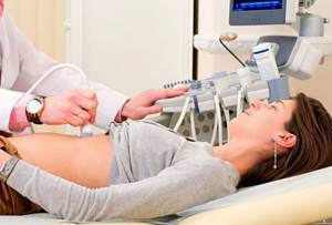
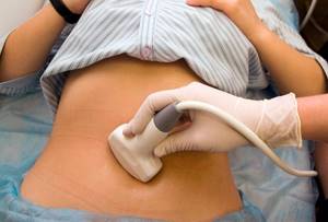
How is an ultrasound of the pancreas performed?
Ultrasound of the pancreas is an absolutely painless and safe procedure. It is performed transabdominally. The patient lies with his back on the couch. The doctor lubricates the area being examined and the transmitting sensor with a special gel for better conductivity of ultrasonic waves. During the procedure, the patient will need to turn over to the left and then to the right side. An ultrasound scanner, which the doctor moves over the skin, transmits images to the monitor of the ultrasound diagnostic apparatus, from which the shape and structure of the pancreas, its location and size can be determined. The whole procedure takes no more than 15 minutes. As a rule, other nearby abdominal organs are examined along with the pancreas: liver, stomach and gall bladder.
Indications
Ultrasound is considered to be the safest and most informative diagnostic method. However, not in all cases it is possible to assess the condition of the pancreas during the study. There are several reasons for this: the deep location of the gland, the presence of nearby organs (the duodenum and stomach are very close to the pancreas and seem to “overlap” it). In addition, it is not possible to give a full assessment of the functioning of the gland when studying obese or flatulent people. Often in such situations, the ultrasound doctor is able to examine only a fragment of the pancreas - its body or head. But in some cases even such information is very important.
As a rule, ultrasound examination is prescribed for:
- acute or chronic pain syndrome arising in the upper abdomen;
- opening of vomiting for no reason;
- obstructive jaundice (yellowing of the skin and sclera of the eyes);
- sudden increase in abdominal volume;
- suspected tumor, cyst or pancreatic cancer;
- high temperature;
- accumulation of fluid in the abdominal cavity (ascites);
- recurrence of pancreatitis;
- suspicions of the development of severe complications after relief of acute pancreatitis (for example, hematomas, abscess, necrosis, cysts, etc.);
- pathologies of the liver and gall bladder, disrupting the functioning of the pancreas and provoking the development of pathological processes in it;
- abdominal injuries.
Decoding the result
The results of the examination are issued immediately after its completion. The study protocol fully describes the structure of the organ, size, presence/absence of various formations. In case of obvious deviations from the norm, the uzist makes a diagnosis, which, in his opinion, characterizes the data obtained. However, the final conclusion is made by the doctor who gave the referral for ultrasound diagnostics.
. The norms for the size of the pancreas in adults according to ultrasound are as follows:
- overall size – 16-24 cm;
- body of the gland – 21-25 mm;
- head width – 32-35 mm
- the thickness of the Wirsong duct is 1.5-2 mm.
Our advantages
- High precision equipment. To perform ultrasound, we use a General Electric LOGIQ 8 scanner. It provides clear visualization of the pancreas, without interference or distortion. This allows you to accurately determine the size of the organ and identify even small deviations from the norm (provided the patient is properly prepared for the examination).
- Qualified personnel . The clinics of the multidisciplinary medical network employ exclusively experienced ultrasound specialists. Doctors have successfully confirmed their qualifications during certification based on the medical education systems of Great Britain and the USA.
- Quick results. The ultrasound procedure takes no more than 30 minutes. The results are deciphered immediately after the examination. Within 15 minutes, the doctor will enter all the results into your medical record.
- A complex approach . The medical network employs experienced specialists of various profiles. If, based on the results of an ultrasound of the pancreas, you need to consult a gastroenterologist, endocrinologist or other doctor, you can get qualified help in our clinic. We will attract the necessary specialists to make a diagnosis and prescribe treatment. You will not need to waste money, effort and time visiting other medical centers.
- Affordable prices. The network of multidisciplinary clinics "Alfa Health Center" has branches in many cities of Russia. In any of our clinics you can do an ultrasound at an affordable price.
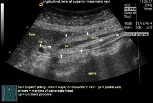
What does an ultrasound of the pancreas show?
Ultrasound of the pancreas shows:
- tissue structure;
- size, shape and contours of the gland;
- location of the gland.
The doctor will also see the following pathologists:
- various neoplasms (cysts, pseudocysts, tumors);
- change in the density of the gland structure;
- inflammatory and infectious processes;
- injuries.
Pancreas thickening on ultrasound
Compaction of the pancreas is not a disease, but a consequence of inflammatory processes occurring in this place. The cause is usually a dystrophic change in tissue and structure, which occurs with chronic endocrine diseases and circulatory disorders.
White pancreas on ultrasound
The white pancreas on ultrasound is detected during an acute inflammatory process - acute pancreatitis. The white color in this case is due to low echogenicity due to the fact that the outflow of enzymes is impaired, blood circulation stagnates and the size of the organ increases. Also, a light or white pancreas is recorded with the development of a disease such as lipomatosis. It occurs most often in older people and obese people. In this case, normal gland tissue is replaced by fatty tissue, the size of the organ increases, due to which the echogenicity also increases, which is recorded on the monitor of the ultrasound machine as a white or light spot.
Is pancreatic cancer visible on ultrasound?
Ultrasound of the pancreas can detect malignant tumors.
- If the doctor sees on the monitor a formation that is well defined and, unlike the pancreas itself, has a clear outline.
- If the lymph nodes located nearby are enlarged.
- The person also complains of sudden weight loss, yellowness of the mucous membranes and skin, vomiting that does not bring relief, and nausea.
In this case, there is a high probability that the formation is of an oncological nature.
Main tasks of ultrasound
There is a certain norm of the pancreas (its size, structure, etc.), deviations from which indicate the development of pathological processes in it and its improper functioning. Therefore, during an ultrasound examination of this organ in women and men, the doctor pays special attention to the following indicators:
- location of the organ;
- configuration;
- gland size;
- clarity of its contours;
- structure of the pancreatic parenchyma;
- level of echogenicity (the ability of the gland to reflect ultrasound waves);
- diameter of the Wirsung and bile ducts;
- the state of the fiber surrounding the excretory ducts.
When performing an ultrasound, all parameters of the pancreas are assessed
In addition, the doctor examines the condition of the vessels located inside and near the organ, which allows him to assess the blood supply to the gland. If an ultrasound examination of the pancreas reveals any abnormalities, the doctor will differentiate between the anomalies of the gland. He faces the difficult task of distinguishing inflammation from a tumor, age-related changes in an organ from chronic pancreatitis, etc.
If the doctor suspects oncology, then an endoultrasound may be required to make an accurate diagnosis. During this procedure, a section of gland tissue is taken with a thin needle (a puncture is made) and its further histological study in the laboratory. This endoscopic examination is carried out under ultrasound guidance.
Contraindications for ultrasound of the pancreas
Due to the fact that the sensor comes into contact with the skin, in case of any lesions of the skin in the epigastric region (dermatitis, purulent inflammation, herpes zoster, various wounds, scabies, etc.), the examination will have to be postponed. It will also not be possible to carry out the procedure if you are severely obese due to the difficulty, and sometimes even impossibility, of seeing the pancreas behind the thick layer of fat. An allergic reaction to the gel used during the procedure is also a contraindication, but is extremely rare.
Pathologies detected during ultrasound
An ultrasound examination of the pancreas allows you to diagnose:
- pancreatitis (acute and chronic);
- necrosis;
- cysts and pseudocysts;
- malignant tumors;
- structural anomalies;
- abscess;
- stones in the bile or pancreatic ducts;
- an increase in nearby lymph nodes, which is a clear sign of the development of inflammatory processes in the body;
- age-related changes;
- ascites.
Each disease requires a certain type of therapy. And to make an accurate diagnosis, ultrasound alone is not enough. It only allows you to confirm or refute the presence of pathological processes in the tissues of the pancreas and gives rise to further, more detailed examination of the patient.
Possible results
All detailed data on the size, shape and condition of the organ are recorded in a specialist’s report. Normal pancreas:
- located next to the spleen and liver;
- has the shape of the letter S;
- looks uniform, has no obvious defects;
- has clear contours;
- easy to visualize: head, body, tail are visible.
The size and other parameters of the pancreas may differ from standard indicators for adults and children of a certain age. But this is not always a reason for making a diagnosis. Diseases are usually indicated by a combination of various deviations from the norm. An ultrasound scan can identify a specific organ defect. Thus, tumors are visualized as oval or round objects with a heterogeneous structure. When the pancreas is inflamed, an ultrasound scan reveals an enlargement of the organ from head to tail. For any deviations from normal parameters, consultation with a specialized specialist is recommended.
Everything you need to know about ultrasound of the pancreas
After reaching the age of 25, every person should undergo an ultrasound of internal organs at least once a year, including an ultrasound of the pancreas. This is especially important for those people whose lifestyle is far from healthy or whose working conditions leave much to be desired. Very often, under extreme living conditions, internal organs wear out faster than a person’s appearance, and cancer problems arise more often than in people of the same age, but leading a more correct and healthy lifestyle.
Why the pancreas?
Because it synthesizes the key hormone (insulin), which makes it possible for energy to penetrate into cells. If its functioning fails, the entire body is doomed to starvation, which will inevitably lead to dire consequences.
Of course, science has not yet been able to find drugs that could completely cure pancreatitis or diabetes. Prevention and timely treatment when the first signs appear are much more important here. The pancreas is the most important component of a healthy body.
Indications for ultrasound examination of the pancreas
If the patient has been prescribed an ultrasound of the abdominal organs, the ultrasound specialist must also examine the pancreas, assess its appearance and size .
However, there are a number of alarming symptoms in which a more detailed study of this organ may be prescribed.
If a person has been suffering from abdominal pain for several weeks with a greater localization in the left hypochondrium.
Feelings of persistent discomfort and heaviness in the stomach after even a small meal.
Indigestion with alternating constipation and diarrhea.
Slight yellowness of the skin and mucous membranes.
Having diabetes of any type.
Very often the condition of the gland depends on the condition of the liver. For any pain in this area, you need to examine both the gland and the condition of the liver itself.
If any of the listed symptoms are present, a person undergoes a mandatory and detailed examination of all organs involved in digestion. The doctor’s task is to promptly recognize dangerous signals, and the patient must properly prepare for the upcoming study.
The biggest problem with ultrasound of the pancreas is air. The organ is in anatomical contact with the intestines and stomach. Air from these hollow organs can significantly complicate detailed examination, distorting visualization and reducing the accuracy of diagnosis.
To prevent this from happening, there are certain rules or preparation for an ultrasound of the pancreas. The most reliable results can be obtained when examining a patient in the morning. It is then, if all the rules are followed, that the research indicators will be the most truthful.
Fasting is necessary for at least 12 hours. Moreover, if you observe fasting during this time, you can also do an ultrasound of the liver. The fact is that most of the air enters the human intestines during the day. Immediately after waking up, a minimal amount remains in the gastrointestinal tract. With minimal “gas contamination,” all the nuances are very clearly visible and you can do an ultrasound of the liver and other organs adjacent to it. This is especially true for people who have increased acidity of gastric juice. Drinks with carbon dioxide will cause excessive gas production, which will negate the results of the study.
Preparation of the subject requires following a diet for 2 days before the examination. You need to avoid foods that can cause fermentation in the intestines. Try to exclude fruits and vegetables, as well as carbonated drinks from your diet.