The brain is the most important human organ, which is protected by the cranium. If its functions are impaired, it is important to find out the extent of the damage in order to take measures to correct the situation. To visualize the bones of the skull, radiography is used - an X-ray of the head is taken. This is an inexpensive and accessible research method that has no alternative. In some cases, this is the only opportunity to identify pathology at an early stage and carry out effective treatment.
The method is based on the different ability of rays to penetrate tissues of different densities. The device records the radiation after passing through the organs on a photosensitive film. The images of bones and dense structures are white, soft tissues with good penetrating ability are dark.
X-ray of the entire skull in two projections 1100
+7 (495) 320-43-41
Indications for radiography of the brain (skull)
The basis for prescribing the study are:
- Injuries, headaches;
- Darkening in the eyes;
- Trembling in hands;
- Nosebleeds;
- Dizziness;
- Pain when chewing;
- Decreased vision and hearing;
- Asymmetry of facial bones;
- Suspicion of brain cancer;
- Fainting;
- Endocrine disorders.
A referral for diagnostics can be issued by a traumatologist, neurologist, endocrinologist, or surgeon. The procedure is not prescribed for pregnant and lactating women. This is the only contraindication.
Possible complications
The danger of x-rays for human health, and in particular for children, is sometimes significantly exaggerated, but it is also wrong to completely deny it. Harm is ionizing radiation that affects the human (child) body with a certain intensity. This radiation, passing through tissues, ionizes molecules, causes a temporary change in the composition of the blood, and changes the structure of proteins. Also, under the influence of radiation, cells can undergo premature aging. The main negative aspect of X-rays is that radiation can provoke malignant degeneration of cells, and therefore cause further development of oncology. However, such cases are extremely rare in medical practice, but the high diagnostic benefit of x-rays is sometimes priceless.
Contraindications for
If we consider that childhood is already a contraindication for x-rays, it is almost pointless to talk about other prohibitions. The main thing you need to know is head x-ray
child is a last resort measure that is used if other types of diagnostics turn out to be uninformative.
How harmful are x-rays?
The radiation dose received by an adult patient is a small part of the annual norm (about 4%). This value is comparable to an hour's stay on the beach. Doctors point out that this approach is incorrect. Indeed, in some cases, the study is carried out for vital reasons, in order to detect a fatal disease. Therefore, the amount of research conducted should be subordinated to the main goal - saving human life. It happens that the diagnosis is also carried out on pregnant women, carefully covering the stomach with an apron.
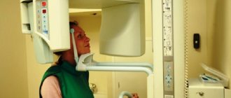
How is the procedure carried out?
Children undergo the examination lying or sitting. There should be no metal objects on the head. Sometimes you need to stand while taking a photo; this is discussed separately. Below the head, the child’s body is covered with personal protective equipment. Pictures are taken in frontal or lateral projection. It is important not to move during the examination. Therefore, soft restraints are used for the smallest children, and sedation is sometimes used. Such measures allow the procedure to be carried out quickly and almost without harm. Sometimes a child calms down thanks to the presence of one of the parents in the office.
Contraindications
Radiography has no significant contraindications unless alternative diagnostic techniques are found with similar information content but less radiation exposure. It is worth discussing the restrictions with the doctor if the child has recently had an x-ray.
Is the procedure harmful?
Ionizing radiation is harmful, especially for a growing organism. Therefore, it is better to do the procedure for a fee and in a clinic with modern equipment. The device used in the SM-Clinic provides a minimum radiation dose: from 0.03 to 0.1 mSv (for children, the permissible radiation exposure is 1 mSv per year). It is also important that the digital technologies used now make it possible to take a picture very quickly, that is, to influence the body with radio radiation for the shortest possible time - up to 1 second. The procedure is practically harmless if it is carried out all the time in the same medical center with the radiation doses received by the child throughout the year recorded in the outpatient card.
X-ray of a child's head
A different approach is used to study the skull of children:
- The child is more likely to receive a higher dose of radiation;
- Exposure to radiation can adversely affect a child's body.
For these reasons, an X-ray of a baby’s head is prescribed only as a last resort, if other diagnostic methods are not informative and the baby’s life is at risk. It is difficult to choose an alternative method, since the bones of the skull have a complex structure, and ultrasound does not recognize all pathologies.
The main reason for prescribing x-rays for a child under one year of age is injury. Radiation of babies is undesirable, but often this is the only way to identify birth injuries that threaten the child’s life. In the case of an X-ray of the head of a newborn, all other organs are protected as much as possible.
What is being researched and why?
Important! An X-ray of the child’s head is performed to assess the condition of the skull, that is, the bone structures, rather than the brain.
X-rays pass differently through different organs and tissues, the images of which in the pictures, due to this, differ in intensity. So, the bone - the hardest tissue - will be bright white, and the soft structures will be gray or almost black. An overview image shows a general image of different structures of the skull: the sella turcica, eye sockets, lower jaw, temporomandibular joint, etc. To clarify the diagnosis or a detailed examination of the pathology, you need to do a targeted study (the tube of the device is directed to a specific area of the skull).
What can you find?
Radiography allows you to see:
- congenital pathologies;
- birth injuries and deformities;
- cysts;
- osteoporosis;
- hematomas;
- neoplasms;
- hernias;
- fractures;
- consequences of inflammatory diseases;
- bone condition.
Headshot during pregnancy
A pregnant woman may need an X-ray of the head bones:
- To provide dental care;
- With dislocations;
- For fractures to determine the location of bone fragments.
When deciding on the need for diagnostics, you should weigh the risks and benefits of the procedure. Conditions in which this issue is resolved already pose a danger to the health of the mother. Refusal to take an x-ray can have serious consequences - lack of medical care will lead to the development of dangerous diseases. Then there can be no question of normal childbirth. In addition, precautions effectively reflect radiation.
According to scientific research, the effect of X-ray radiation is especially unfavorable in the early stages of pregnancy. The result may be termination of pregnancy or pathology of fetal development.
Method of performing an X-ray of a child’s skull
Children from one year old are seated in a chair and secured. There should be no metal pins or headbands in your hair - this can distort the picture of the disease. The body must be reliably shielded with protective equipment. The thyroid gland is covered with a protective collar.
If the baby's parents are present during the x-ray, they also need a protective apron. The time of the procedure and the quality of the image depend on how immobile the regime is observed. After the procedure, to neutralize the negative consequences, the child should drink plenty of fluids.
Features of an X-ray of a newborn's skull
A very young one-month-old child who is unable to stand and sit independently may also be sent for an X-ray of the skull if there are special indications. Such patients are placed on the work table of the X-ray machine, areas of the body that are not to be photographed are covered with a lead apron, and the limbs are secured with special belts.
Where to get a head x-ray
If you need to take a head photograph, contact the Central Clinical Hospital of the Russian Academy of Sciences. We guarantee prompt and accurate diagnosis through the use of advanced digital equipment that provides the lowest level of radiation. Experienced doctors interpret X-ray results. You can make an appointment on the Central Clinical Hospital website online or by phone. You can find out how much the procedure costs at the clinic. Also, current prices are indicated on our website.
You can find out prices for brain x-rays and make an appointment online on our website or by calling the clinic.
Are there any differences from skull x-rays in adults?
There are no fundamental differences between X-rays of the head of a child or an adult. Of course, it is easier for adults to explain what this procedure is, why it is needed and how to behave during the diagnostic process. It is difficult to get children to remain still and hold their breath. They may also become overly anxious, especially if they see their arms and legs being restrained with straps.
However, many years of medical practice show that X-rays can be taken for both children and adults, obtaining quite informative images.
Indications and contraindications for x-rays in infants
Do children under one year old get x-rays? Yes, it can be used to make an accurate diagnosis, but such an examination method is never used as a method of preventive detection of diseases. For the latter case, there are safer methods, such as ultrasound.
When do X-rays take place on infants?
- The need to identify bone fractures associated with injury (falling from a bed, changing table, etc.). This may lead to the need for an X-ray of the head of a child under one year old due to the risk of developing skull fractures.
- Identification of birth injuries.
- If doctors suspect congenital or acquired disorders in the musculoskeletal system, for example, dysplasia of various joints or rickets.
- Suspicion of foreign bodies in the digestive system or in the trachea and bronchi.
- The need for comprehensive preparation for surgical intervention, for example, during operations to correct heart defects.
- Identification of the level and causes of intestinal obstruction.
As a rule, following strict indications and limiting the amount of radiation exposure during the year can reduce the risk of a child developing negative consequences from the procedure.

Carrying out an X-ray examination of a baby
How are x-rays done for children under one year old?
- Firstly, such a procedure should always be carried out in a specialized office.
- Secondly, during the examination, one of the parents must be next to the child to hold the baby and calm him down in preparation for the x-ray.
The basic rule for a successful X-ray examination is maximum immobility. If the child moves, the images may be of poor quality, which will lead to the impossibility of their interpretation and the need for a repeat procedure. In this regard, the baby’s mother or father must hold him well.
Preventing exposure to other areas of the body that are not being screened should be a priority. To solve this problem, lead plates or linings are used that do not transmit ionizing radiation and protect the body. In addition, for children under one year of age it is very important to use digital devices, in which the level of radiation exposure is significantly lower.
The duration of the procedure is 5-20 minutes, depending on the preparation and the child’s reaction to the need for examination. As a rule, a properly organized x-ray examination does not lead to the development of negative consequences from x-rays.

Consequences of x-rays for a child’s body
The child's body is more sensitive to ionizing radiation than the adult body. In this regard, the risk of developing negative consequences of X-rays during studies of the brain, kidneys, lungs and other organs is much higher in childhood.
By exerting their biological effects, X-rays can lead to various disorders in the cells of the body - both somatic and genetic, which increases the risk of developing benign and malignant neoplasms in the future, and also affects the risk of other pathologies of internal organs. At the same time, in a child’s body, organs are located next to each other, which makes it difficult to limit exposure to ionizing radiation.
It is important to note that the peculiarities of the location of red bone marrow in children (it is located in the bones of the limbs and skull) can also cause negative consequences associated with inhibition of its work and a decrease in the production of blood cells.
Necessary X-ray precautions for children
What needs to be done to reduce the risk of negative consequences?
- X-rays should be performed on children under one year of age only if indicated. Under no circumstances should you use it or fluorography as a preventive measure.
- Research must always be carried out using modern equipment and in compliance with all safety regulations.
- It is important to use protective shielding panels that protect the child’s organs and tissues from ionizing radiation.
- X-rays for children under one year of age should be performed a minimum number of times. If there is a need for it, then the number of pictures in one year should not exceed 1-2.
X-rays for children under one year of age pose a serious danger to their health. However, proper organization of the examination and compliance with all safety regulations can minimize the risk of developing negative consequences. It is important to remember that x-rays should only be used when absolutely necessary. In all other situations, the choice should be made in favor of safe studies, such as ultrasound, etc.
Anton Yatsenko, pediatrician, especially for Mirmam.pro