Baby teeth and molars - what's the difference?
The differences between temporary and permanent are the following characteristics:
- The roots of baby teeth are shorter, widely spaced and angled. This is explained by the need for the formation of molars;
- the enamel of temporary teeth is bluish, it is much thinner, and therefore more susceptible to the development of caries;
- The child’s milk teeth are smaller than the molars;
- The number of some teeth is not equal to the number of others. There are fewer temporary ones, because some elements of the series immediately grow as radicals;
- the tissue of baby teeth is poorly mineralized;
- temporary teeth grow vertically, and the crowns of permanent teeth are directed outward (towards the lips and cheeks).
Causes of occlusion pathology
The formation of the dental system is influenced by a number of external and internal factors. If it is possible to identify such a causative factor and eliminate it in time, then the correction of violations is much easier and faster. We have combined the most significant reasons into one list:
- Heredity. The presence of occlusion pathology in one of the parents increases the likelihood of a defect forming in the child by 40%. If a pathology in the structure of the dental system is detected in both parents, then you should not expect a miracle. It is better to contact an orthodontist in a timely manner and artificially “direct” the growth of teeth and jaws in the right direction.
- Bad habits. The child actively tastes any objects. There is nothing wrong with this until holding something in your mouth and sucking becomes a habit. Absolutely everything is used: the edge of the blanket, fingers and even lips. Pacifiers do not cause harm only in cases where the child weaned off them in a timely manner.
- Problems of the musculoskeletal system. The bite affects the condition of the spine, primarily the cervical and thoracic regions. And pathology of the musculoskeletal system, for example, flat feet, is a significant factor that can disrupt the normal development of the bones of the facial part of the skull.
When is an x-ray indicated in childhood?
An x-ray of the child’s baby teeth is scheduled the day before treatment. The doctor resorts to this measure to make sure that the upcoming therapy will not harm the process of formation of permanent teeth.
Specific indications for radiography include:
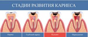
- the presence of carious lesions and other destructive processes;
- the appearance of jaw anomalies;
- diagnosing abscesses;
- the need to control impacted teeth and the natural change of temporary elements to molars;
- Carrying out bite diagnostics;
- inappropriate condition of the root system of temporary teeth.
More often, the procedure is resorted to in case of delayed growth of permanent elements of the series.
Signs of occlusion pathology
In most cases, it is impossible to determine the “correctness” of the bite visually. This can be done by an orthodontist, who is highly advisable to see the child annually, from the eruption of baby teeth to the completion of the formation of a permanent bite. A specialist can identify an emerging problem, and even the prerequisites for the fact that the bite may be formed incorrectly.
For example, crowding of baby teeth significantly complicates the process of eruption of permanent teeth. Molars are larger than primary teeth, so they generally need more space in order to occupy the correct position in the dentition.
When examining a child’s teeth, parents are unlikely to be able to assess the symmetry and uniformity of jaw development and the correct placement of teeth in the dentition. But there are other manifestations, the presence of which may indicate problems with occlusion or a predisposition to the development of orthodontic anomalies:
- Incorrect breathing. We can breathe through our mouths if, having caught a seasonal ARVI, we suffer from a runny nose. This is fine. It is a completely different matter if the child breathes through his mouth all the time out of habit or due to the presence of enlarged adenoids.
- Snore. Children can snore in their sleep only if there are serious disorders in the development of the dental system. This feature, unusual for childhood, should not be left unattended. Not only can you miss the best time to correct your bite, but during sleep apnea the brain does not receive enough oxygen, which can affect the mental abilities of a growing person.
- Certain facial features. What matters is the structure and proportionality of the lower third of the face. The chin should not be too massive, nor, on the contrary, sloping back. The upper jaw should also not be the most protruding part of the face, as this indicates its hypertrophy.
- Posture disorders. Changes in bite due to scoliosis and other disorders in the musculoskeletal system can develop as a compensatory reaction of the body, which needs to develop coordination of movements and the ability to maintain an upright body position at any cost.
Types of x-rays when examining children
Depending on the goals of the study, there is a need to obtain one or another type of x-ray. Among them:
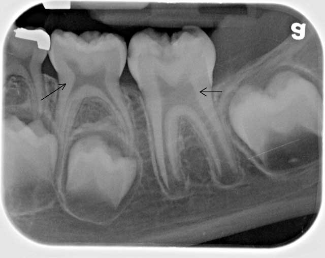
- targeted radiographs - show one or a pair of teeth and nearby soft tissues;
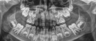
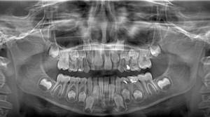
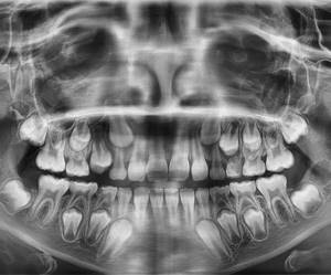
- panoramic image of the jaw - displays a picture of the dentition along with the rudiments of the radical elements;
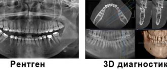
- 3D images are three-dimensional images of the entire jaw (upper, lower row) or a section of it.
The difference between panoramic and targeted radiographs
The difference between a panoramic X-ray and a targeted X-ray is the area of study. In the first case, the object of study is the jaw row, in the second - a specific area of the child’s gums, jaw or oral cavity.
Sight shot
Panoramic shot
In the process of targeted radiography, the beam tube is brought closer to the soft tissues. A successful shot shows one or two elements adjacent to each other.
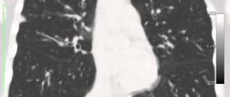
3D modeling in negative
We are talking about a procedure carried out using innovative equipment that allows one to obtain a 3D image of the jaw row or its area. Such an image helps to determine the location of pathological canals, areas of the root system, and fillings. A study is scheduled on the eve of endodontic therapy and implantation.
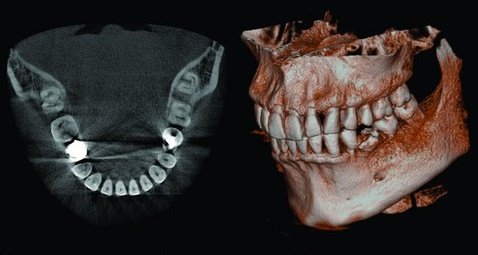
3D
What unformed root tips look like on an x-ray
In unformed tips, the length of the root is almost normal, the walls are parallel to each other. The wide root canal ends in a funnel-shaped extension. In the area of the apex, the periodontal fissure merges with the growth zone, which incompetent specialists sometimes identify as a pathology.
Throughout the entire length of the root and at the apex, a compact plate of the socket wall is differentiated. This picture is typical for the upper central and lateral incisors in 8-year-old children, for the lower central incisors in 6-year-old children, as well as for the lower lateral incisors and first lower molars in 7-8 year old children.
Features of the procedure
To x-ray the jaw of a child with baby teeth, a film or digital device is used. The equipment is placed in a specially designated room, where the photo laboratory is also located.
The algorithm of action for the specialist and the patient is as follows:
- Before the procedure, the child needs to remove metal items (watches, jewelry, glasses, etc.).
- A special protective apron is put on the patient’s body, into which lead plates are sewn. Thus, the child’s internal organs are protected from the adverse effects of x-rays.
- The subject is asked to press a plastic plate with his teeth. The patient's lower jaw should be pressed tightly against the septum of the device. During the examination, the patient is required to remain motionless.
- The plates of the X-ray equipment begin to move around the child's head. This process takes no more than 20 seconds.
At the end of the examination, the panoramic image is printed on paper or transmitted electronically.
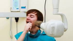
The principle of intraoral radiography (sighting) is somewhat different. The patient is seated on a chair and a protective apron is used. After the child opens his mouth, the specialist installs a film or matrix inside the examinee’s oral cavity. The doctor then places the machine's tube near your mouth. The duration of the procedure is no more than 60 seconds.
The result of the x-ray examination is saved on the computer. This makes it possible to further monitor the dynamics of the clinical picture.
The issue of X-ray safety for children
Any X-ray radiation, including in the dental industry, carries a certain degree of danger to the patient’s health. Research in the field of medicine confirms the possibility of developing acute radiation sickness when exposed to increased radiation. However, X-ray equipment is not enough to provoke it.
X-raying of a child’s primary teeth can be safe if the examination schedule and radiation safety standards are followed.
The permissible annual exposure rate, according to regulatory documentation, is 5 mSV (millisievert). In the case of small patients, as well as pregnant and lactating women, this figure is halved.
Dental radiography is characterized by minimal mZV indicators. With digital X-rays, the amount of single exposure is 0.01-0.03 mSv. In this case, the permissible single dose is 0.1 mSV. The development of radiation sickness is discussed only when this figure increases by (0.7 ZV).
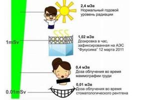
X-rays are often prescribed periodically, for example, as part of a long therapeutic course. How often can dental x-rays be taken? According to WHO, if 5-6 such procedures are carried out during the year, the radiation background of a small patient will not be disturbed. However, one should not lose sight of the factor of the presence of its own background radiation in the area in which the child lives. For example, for Moscow this figure is 20 µSV.
There is no official evidence that chronic doses of irradiation to the oral cavity (for example, long-term therapy) result in the development of cancer.
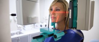
How do you do an X-ray of a child's head?
Many mothers are afraid when their children (especially under the age of 3 years) are prescribed x-rays. Almost everyone knows about the harmful effects of ionizing radiation, so such anxiety among parents is completely justified. It must be said that when photographing the head, the child’s body is reliably protected by a lead apron or vest, and the radiation exposure received during the procedure is not so great as to seriously affect the growth and development of the child’s body.
The quality of the images largely depends on how still the child was during the scan. To minimize his movements, fixing straps are used. To prevent the child from worrying, relatives are allowed to be in the office. If the child is small or very restless, he is given sedatives.
Is it possible to refuse x-rays for a child?
The child's parents have the right to refuse x-rays. However, responsibility for making such a decision rests entirely with them. Such diagnostic methods are not always able to provide a complete picture of the condition of the patient’s oral cavity.
Without X-rays, the doctor cannot arrange prosthetics. If you refuse the procedure, the planned treatment becomes impossible.
Radiography of baby teeth for children is included in the list of the most informative diagnostic methods. This procedure helps to assess the condition of the row elements, nearby soft tissues and identify the rudiments of permanent teeth without harm to the child. For diagnostic purposes, panoramic, targeted and 3D images are created. Digital X-ray is recognized as the most efficient and safe diagnostic tool.
Contraindications
If there is bleeding
Bleeding in the mouth, caused, for example, by problem gums, can affect the quality of the X-ray image. First, the doctor must prescribe hygiene procedures that can stop the bleeding, and only then send for x-rays.
If you feel unwell
If your child is sick, has a fever, or simply feels unwell, you should not take him for an x-ray. It is better to postpone this procedure until complete recovery.