What will an x-ray show?
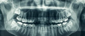
The images taken by the X-ray machine visualize the tongue, larynx, nasopharyngeal vault, nasal cavities and septum, and vocal cords. From the images, the doctor can determine:
- Damage to bone tissue - fractures, cracks.
- Deviation of the nasal septum.
- Neoplasms of various nature - tumors, cysts, polyps.
- Inflammatory processes - otitis media, sinusitis, frontal sinusitis, sinusitis.
- Foreign bodies trapped in internal cavities.
- Accumulations of liquids.
- Pathological proliferation of lymphoid tissue - adenoids.
- Damage to the vocal cords.
How detailed the structure and condition of organs will be visible depends on the quality of the image and the number of pixels on it. The more there are, the more accurately and in detail even small elements will be visible. This is provided by digital X-ray machines. The quality and detail of the images is one of the reasons why you should choose an examination using modern equipment.
How is an ENT x-ray performed?
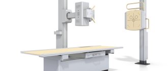
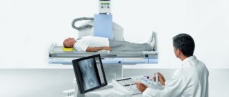
X-ray examination of the ENT region is carried out in a special diagnostic room using an X-ray machine. The designated area is examined according to the same rules as other parts of the body. The patient is asked to take a sitting or reclining position. The picture is taken in two projections (frontal and lateral). Less often, it is necessary to take a picture with additional overhead projections.
The procedure itself requires very little time. Preparation for the image lasts no more than 20 seconds (positioning the patient, fixing the installation). It takes even less time to take a photo (2-5 seconds). It takes a specialist a few minutes to print and decipher the result.
X-ray is a very fast, painless and informative way to diagnose serious pathologies.
Indications for X-ray
An otolaryngologist prescribes an x-ray of the nasopharynx when it is necessary to identify the cause of the patient’s complaints. But this is not the only diagnostic method - in addition to X-rays, rhinoscopy can be used - examination of the nasopharynx using special instruments. It would seem that a simple examination is preferable to an x-ray with its harmful radiation. However, this is not quite true. Even if we don’t say that the radiation exposure from a digital X-ray machine is minimal (and this is the second good reason to choose a modern clinic), X-rays have their advantages. It is not always possible to conduct an examination - it happens that the patient has a strong gag reflex or cannot open his mouth wide enough. When a routine examination is insufficient to make a diagnosis, an x-ray is prescribed.
Main indications for x-ray of the nasopharynx:
- Injuries to the head, nose, bridge of the nose, in which there is a risk of fractures or cracks in the bones of the skull.
- Entry of foreign objects.
- Difficulty breathing, constant runny nose, snoring.
- Prolonged headaches.
- Suspicion of sinusitis or latent otitis media.
- Adenoids.
- Pain in the sinuses.
- Increased body temperature with other symptoms that indicate problems in the ENT organs.
- Frequent nosebleeds for no apparent reason.
- Purulent discharge from the nose, ears.
- Suspicion of a tumor or other neoplasm in the nasopharynx.
Interpretation of results
The radiologist who conducts the study interprets the results. He draws up a report, which the patient then gives to the attending physician. Fractures and bone displacements are very visible on x-rays. Round formations near the walls of the sinus are interpreted as cysts, and neoplasms on an x-ray form shadows of different shapes. Also, using an X-ray of the nasopharynx with contrast, it is easy to identify the degree of hypertrophy of the tonsils, since the study allows you to study the lumen of the nasopharynx and draw conclusions about the size of the adenoids. However, do not forget that the final diagnosis is made by the attending physician.
Contraindications
X-ray using a digital device has significantly less negative impact on humans, but still has limitations in its use. Main contraindications for x-rays:
- Pregnancy – due to the risk of harm to the developing fetus.
- Age below 16 years.
- The patient's serious condition.
The decision whether to conduct an examination or not is made by the attending physician. For example, an x-ray can be done for children if the cause of the complaints has not been established by other methods.
If X-rays are prohibited, alternative diagnostic methods are ultrasound and MRI.
Contraindications for
Absolute contraindications to radiography of the nasopharynx are pregnancy and age under 15 years. However, this study is very often prescribed to young children who cannot remain still for a long time for a standard examination or more modern diagnostics using MRI.
. The basis for conducting an X-ray examination for a child can only be a doctor’s indication. However, it is worth noting that of all types of x-ray examinations, x-ray of the nasopharynx gives the lowest dose of radiation.
Advantages and disadvantages of the procedure
The advantage of radiography of the nasopharynx is the efficiency of the method, since it is necessary to remain motionless for a few seconds to take the picture. Modern X-ray equipment provides a radiation dose comparable to what you would receive when traveling on an airplane, so you should not be afraid of your next x-ray of the nasopharynx. However, even under such conditions, doctors prescribe radiography when it is really necessary, especially when it comes to the health of the child. The disadvantage is that X-rays of the nasopharynx without contrast are not very informative, so this procedure is performed only if there are no other options.
How is an x-ray of the nasopharynx performed?
There is no need to prepare for the examination - all you need to do in the radiologist’s office is remove all metal objects from your head and neck and put on a protective belt.
X-rays of the nasopharynx are often done in frontal and lateral projections - such angles provide the most informative view. The patient lies with his head leaning against the device sensor, and the doctor checks the correct positioning. He will pay attention to the fact that the line that could conditionally connect the patient’s eyes and the device runs strictly perpendicular to the cassette (with an angle of 90°C).
The patient must remain completely still while the image is taken. To be sure of this and to avoid accidental movements, the head and chin can be fixed in a certain position.
The whole procedure lasts 5-10 minutes, and after analyzing the images, the radiologist gives an opinion where he describes the condition of the nasopharynx and the detected pathologies. The diagnosis is made by an ENT doctor based on the conclusion of a radiologist and the results of other examinations.
How to prepare for the procedure?
X-ray of the nasopharynx, which shows the condition of many organs in a standard manner, does not require preparation. However, before more detailed contrast radiography, which shows the presence of adenoids in the nasopharynx, it is necessary to inject a substance into the trachea that will color the internal organs and give them a clearer outline. It is also worth warning your doctor about already diagnosed pathologies of the nose and nasopharynx, for example, a broken nasal septum.
For special indications, an X-ray of the nasopharynx can be done even for a child, but the procedure will be safe only in a specially equipped diagnostic room under the supervision of a qualified radiologist. Immediately before the x-ray procedure
nasopharynx, the patient is asked to remove metal objects and anything that may interfere with the examination. Then a protective apron made of leaded rubber is put on it, which will reduce the radiation exposure during the study. The doctor then positions or sits the patient in a comfortable position and adjusts the equipment so that the results of the procedure are clear and understandable. After the patient has taken the required position and the equipment is adjusted, the doctor gives the command not to breathe or move for several seconds. During this time, the nasopharyngeal cavity is photographed.
X-ray of the nasopharynx in lateral projection
X-ray of the nasopharynx in lateral projection is performed to diagnose adenoids. In this case, the patient is positioned in such a way that the beam of the X-ray equipment is directed at the nasopharynx on the right, then on the left side. During the examination, the patient must follow the doctor's instructions so that images are taken while inhaling and exhaling. Also, an x-ray of the nasopharynx in a lateral projection, taken at the moment of pronouncing the sounds “u” and “i”, allows you to see and record the vocal cords on the x-ray.
X-ray of the nasopharynx in Moscow
You can consult an otolaryngologist and undergo an X-ray examination every day at the diagnostic and treatment center. The clinic uses Brivo XR575 Premium, a modern X-ray machine that meets the highest quality standards and expectations - minimal radiation dose, high speed, digital image format and high detail. The interpretation of images, diagnosis and treatment are carried out by doctors with many years of experience - we know how important accuracy and accuracy are when it comes to health.
Children's and Children's Center "Kutuzovsky" is a clinic where they will help restore and maintain the most precious thing - health.
Make an appointment for a consultation and appointment with a doctor.
Features of X-ray diagnostics of ENT organs
Some diseases in otolaryngology require deep and accurate diagnosis. For example, the location of the maxillary sinuses does not allow a thorough examination without the use of special diagnostic equipment. radiography
is often chosen as an effective diagnostic technique .
X-ray
– this is a fairly simple, extremely safe, completely painless and quick way to obtain all the important information about a specific area of the ENT organs.
Advantages of the method:
- availability
- non-invasiveness
- high resolution
- relatively low radiation exposure
- availability of a document (x-ray), which makes it possible to compare with previously taken photographs
- possibility of research for preventive purposes
- lack of special training for most studies
For adults, X-ray examinations are performed both according to indications and for preventive purposes (chest x-ray).
For children of any age, X-ray examinations are performed only when indicated.
The decisive factor that pushes a doctor to prescribe x-ray examinations for children is the inability to make a diagnosis in any other way.
The image obtained during an X-ray examination is a radiograph - a specific document reflecting the condition of the area of the body being examined at the time of the examination.
Depending on the type of X-ray machine, the radiograph can be obtained on film or recorded on digital media as a file.
The main document reflecting the result of an x-ray examination is the x-ray examination protocol with the conclusion of a radiologist
and recommendations.
A patient who has undergone an X-ray examination is given a radiograph/radiographs on film or digital media.
(disk, flash drive, etc.) and an x-ray examination report with a conclusion.
The result is issued within 24 hours after the study (Order of the Ministry of Health of the RSFSR No. 132 of 08/02/1991 “On improving the radiology diagnostic service”).
Important!
X-ray diagnostic or therapeutic procedures involving irradiation of patients are carried out only as prescribed by the attending physician and with the consent of the patient, who is previously explained the benefits of the proposed procedure and the associated health risks. The final decision on whether to perform the appropriate procedure is made by the doctor.
When performing medical x-ray radiological procedures, at the request of the patient, he is provided with information about the expected or received radiation dose and its possible consequences. (clauses 4.17 and 4.19 Basic sanitary rules for ensuring radiation safety (OSPORB - 99/2010)).
Quality and safety of diagnostics in ViTerra
It is no secret that the latest generation of medical equipment models compare favorably with the medical equipment of previous years.
Modern X-ray machines installed in clinic No. 1 ViTerra Belyaevo allow obtaining highly accurate results and minimize the harmful effects of X-rays on the patient’s body.
Due to the use of top-level medical equipment, the study in question was completely safe and effective.
Today, X-ray examination of the ENT organs does not cause any concern for patients and is considered one of the best diagnostic methods among specialists at various levels.


