Despite the fact that today there are such diagnostic methods as computer and magnetic resonance imaging, radiography also does not lose its relevance. Several years ago, an improved examination method was created - digital x-ray. This diagnostic method also projects images of bone fibers and organs using X-rays.
In this case, the image is processed not in a classical way, but digitally, due to which it is clearer and allows you to view the tissues being examined in detail. Not only does digital radiography offer multiple benefits, but it also allows images to be stored on a computer for long periods of time.
What is it: the concept of the method and its essence
Digital radiography is based on the passage of x-rays through the body tissues being examined. An X-ray tube acts as a radiation source. The examination result is stored on film, if lost, the image cannot be restored. This method uses an electronic sensor to convert the image into a digital signal. Subsequent recording of the result on film or disk is possible.
Computer diagnostics is carried out in several stages:
- Search for tissues to be examined.
- Configure the necessary parameters.
- Recording the result.
- Evaluation of the resulting image.
- Archiving.
The procedure takes no more than 15 minutes. The technique allows you to increase the quality of diagnostics and expands the possibilities for storing and copying images.
X-ray examination performed using digital equipment has a number of features:
- specialized equipment can be portable or stationary;
- taking up to 200 pictures per hour;
- wide possibilities for storing and transmitting results;
- image reproduction without the need for printing;
- high image quality.
Taste and color
The Mars Bioimaging scanner allows you to obtain color volumetric images of bones and soft tissues. The resulting three-dimensional images are clearer and more accurate than conventional monochrome X-rays, which will help doctors in diagnosis.
When taking traditional X-rays, the rays pass through tissue and are recorded on special film. Such an X-ray is essentially a projection of a three-dimensional object onto a plane, which is why doctors often needed to take at least two pictures in different projections in order, for example, to determine the severity of a fracture, delineate the boundaries of a cancerous tumor, or diagnose a vascular abnormality.
The principle of obtaining an image did not change; the technology affected only the method of recording the results. The new development is based on the fact that X-ray waves of different lengths do not attenuate equally quickly when passing through different materials. A sensor that measures the attenuation coefficient provides information about the properties of the medium. Then (and this is a fundamental difference from traditional analog X-rays) algorithms come into play, generating a full-fledged three-dimensional color image. Whereas, using the traditional technique, poorly transmitting bone causes the same darkening in the image as a layer of fat twice as large, then the new method allows us to distinguish between these two cases, since different wavelengths in bone and fat tissue are attenuated differently. As a result, the “color” image shows bones, muscles, fat layers and disease markers in detail, and the image itself is more reminiscent of a model from electronic anatomy atlases.
Types of digital radiography
Digital radiography allows you to capture images using several methods. There are the following main types of computer diagnostics:
- indirect radiography – optical transfer of the resulting image from a fluorescent screen to a special matrix;
- use of stimulated phosphors with image scanning;
- direct method - use of semiconductor detectors.
The most common is a digital radiography system, which includes a special screen and matrix that allows you to convert an analog signal to a digital one. The technique allows you to clarify the condition of the chest, heart, and blood vessels.
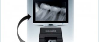
The use of phosphor plates is the next most frequently used method. With this diagnosis, an image of the organs being examined is formed on a special screen. The photo is hidden and can be stored for several hours. An infrared laser scans a hidden image. This equipment can be used in conjunction with an analog radiography apparatus, which improves the quality of visualization.
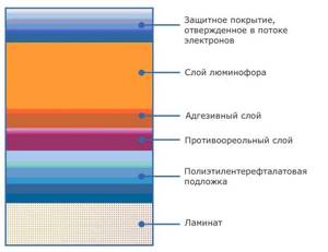
Direct digital examination is distinguished by the high quality of the resulting images. The method belongs to the category of developing diagnostic techniques performed using computer equipment.
What clinical situations can digital x-ray detect?
Doctors refer patients with suspected various pathologies for an X-ray examination. The diagnostic procedure makes it possible to increase the effectiveness of advisory and therapeutic assistance.
Studying the lungs using X-rays allows us to exclude serious diseases: tumors, tuberculosis, and cancer. In some cases, additional diagnostics may be required to clarify the diagnosis.
Using special equipment, a specialist can perform a survey designed to identify the following conditions that require surgical correction:
- perforated ulcer;
- intestinal obstruction;
- stone formation.
When using contrast agents on digital X-ray images, it is possible to identify tumor neoplasms, functional changes in the digestive organs and urinary system.

What can be detected with digital x-ray
Doctors prescribe digital radiography for a variety of pathologies, since it can be used to visually assess the condition of almost all bone fibers and tissues.
- During the diagnosis of the brain, the presence and condition of metastases can be considered. An examination is also often prescribed to assess the condition of the brain after a stroke;
- if X-rays of the lungs and respiratory tract are taken, lung cancer, fibrosis, pneumonia and bronchitis can be detected;
- examination of the abdominal area helps to detect the presence of neoplasms, metastases, abscesses and destructive changes in tissues;
- An X-ray of the spine is usually done if the patient has suffered a serious injury or if a hernia, infection or oncology is suspected.

Doctors assure that such an examination allows you to obtain complete and objective information about the condition of internal organs and bone fibers, therefore it has a huge number of indications for carrying out
Indications and contraindications for the study
Radiation research methods are widespread in many medical fields: traumatology, pulmonology, otolaryngology, neurology, gastroenterology, dentistry.
Indications for digital x-ray examination are the following situations:
- assessment of the condition of bone and joint tissue;
- identification of traumatic injuries;
- diagnosis of tumor formations;
- determination of the focus of the inflammatory process;
- detection of pathologies and developmental anomalies;
- identification of degenerative-dystrophic conditions;
- detection of foreign bodies;
- analysis of the quality of therapy provided;
- preparation for surgical procedures.
There are no absolute prohibitions on taking x-rays. In case of urgent need, the method allows you to examine infants and pregnant women. The possibility of using this study is determined by a medical specialist.
Colorless story
X-rays were discovered by Wilhelm Roentgen in 1895. The scientist experimentally established that radiation can pass through materials, in particular through human tissue, and the denser the medium, the greater the proportion of radiation it absorbs. One of the first photographs taken by the researcher was an X-ray of his wife Bertha's hand, which clearly showed the light silhouettes of bones - bones passed less X-rays than less dense muscles and skin.
Roentgen himself assumed that the rays he discovered would be useful in industrial production, and not in medicine. However, over time, it was doctors who found the most useful application for the discovery. By 1900, hospitals in the developed world were using X-ray machines relatively widely. The device was especially useful in the treatment of gunshot wounds and foreign bodies entering the body, as well as in the treatment of tuberculosis.
Advertising on Forbes
How is the research procedure carried out, features of the study
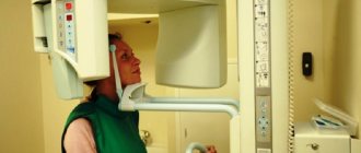
No special preparation is required for the examination. Digital radiography is carried out in the same way as analog diagnostics. Specialized equipment can be installed in clinics and inpatient departments.
To obtain a high-quality image that ensures the accuracy of the result, you must follow the doctor’s instructions:
- remove all metal objects;
- do not move;
- hold the breath.
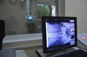
The resulting image is assessed without the participation of the patient. The radiologist examines the resulting image and draws up a conclusion about the detected changes. Conclusions about the patient's condition are provided in writing the next day after the diagnosis.
The examination results do not contain a diagnosis. The conclusion contains conclusions about the shadow pattern of the resulting image. Further diagnosis and treatment strategy are prescribed by the specialist who recommended the diagnosis.
Contraindications
Digital radiographic examination is considered one of the most gentle, and if there is an urgent need, it can be carried out during pregnancy. The examination can also be carried out in childhood. But experts warn that even despite the minimum concentration of radiation, x-rays are performed only if there are absolute indications, since in any case they are harmful to health.
Prescribing digital radiography on your own is strictly prohibited; the examination is carried out exclusively as prescribed by a specialist. The doctor decides on the need for such an examination after conducting a physiological examination, taking an anamnesis and preliminary laboratory tests. Fluoroscopy is often prescribed to confirm the diagnosis or if the doctor has doubts about the patient’s condition.
Pros and cons of digital radiography
Advantages of digital radiography:
- obtaining a high-quality image that allows you to examine the area under study in detail;
- simplicity and speed of obtaining results;
- ease of storing and moving archives;
- the ability to examine a larger number of patients compared to the analogue method;
- possibility of remote consultation;
- reduction of survey costs;
- environmental Safety.
The main disadvantage of this method is the high cost of digital equipment necessary for conducting an X-ray examination. During diagnosis, the patient is exposed to x-rays, which increases the radiation exposure to the body.
Collaboration at the particle level
The new equipment at Mars Bioimaging is called a “spectral computed tomography apparatus.” It is based on the Medipix3 chip, originally developed at CERN for the Large Hadron Collider. Medipix is able to recognize a particle that hits every pixel of the sensor. Thanks to this, it provides high image clarity and contrast.
The Medipix3 X-ray machine has been enhanced with computational algorithms that convert data from the detector into the final image. The colors represent the different energy levels of the X-rays detected by the detector.
“Promising research results suggest that spectral imaging, when used in clinics, will provide more accurate and personalized treatment,” said Anthony Butler, one of the creators of the device.
Clinical trials of the new device will take place in the coming months: the scanner will begin working in one of the departments of a New Zealand hospital.





![Table 4. Major muscular dystrophies accompanied by increased CPK levels [41]](https://ddpskov.ru/wp-content/uploads/tablica-4-osnovnye-myshechnye-distrofii-soprovozhdayushchiesya-povysheniem-urovnya-kfk-41-330x140.jpg)
