- home
- Functional diagnostics
- ECG of the heart
An ECG or electrocardiogram is a method for studying the state of the heart muscle based on data on its electrical conductivity. For people far from medicine, this does not sound very clear. It is important for an ordinary patient to know that this simple and painless diagnostic method allows one to accurately and quickly identify any cardiac dysfunction.
Ease of execution and simplicity made it possible to include a cardiogram in all preventive programs as a screening for cardiovascular diseases (along with measuring blood pressure).
Where in Khabarovsk can you get an ECG?
If you are interested in where in Khabarovsk you can get an ECG for a fee, then contact the medical office. We can undergo a comprehensive examination of the state of the cardiovascular system, which includes an ECG, Holter blood pressure monitoring and ultrasound of the heart.
You can sign up for the procedure by calling 8. We are located at Khabarovsk, st. Panfilovtsev, 38. The administrator will select a convenient day for you to visit a specialist and coordinate the appointment time.

In our center, you can do an ECG directly on the day of treatment, since the method does not require any preliminary preparation.
Recording an ECG in children and pregnant women
Electrocardiography is performed on pregnant women and even newborn children. There are no contraindications to the study, but certain difficulties will arise with people with serious mental disorders. The difference in recording an ECG in an adult and a child is only in the size of the electrodes, which can also be purchased on the Avicena-med medical consumables resource.
Some nuances arise when taking an ECG in people with dextracardia. In such patients, the heart is on the right side, so the results of the study will be exactly the opposite of normal. If the patient is aware of his peculiarity, he is obliged to warn the medical professional.
How much does a cardiogram cost?
The price of an ECG is very low, so any patient can afford to have a cardiogram. The table below shows the cost of different types of examination:
| Code | Name of procedure | price |
| 8.01 | Holter monitoring 24 hours | 2200 |
| 8.03 | Electrocardiography with stress | 1000 |
| 8.04 | Electrocardiography | 700 |
The medical office often holds promotions during which you can get examined for a lower price. Discounts are available on all procedures, including ECG.
How to wear a halter correctly
During the diagnostic period, it is necessary to ensure that the device is not overcooled and does not come under the influence of a magnetic field. You need to follow your doctor's recommendations and ensure that the electrode is firmly attached to the body. In rare cases, the cardiologist decides to use a portable recorder for diagnosis, recording data “on demand”.
It is very important to understand how to remove a halter correctly. You need to check with your doctor in advance about the order in which you should proceed. In most cases, it is enough to simply peel off the sensors and remove the device. Even when the battery is disconnected, the received data remains in memory.
What does ECG mean? Types of electrocardiogram
Cardiac electrocardiography (ECG) is an assessment of the activity of the heart muscle by recording electrical signals. A normal ECG is done in 12 leads, that is, 12 electrodes are attached to the patient’s body - 6 chest and 6 on the limbs (right and left arms, as well as the left leg).
The method helps:
- detect heartbeat rhythm;
- assess the blood supply to the heart at rest;
- find out the conductivity of the heart muscle;
- diagnose chamber enlargement.
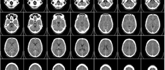
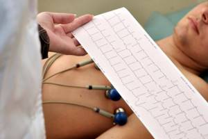
As a result of the examination, the equipment produces a paper tape with a curve depicted on it, repeating the work of the heart muscle. A non-specialist will see on it only teeth of varying amplitudes, which are repeated at equal intervals. The cardiogram is deciphered by a cardiologist. With its help, you can identify deviations from the norm and diagnose dangerous diseases that require immediate treatment.
An ECG of the heart can be done without any fear for both children and adults. This method is:
- painless, does not cause discomfort;
- safe for well-being and health.
Even a pregnant woman is not prohibited from having an ECG done; it will not cause harm to the growing fetus.
Classic ECG
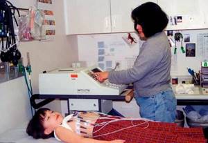
Everything described above related to the so-called classical cardiogram. During data acquisition, the patient lies on the couch without moving. He should try to calm down and think about something pleasant and relaxing. The doctor or nurse places the electrodes on the body according to the diagram and turns on the device. The electrode attachment points are pre-wetted with a special gel for better electrical conductivity.
No more than 5 minutes are allotted for conducting an ECG of the heart. The doctor sees pathological changes on the graph immediately. It takes a specialist 10 minutes to describe the heart cardiogram, or rather decipher the results obtained. This is the case if the results are read and signed manually. If the processing method is computerized, everything happens even faster.
Holter ECG monitoring
24-hour Holter ECG monitoring is a more complex diagnostic technique that involves round-the-clock recording of cardiac parameters. This is done in order to identify pathologies that occur during exercise, sleep, or after taking medications.
The bottom line is that sensors are attached to the patient’s body, connected not to a stationary one, but to a miniature portable cardiograph. In this form, a person can go home and engage in his usual activities - running, sleeping, eating and taking medications. The only thing contraindicated during the examination is water procedures. At the same time, it is very important to keep a detailed diary of your activity, where you need to enter all actions. At the end of Holter monitoring, the doctor will correlate the data obtained from the cardiogram with the patient's records.
ECG with stress
During an ECG, measurements of electrical activity occur during inhalation and exhalation, which makes it possible to obtain results from different angles and at different positions of the heart in the chest. If this is not enough to make a diagnosis, it is recommended to do a stress ECG. We are talking about the Martin-Kushelevsky functional test, which is carried out to detail data on the work of the heart.
In order to see how the heart reacts to the load, the subject is asked to do 20 squats in half a minute. The ideal time is morning. It is advisable not to eat before visiting a specialist. The method is used as a supplement to the classic cardiogram.
As a result of these activities, you can see how the heart reacts to physical activity and how quickly normal heartbeat is restored after its cessation. A stress ECG is used both during general medical examination and to monitor changes in dynamics during the treatment of pathologies of the cardiovascular system. At the decision of the cardiologist, the ECG of the heart can be supplemented with echocardiography data.
You can get an ECG with stress in Khabarovsk at the medical office.
Indications for the procedure

A doctor of any specialization can prescribe an ECG for a child or an adult. More often, this is the responsibility of a cardiologist and therapist, who during an examination suspected disturbances in the functioning of the heart muscle.
In addition, sometimes it is recommended to do an ECG as a routine procedure. This is necessary in the following cases:
- examination before entering school;
- in adolescence, when the body's systems are being restructured;
- School graduates also undergo an ECG of the heart to receive a certificate in form 086/u - for admission to secondary and higher educational institutions;
- before passing the driving test;
- when undergoing any medical examination.
Medical indications for examination may include patient complaints of deteriorating health and poor health:
- Periodically occurring pain in the chest, abdomen, back, neck (suspicion of coronary heart disease);
- The appearance of shortness of breath and difficulty breathing during physical activity and at rest;
- Increased blood pressure and interruptions in heart function;
- Swelling of the legs, general weakness, fainting;
- Previous history of stroke or myocardial infarction;
- Audible heart murmurs, history of diabetes mellitus, rheumatism.
Taking a cardiogram is recommended when preparing a patient for surgery, when registering for sanatorium treatment, to issue a certificate confirming good health for active sports.
It is recommended to do an ECG annually after 40 years of age in order to timely track negative age-related changes in heart function and begin appropriate therapy.
Who should not have a cardiogram?
Since electrocardiography is the easiest and safest diagnostic method, it is often abused by both patients themselves and persons with medical education. Today it is believed that any deviations from the norm visible on the cardiogram should be interpreted with great caution. The doctor often runs the risk of being misled and starting to treat the ECG result rather than the patient himself.
In medicine, cardiologists use a different tactic. Any changes in the cardiogram are interpreted only in conjunction with the patient’s complaints and the results of other examinations - blood tests, blood pressure readings and ultrasound of the heart. For a cardiologist and a specialist in functional diagnostics, it is very important to separate the concepts of pathology and individual norm.
Based on this, a completely healthy patient without any complaints about his health does not need an annual ECG, except if required by his type of activity.
Frequency of the study
During an ECG, sensors attached to the chest do not have any effect on the body. Their only task is to capture and transmit cardiac impulses. That is why an ECG cannot harm a person, even if the study is carried out daily.

Doctors consider the following frequency of ECG to be justified:
- Healthy population under 40 years of age . For such people, one study per year is enough.
- People with chronic illnesses . For patients suffering from cardiovascular diseases, an ECG schedule is prescribed by the attending physician. It is generally recommended that the survey be conducted quarterly.
- Workers associated with risk . Doctors advise people whose profession is associated with risk (for example, the Ministry of Emergency Situations) to undergo examination every six months.
- People who have crossed the 40-year mark . This population needs cardiac testing every 3-6 months. The frequency of an ECG depends on the state of health and is determined solely by the doctor.
An ECG is mandatory for pregnant women. Expectant mothers experience hormonal changes. These disturbances affect the blood circulation process. Therefore, the ECG cannot be ignored. The frequency of the study depends only on the woman’s condition and the course of pregnancy.
How to prepare for the procedure?
Electrocardiography does not require any special preparation. The patient can undergo examination immediately after being appointed by a specialist.
Before visiting a doctor, you should think about your appearance. To attach the sensors, you will need to expose your chest, forearms and lower legs, so it is best to choose clothing that can be easily removed or unfastened.
Some patients are interested in whether it is possible to do an ECG after eating or drinking alcohol? Doctors do not recommend eating later than an hour before the procedure, but it is better to avoid alcohol altogether the day before.
What should you not do before an ECG?
Before performing an ECG of the heart it is not recommended:
- overeat;
- smoke;
- attend training or experience heavy loads;
- drink alcohol or strong coffee.
Anything that may affect the result of the examination is best done after it.
Poor electrode attachment can also distort the results. This happens if you don't degrease the skin enough. Doctors recommend not using fatty creams and body lotions on the eve of an ECG, and then the specialist will immediately notice any changes in the functioning of the heart.
On the eve of the study
Patients who are prescribed an ECG must begin preparing for the examination the day before diagnosis with the following activities:
- Complete peace of mind . On the eve of the ECG, it is necessary to completely protect yourself from stressful situations if possible. Experiences and high anxiety can distort the results of a cardiogram.
- Full sleep . Before the study, be sure to get enough sleep. In a tired body, the heart functions in an enhanced mode.
- Quitting alcohol . It is strictly forbidden to drink alcohol before an ECG. Alcohol causes the blood to thicken, causing the heart to work much harder.
- Proper nutrition . You should not overload the body with too heavy dinner and breakfast before the ECG. It is advisable to exclude fatty and heavy foods from the diet. Otherwise, the body will devote all its reserves to digesting food, and the heart will be forced to work “for wear and tear.”
- Refusal of physical activity . In order for the cardiogram to show reliable results, doctors advise not to exercise on the eve of the ECG, and to do without exercise or jogging in the morning.
- Elimination of thermal procedures . It is not recommended to visit saunas or steam baths before taking an ECG. Doctors advise against taking a bath. Any thermal procedures can increase blood circulation and heart activity.
- Taking medications . The day before the examination, it is necessary to completely avoid taking medications, except those that are prescribed on a regular basis. The doctor must know about the latter.

How is a cardiac cardiogram taken?

The algorithm for taking an ECG is as follows:
- To obtain data, the patient undresses to the waist, freeing his chest from clothing. It is also necessary to expose the skin on the wrists and ankles.
- The patient lies down or sits on the couch.
- The nurse places the suction cups. The application of electrodes during ECG occurs only after treating the surface of the skin with a conductive solution or water to improve the quality of the received signals.
- The device turns on and the specialist takes a cardiogram. How long does the manipulation last? On average about 5 minutes.
It is important that the functional diagnostic room is warm, since physiological tremors can distort the examination results.
How to install electrodes?
The reliability of the results obtained depends on the correct installation of the electrodes. Medical personnel working in the ECG room undergo special training, so they do not have the right to confuse the algorithm for applying branches.
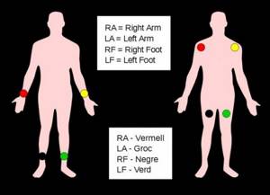
There are a total of 12 leads (standard, amplifying and chest leads), which are recorded by applying 10 electrodes (with a small-channel cable, 4 electrodes are installed on the limb, and the chest leads are recorded alternately with the fifth. The order of installing the electrodes is as follows:
- A green electrode is attached to the right hand;
- A yellow electrode is installed on the left hand;
- A green electrode is attached to the left leg;
- A black grounding electrode is installed on the right leg;
- A green chest electrode is placed in the 5th intercostal space to the right of the sternum;
- At the same level, but to the left of the sternum, a yellow chest electrode is attached;
- A green chest electrode is attached along the left parasternal line, at the level of the 5th rib;
- A brown electrode is attached at the apex of the heart or in the 5th intercostal space along the midclavicular line;
- A black chest electrode is installed along the anterior axillary line at the same level;
- A purple chest electrode is attached along the midaxillary line in the 5th intercostal space.
All electrodes can have different sizes and configurations, which depends on the type of study (one-time recording, daily monitoring or stress test) and the features of the device. In addition, there are disposable and reusable electrodes. You can choose the appropriate option in the online store, but keep in mind that disposable electrodes are designed for modern models of electrocardiographs.
How to read ECG results?
The most important thing in electrocardiography is the interpretation of the results. Any medical practitioner can buy an ECG device and is treated by competent cardiologists who are ready for medical cases of any complexity.
Decoding a cardiogram is a complex issue, requiring specialized education, extensive experience and great passion for one’s work. You should not try to do this on your own, calling on your neighbor-nurse, books and the Internet for help. Trust the professionals in your field to get the correct diagnosis.
General concepts about describing an ECG
During an ECG, the work of the heart is examined by recording bioelectrical parameters. Its results are reflected by an electrocardiogram - a graphic image of the received impulses, formed in graphics on a paper tape. Each graph represents the characteristics of the work of different parts of the heart muscle. The cardiac cardiogram consists of parts or leads - I, II, III, aVR, aVL, aVF.
What signs on the ECG may indicate the presence of cardiac pathology:
- incomplete blockade of the right or left bundle branch;
- left ventricular hypertrophy;
- sinus rhythm disturbance and others.
What diseases can be detected using a cardiogram?
Although an ECG cannot serve as the only criterion for making a diagnosis, its results allow one to suspect a wide range of pathologies. Based on the ECG findings, the cardiologist may prescribe other examinations and laboratory tests.
Most often, the following syndromes and diseases are detected on the cardiogram:
- atrial fibrillation;
- heart block;
- extrasystole;
- atrioventricular block;
- ventricular or atrial hypertrophy;
- tachycardia;
- myocardial infarction;
- coronary heart disease (CHD).
Depending on the severity of a particular pathology, doctors may assume an acute or chronic course of the disease.
Decoding indicators
ECG is assessed according to several criteria:
- The rhythm is correct and regular. No extraordinary contractions (extrasystoles).
- Heart rate. Normally - 60–80 beats/min.
- Electrical axis - normally R exceeds S in all leads except aVR, V1 - V2, sometimes V3.
- Width of the ventricular QRS complex. Normally no more than 120 ms.
- QRST - complex.
QRST - normal complex
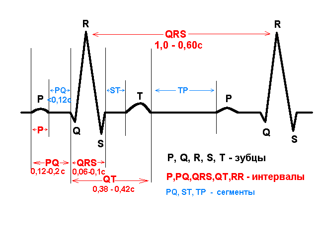
Brief designation of the main elements of the film:
- P wave - shows atrial contraction;
- PQ interval is the time the impulse reaches the atrioventricular node;
- QRS complex - shows ventricular excitation;
- T wave - indicates depolarization (restoration of electrical potential).
Video about ECG norms from the Mass Medika channel.
How is an ECG done for children?
Giving an ECG to a child can be challenging. It is difficult, and sometimes impossible, to make a very young patient lie quietly for several minutes. Children's crying, hysteria and panicky fear of doctors will certainly affect the results of the cardiogram, distorting the picture.
The main role in preparing the child is played by parents, who should, if possible, explain to the child what needs to be done without frightening or exciting him.
Taking any sedatives (for example, valerian) before an ECG is unacceptable!
Preparing for an ECG
On the day of the ECG diagnosis, you need to get a good night's sleep. It is not advisable to perform any physical exercise (exercise, jogging, etc.). If the ECG at the clinic is scheduled for early morning, then you need to avoid a heavy breakfast. During the day procedure, you should limit yourself to a light snack 2 hours before the start of the session.
It is important to avoid tea, coffee, energy and other drinks on the day of electrocardiography, as they stimulate the activity of the heart, as a result of which the results may be distorted. The use of medications that have a vasoconstrictor effect (including some eye drops, cold remedies, etc.) is excluded. Smoking is strictly prohibited 2 hours before the cardiac ECG.
You should not apply various care products (lotions, creams) to your body, as their components form a greasy film on the surface, which in turn negatively affects the contact of the electrodes with the skin.
Reviews about taking a cardiogram in a medical clinic
Our doctors have unique experience in conducting diagnostic procedures and a high level of professionalism in assessing the results obtained. We perform an ECG of the heart to prevent dangerous diseases, as well as to monitor the effectiveness of treatment over time. We work with children and adults. Our patients are always satisfied with the results of our cooperation.
Many thanks to Olga Yuryevna! I took my mother to see a therapist. They took a cardiogram, immediately did an ultrasound of the heart and immediately received a full consultation with a doctor with recommendations and treatment. Mom and I were satisfied. Thank you for your attentiveness, sensitivity and high professionalism! Take care of such doctors.
Anonymous patient, July 06, 2012
Why does an ECG help us judge the condition of the heart?
The human heart is essentially a pump that keeps blood moving.
To do this, the heart muscle either contracts or relaxes. The rate of contraction of the heart forms the heart rhythm. A healthy heart contracts at regular intervals, and the duration of this interval should also fall within the norm, which depends on age. A normal resting heart rate usually ranges from 60 to 100 beats per minute. In children under 12 years of age, higher values will be normal (for example, under the age of 1 year, the average value is 130 beats per minute). With age, the heart rate slows down: in older people, the normal heart rate is 60-65 beats per minute. The contraction of the heart muscle is regulated by electrical impulses generated in the heart itself. The main role belongs to a group of cells that form the so-called sinus node, located in the right atrium. It is he who generates impulses that normally set the contraction of the heart. In this case, the bioelectric mechanism of the heart is designed in such a way that the muscles of the atria contract almost simultaneously, pushing blood into the ventricles of the heart, and the muscles of the ventricles also contract simultaneously, but with a delay relative to the atria of about one tenth of a second. This time is necessary for the ventricles to fill with blood. Any violation of this complex and precise mechanism of generation and propagation of electrical impulses leads to disturbances in the functioning of the heart, manifested in the form of symptoms of heart disease. Conversely, heart pathology affects the accuracy of the bioelectric mechanism. Therefore, when symptoms of heart disease occur, first of all, an ECG is done.