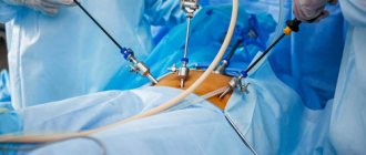
Hysterosalpingography is one of the methods for gynecological examination of female reproductive health. During the procedure, the area being examined is exposed to x-rays, thanks to which the diagnostician (in this case, a radiologist) receives information about the condition of the fallopian tubes and can assess their patency.
Why might such a question as the degree of patency of the fallopian tubes in the female body even arise? The fact is that this factor directly affects a woman’s ability to bear children. And the number of patients diagnosed with infertility, unfortunately, has tended to increase over the past decades.
The uterus and fallopian tubes: what they are responsible for in the body
The uterus is an unpaired hollow internal organ, one of the main functions of which is the development of the embryo and gestation of the fetus. Anatomically, the organ is represented by the bottom, body and neck, with the neck being at the bottom and representing a narrowing of the organ, and the bottom is located at the top. The place where the uterus meets the cervix is called the isthmus. The lower part of the cervix passes into the vagina, and the upper part lies above it. The body of the uterus has anterior and posterior surfaces. In front of it is the bladder, behind it is the rectum.
Content:
- The uterus and fallopian tubes: what they are responsible for in the body
- What is hysterosalpingography
- In what cases is a patient prescribed an examination?
- What contraindications does the procedure have?
- Preparation requirements for hysterosalpingography
- Examination technique
- Diagnostic results: what to do next
- Possible complications and consequences of the procedure
The size of the organ and its mass change throughout a woman’s life. The length of the uterus in an adult woman reaches 7-8 centimeters. The weight of a nulliparous woman does not exceed 50 grams, and that of a woman who has given birth does not exceed 80-80 grams.
Fixation in the pelvic area occurs due to the left and right broad ligaments, round ligament and cardinal ligaments of the uterus.
The organ, which has the shape and appearance of a pouch, is formed by three-layer walls. The inner layer is a mucous membrane covered with ciliated epithelium. From the outside, the organ is covered by a serous membrane, and in the middle the wall is formed by muscle tissue.
It’s not for nothing that the fallopian tubes got their name – these organs look like hollow tubes. There are two of them in a woman's body. The length of each tube ranges from 10 to 12 centimeters, the lumen diameter is up to 2-4 millimeters. The uterine, or fallopian, tubes are directly involved in the reproductive process - through them the egg from the ovary passes into the uterus, in it the moment of fertilization of the egg by the sperm occurs, and the embryo moves through the tubes to the uterus. The organs are located on the sides of the fundus of the uterus, their narrow end opens into its space, and their wide end opens into the abdominal cavity, that is, the tubes connect the abdominal cavity and the uterus.

Their structure is presented:
- funnel;
- ampoule;
- isthmus;
- uterine part.
The structure of the wall of the fallopian tube is similar to the wall of the uterus: it also contains three layers (serous, muscular, mucous).
The ability to diagnose tubal patency, as well as check the condition of the uterus, gives women who cannot conceive or carry a baby a chance to become pregnant and give birth to a healthy child.
What is an X-ray machine?

An X-ray machine is equipment that is used in medicine to obtain analytical data about a patient’s condition. Thanks to X-ray radiation, an image is formed that allows one to assess the condition of internal organs, the condition of bone and muscle tissue, and detect pathological changes.
The X-ray radiation generated by an X-ray tube is often used in other installations. In particular, the X-ray tube is built into a tomograph, introscope and other systems that allow a comprehensive examination of the object.
There are more than 10 different types of X-ray machines, and each one performs a specific task. The invention of this equipment made it possible to make significant advances in the field of medicine and save hundreds of lives.
What is hysterosalpingography
The procedure is often prescribed specifically to diagnose the causes of infertility. There are two types of hysterosalpingography, depending on the method of conducting the study:
- echohysterosalpingoscopy;
- X-ray hysterosalpingography.
Each of the techniques is a method of obtaining an image of the fallopian tubes and/or uterus, their external and internal state. In the first case, the patient is examined by an ultrasound diagnostician using a special scanning machine with an attachment. Before this, a saline solution is injected into the woman's uterus. This method is more suitable for studying the functional characteristics of the uterus. The classic type of examination is x-ray with contrast. It, unlike an ultrasound examination, visualizes the patency of the fallopian tubes.
How does an x-ray work?
The X-ray machine is equipped with the following parts and components:
- emitter tube generating X-rays;
- a generator that provides the emitter with the necessary parameters of electrical energy, with its help the current of the X-ray tube is regulated;
- a device for converting radiation into an image - a detector in digital devices or film in analog devices;
- X-ray equipment control unit;
- tripods that allow you to control the installation.
The principle of operation is quite simple: X-rays penetrate the body, internal tissues absorb them. Based on the degree of absorption, an image is formed that can be displayed on a monitor or on a special film. If necessary, a contrast agent is introduced to obtain a clearer image.

In what cases is a patient prescribed an examination?
The most common indication for hysterosalpingography is infertility. If a woman who does not have previously diagnosed reproductive health disorders cannot become pregnant within a year, with one regular sexual partner, without using contraceptive methods, a gynecologist or reproductive specialist can refer her to this type of study.

Other indications for the procedure:
- presence of previously diagnosed uterine pathologies;
- malformations of the uterus and fallopian tubes;
- suspicion of cancer;
- the likelihood of tuberculosis of the genital organs;
- a history of pregnancies that ended in miscarriage or fetal death.
Scope of application of the X-ray machine
X-ray machines are mainly used in medicine. In 1895, William Roentgen discovered radiation that can penetrate any substance. Designers from around the world immediately created a huge number of devices that allow for a comprehensive examination. But X-rays are used not only in medicine; the scope of application of the device is as follows:
- Veterinary medicine. Allows you to examine organs and tissues of animals, promptly find and eliminate pathologies.
- Industry. Using the device, it is easy to control the quality of materials, detect defects, cracks and cavities.
- Art history. With the help of such research, you can study the paintings of artists, confirm authenticity, discover hidden layers of paint, and so on.
- Also, with the help of X-ray inspection units, the safety of passengers at airports and postal correspondence is ensured. X-raying things allows you to identify dangerous objects.
What contraindications does the procedure have?
Due to the fact that this diagnostic method involves the introduction of a contrast agent or saline solution into the uterus and tubes, and the classical examination, moreover, is associated with X-ray irradiation, hysterosalpingography is not prescribed:
- at the slightest suspicion of pregnancy;
- with hypothyroidism;
- in the presence of diagnosed renal or liver failure, liver cirrhosis;
- for acute inflammatory processes in the genital organs, inflammation of the vagina and vulva;
- with uterine bleeding;
- if you are allergic to contrast agents;
- in the general serious condition of the patient;
- for infectious diseases of the genital organs;
- with thrombophlebitis and acute heart failure.
Types of X-ray machines
X-rays themselves are not used very often. However, emitters are used in other equipment, which allows for a comprehensive examination of the patient. Therefore, a large number of types of X-ray machines have appeared.
Here are the main ones:
- General purpose X-ray diagnostic apparatus. This is just a device in its purest form, which performs one function - the formation of one image.
- Angiograph. Provides examination of blood vessels, helps identify aneurysms, vascular malformations, thrombosis and a number of other diseases.
- Dental X-ray machine. This is a device that provides detailed data to dentists. Used in dental clinics to determine pathology and control the installation of pins and filling canals.
- X-ray mammograph. Allows you to diagnose the condition of the mammary gland, determine pathologies, including cancer.
- Fluorograph. A device that allows you to assess the condition of the respiratory system, examines the lungs and helps to detect tuberculosis and other pathologies in a timely manner.
- X-ray computed tomograph. Allows you to conduct a comprehensive examination and detect pathological structures throughout the body. If necessary, contrast enhancement is used to provide a clearer image.
- X-ray therapy apparatus. Allows treatment of patients with cancer. With the help of radiation and radiotherapy, cancer cells are eliminated, metastases are destroyed and tumor development is stopped.
- Flaw detection X-ray machine. It is used for industrial control and allows you to determine defects and damage to products.
- Pantomographic dental apparatus. Used to obtain a complete panoramic image of the dental system. Used to create mouth guards, dentures and eliminate oral pathologies.
These are the main types of X-ray machines, but there are others that are not used as often.
Preparation requirements for hysterosalpingography

The diagnostic procedure is mainly prescribed for the first half of the menstrual cycle. It is best to carry it out in the first few days after the end of menstruation, but hysterosalpingography is allowed within two weeks after its completion. Such requirements are determined by the structural features of the female body - during this period the endometrium of the uterus is thinnest and the cervix is soft, so inserting a catheter requires practically no effort, and the doctor has a better view. In this case, it is imperative to wait until the release of menstrual blood stops completely, since the presence of blood clots in the vagina and uterus makes diagnosis very difficult.
As for determining the patency of the fallopian tubes, the procedure for this purpose is recommended to be carried out during the second phase of the cycle.
Also, as a preparation, the doctor usually directs the woman to undergo general blood and urine tests and a smear for flora. Before conducting the examination, it is necessary to make sure that there are no pathogenic microflora in the vagina, since catheterization of the uterus in this case can lead to infection entering its cavity.
X-ray examination of the pelvic bones

To most accurately determine pathologies of the pelvic bones, radiography is most often used. This is a simple technique, the result of which is highly informative and accurate. The main advantages of x-rays are:
- accessibility – there is an X-ray machine in almost every clinic;
- painlessness;
- relative safety;
- efficiency - together with decoding, this diagnostic procedure lasts approximately 10 minutes;
- simple preparation for a pelvic x-ray (how to do it will be described below).
Examination technique
During the procedure, the patient lies on a couch or a special gynecological chair. The X-ray machine is located above it, and if echohysterosalpingoscopy is planned, the doctor uses a special vaginal sensor.
Before inserting a catheter into the genitals, the doctor disinfects the cervix, vagina and external genitalia with an antiseptic solution. The catheters and devices used in the process must be strictly sterile, and a special condom is placed on the ultrasound sensor.
Before inserting the catheter, the doctor examines the patient using a speculum. After this, a soft catheter is inserted into the cervix, through which a contrast agent is injected with a syringe. The substance enters directly into the uterine cavity and gradually passes into the fallopian tubes. At this time, the doctor takes a series of x-rays.
Echohysterosalpingoscopy of the uterine cavity follows a similar pattern: the doctor injects a saline solution into the uterine cavity, after which he carefully inserts the ultrasound device sensor.
The procedure usually does not cause severe pain or discomfort. The sensations during it are reminiscent of the pain syndrome in the first few days of menstruation. In this regard, most often, the use of anesthesia is not required. However, if the patient knows that the first days of her menstruation are very painful, and if there are no contraindications to the use of anesthesia, the doctor may administer local anesthesia before starting the diagnosis. General anesthesia is not used for this procedure.

If there is a possibility of spasms of the fallopian tubes or uterus, the doctor suggests taking antispasmodics, for example, the drug “No-shpa”. Most often, in such cases, an injection is given so that the active substance enters the blood faster.
How is the examination carried out?
Typically, x-rays are taken in a direct (antero-posterior) projection. The examination is carried out in a strictly horizontal position of the subject. The patient lies down on the work table of the X-ray machine, extends his legs and rotates them inward by approximately 15° (provided that he does not suspect a fracture or dislocation of the hip joint). A special cushion is placed under the knees of the person being examined. The elbows are located on the sides of the body, the hands are placed on the chest.
During filming, the patient must remain motionless and after taking a deep breath, do not breathe for several seconds. The quality of the pictures depends on this.
In addition to X-rays of the pelvis in a direct projection, other images can be taken to clarify the diagnosis:
- in the posterior axial projection of the pelvic inlet;
- in the posterior oblique projection of the pelvis.
Diagnostic results: what to do next
The obtained images of the uterus and fallopian tubes, or images of echohysterosalpingoscopy, are interpreted by a diagnostician. He draws up a conclusion on them, in which he displays the most objective and reliable information.
X-rays show the filling of the fallopian cavity and fallopian tubes with contrast material. If the drug passes freely through the tubes and is visualized in the abdominal cavity, then everything is in order with the patency of the fallopian tubes. If the image clearly shows that the fluid did not pass and stopped at a certain level, this confirms the presence of obstruction. The alternation of dark and light areas in the pipes in the image indicates that they have adhesions.
In addition, the peculiarities of the distribution of contrast throughout all examined cavities and organs makes it possible to see neoplasms, polyps, and foci of inflammation.
Also, the diagnostician draws conclusions about the size of the uterus and fallopian tubes, the features of their structure and location, and the structure of the inner wall of the uterus from photographs or the monitor of an ultrasound machine. For example, its uneven relief may indicate the presence of adhesions, inflammation, polyps or fibroids.
If the results of the examination give reason to suspect the presence of uterine cancer, it is necessary to order additional examinations, including taking tissue for a biopsy.
The diagnostic doctor's conclusion, along with the photographs, is transmitted to the attending physician, who referred the woman for examination.
After hysterosalpingography, the patient may experience mild vaginal bleeding for several days. Pain in the lower abdomen that appears during the examination usually goes away after 20-30 minutes. In the next 3-4 days, you should avoid sexual intercourse, visiting a bathhouse, sauna, or taking a bath.
Decoding the results
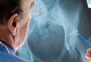
Immediately after the x-ray, the radiologist begins interpreting and describing the images. To do this, he needs to assess the following qualitative and quantitative indicators:
- size of the neck-shaft angle;
- the magnitude of the Wiberg angle and the degree of change;
- the angle of the femoral neck (to identify antetorsion, inclination);
- value of the width of the sacroiliac joint;
- the width of the gap between the bones of the hip joint.
The following additional pathological signs are also accepted for analysis:
- deformation of the femoral head;
- dislocations and subluxations of the hip joint
; - rotation of joint bone fragments;
- displacement of the femur in width or length;
- expansion/constriction of the symphysis.
When interpreting, the radiologist does not make a specific diagnosis. He describes the pathological signs that are visualized in the images, and the attending physician correlates them with other pronounced clinical signs and makes a conclusion about the presence of a particular disease.
Normal indicators
The X-ray image should show a symmetrical image of the two halves of the pelvis, the sacrum, the intervertebral foramina of the sacrum, as well as the branches of the pubic and ischial bones. The bone substance should be clearly visible, the contours of the two acetabulums and the neck of the femur should be visible.
There are certain indicators of the normal condition and structure of the hip joint, with which the actual data are compared when deciphered. For example, the Wiberg angle should normally be located between the line of the center of the femoral head and the superior-outer edge of the acetabulum. The normal angle is approximately 30 degrees. The angle of inclination of the entrance to the acetabulum is also normal - 31-42 degrees. The neck-shaft angle should normally be from 115 to 140 degrees.


