Having a hysterectomy is a difficult decision for both the woman and her doctor. However, in some cases, hysterectomy remains the only correct solution.
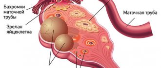
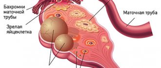
The uterus is a hollow organ located in the woman’s pelvis, which consists of 3 layers:
- epithelial, represented by the endometrium;
- muscular (myometrium);
- serous (perimetry).
The uterus maintains its position thanks to ligaments, when weakened, prolapse or prolapse of the organ occurs. From the bottom up in the uterus, the cervix, body and fundus are distinguished.
Indications for hysterectomy
Hysterectomy is a serious operation leading to complete loss of the organ being removed, which requires strict indications. The reasons for radical surgery may be:
- Adenomyosis is the growth of the endometrium into the muscular layer of the uterus, which can be accompanied by severe pain and bleeding.
- Malignant neoplasms of the endometrium. Hysterectomy is used as one of the options for solving the problem, but it is far from the only one. The selection of treatment is carried out individually based on the morphology of the tumor. Oncology today is the fastest growing industry, so they try to resort to radical operations as rarely as possible. However, there are situations when complete removal of the uterus cannot be done.
- Uterine fibroids. Multiple fibroids in combination with cervical pathology create a high risk of necrosis and hemorrhage.
- Endometriosis is the growth of the endometrium outside the uterus.
- Heavy bleeding
- Prolapse or prolapse of the uterus, which poses a threat of permanent infection.
Indications for laparoscopy
Diagnostic laparoscopy is performed for visual examination and collection of biomaterial for examination.
Laparoscopy provides full visual and contact access to the internal organs, so it is often used for these procedures:
- Surgeries for ectopic pregnancy;
- Surgeries for ovarian cysts;
- Acute inflammatory processes in the body (tubovarial tumors, purulent tumors of the ovaries, fallopian tubes);
- Oncological (malignant) tumors of the uterine appendages (usually the ovaries);
- Correction of developmental defects of the reproductive organs;
- Research into the causes of infertility;
- Creation of artificial obstruction of the fallopian tubes for the purpose of contraception (surgical sterilization);
- Treatment of genital (external) endometriosis;
- Removal of pelvic organs (uterus, fallopian tubes, ovaries, etc.);
- Identifying the causes of pain in the lower abdomen of unknown etiology (chronic pelvic pain).
The capabilities of laparoscopy are expanding every year, which increases the scope of its application. This allows us to treat a wide range of gynecological diseases in a less traumatic way.
Types and methods of operations
In gynecology, vaginal or laparoscopic access is predominantly used. This method of surgical intervention has a number of advantages for both the doctor and the patient. Laparoscopic hysterectomy minimizes invasiveness, trauma, blood loss, and provides good visualization for the surgeon during surgery. The patient receives the opportunity for early rehabilitation and minimal cosmetic defects that heal quickly.
The method and type of hysterectomy depends on the reason for removal, as well as the goals and objectives of surgical treatment.
Methods for removing the uterus:
- Vaginal;
- Laparotomy (through one incision on the anterior abdominal wall);
- Laparoscopic (access through small incisions in the abdomen).
Laparotomy is usually used when there are contraindications or when it is impossible to perform the operation via vaginal or laparoscopic access. With this method of intervention, a cosmetic defect in the skin of the abdomen remains.
Preference is given to laparoscopic access in several cases:
- the patient has an anatomical obstacle in the form of a narrow vagina;
- severe obesity;
- removal of the uterus along with appendages;
- the need for revision of the abdominal organs, subdiaphragmatic space, lymph nodes (most often for oncological reasons for removal of the uterus);
- expected adhesive process in the abdominal cavity;
- availability of specialists of the appropriate level and the necessary endoscopic equipment.
Recently, more and more surgeons prefer vaginal access with laparoscopic assistance, which allows combining the advantages of two methods at once.
Types of hysterectomy:
- subtotal - partial removal of the uterus while preserving the cervix. In other words, supravaginal amputation of the uterus. This type of resection is performed more often at a young age in the absence of pathology from the cervix;
- total - removal of the uterus along with the cervix;
- radical - removal of the uterus along with the appendages, the upper part of the vagina, surrounding lymph nodes and tissue.
A little about the history of the development of laparoscopy
In our country, as well as throughout the world, the development of laparoscopy continues. Unfortunately, in the outback, such operations still remain the exception and not the rule, although laparoscopy has existed in the world for more than 100 years.
The first experience of laparoscopy was described back in 1910, and until the mid-twentieth century, laparoscopy was of a diagnostic nature, it developed, more and more complex equipment was created, and safe lighting systems were developed.
But after 1950, laparoscopy began to be used for surgical interventions, first to stop intra-abdominal bleeding, puncture cysts, and then to separate adhesions and collect eggs. It was from this moment that a breakthrough occurred in the treatment of infertility. Before the advent of laparoscopy in gynecology, this diagnosis sounded like a death sentence for married couples. Currently, all possible operations in gynecology are performed using laparoscopic access, including for malignant diseases of the pelvic organs.
Preparation
Preparation for surgery is an important stage, which largely ensures the success of the upcoming manipulation. Standard preparation includes general clinical examinations, colposcopy, cytology smear, examination for sexually transmitted infections, reconciliation or discontinuation of medications that the patient is taking.
In the case of a large uterus, hormonal drugs are usually prescribed for 2-3 months to reduce it. Particular attention is paid to patients who are at risk for thromboembolic complications. Such women are prescribed drugs that improve the rheological properties of blood a few days before surgery. All patients are given a cleansing enema on the eve of the intervention. The operation is performed on an empty stomach in the first half of the day.
Execution technique
The surgery is performed under general anesthesia. Already in the operating room, antibiotics are administered to prevent surgical infection. The technique used depends on the chosen access and the extent of resection.
In general, the operation can be divided into several stages - preparation of the uterus, intersection of ligaments, vascular hemostasis, organ cutting and closure of the resulting defect.
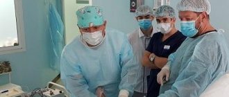
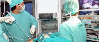
Laparoscopically assisted vaginal hysterectomy, which combines the advantages of several approaches, is of some interest. During the operation, three trocars are used, which are inserted through small incisions on the anterior abdominal wall. A uterine manipulator is inserted through the vagina, with the help of which the surgeon coordinates the position of the uterus in the pelvis, which facilitates the intervention, reduces the duration and prevents complications. Then the round ligaments are crossed on the right and left, and the bladder is separated.
With a subtotal hysterectomy, the surgeon cuts the fallopian tubes and the ovarian ligaments without affecting the cervix, while a total resection involves the removal of all parts of the organ. The next stage is hemostasis. The uterus is abundantly supplied with arteries and veins that feed it. The vessels are coagulated at the level of the internal os, where after hemostasis the body of the uterus will be cut off from its cervix. The resulting defect is closed using peritoneal sheets during subtotal hysterectomy. This process is called peritonization. Whereas with total resection, sutures are placed on the vaginal wall. The uterus is removed using a morcellator, a device for grinding tissue. The organ is removed through the abdominal cavity or through the posterior vaginal fornix. At the end of the operation, the obstetrician-gynecologist carefully checks the pelvic cavity and monitors hemostasis.
For women at risk for developing prolapse, after removal of the uterus, the posterior vaginal vault is strengthened. This manipulation is called culdoplasty.
How is laparoscopy of an ovarian cyst performed?
At the Yusupov Hospital, laparoscopy of ovarian cysts is performed under general anesthesia. On the day of the operation, the patient is consulted by an anesthesiologist who assesses the woman’s condition and decides whether there are any contraindications to surgery. The use of tracheal intubation allows you to regulate breathing and maintain the required depth of immersion in anesthesia. Before laparoscopy, the doctor administers premedication, which consists of tranquilizers that have a sedative and hypnotic effect. Sometimes mask anesthesia is used instead of injection drugs.
On the operating table, the patient is in the Trendelenburg position - this is a forced position of the body when the head tip tilts down 30º. This position provides access to the ovaries by moving the intestine toward the diaphragm. After processing the surgical field, a puncture is made in the abdominal wall, through which carbon dioxide enters the cavity. Thanks to the gas, the space between the internal organs increases. This allows for better visibility during surgery. Then a laparoscope, a special instrument with a light source and a camera attached to the end, is inserted into another hole. After advancing the instrument to the ovaries, 2 more incisions are made in the lateral sections of the abdomen, which are intended for the introduction of additional instruments.
If there is a need for wide access, then all instruments are removed and surgeons begin laparotomy.
Depending on the type and location of the cyst, removal can be done in different ways (at the discretion of the doctor). Once completed, a thorough check is carried out, after which the instruments are removed and the carbon dioxide is sucked out. Skin sutures and an aseptic bandage are applied.
The patient is transferred to the ward if there are no breathing problems or other complications.
Rehabilitation period
All patients, regardless of the chosen access, are prevented from infectious postoperative complications with the help of antibacterial drugs, and are also prevented from thromboembolic complications. The rehabilitation period largely depends on the chosen approach and the degree of tissue trauma.
The postoperative period after surgery via vaginal access includes activating the patient on the 2nd day after the intervention, using pneumatic cuff compression or wearing compression garments for at least 2 months after surgery to prevent thrombosis. Sutures are treated throughout the entire period of hospital stay. It is necessary to thoroughly toilet the perineum after each urination. Discharge is carried out on the 2-3rd day after surgery. The patient is advised to follow a diet to avoid constipation and limit heavy lifting. Gynecological examinations using a speculum are also excluded for 6 weeks after surgery.
The rehabilitation period after laparotomy lasts longer than with other types of surgical interventions. The basic principles of postoperative management of such patients are adequate pain relief, gentle stimulation of the intestines and careful treatment of the postoperative wound. As with the vaginal approach, patients are allowed to stand up on the 2nd day after surgery. To prevent thromboembolic complications, in addition to medicinal methods, they use bandaging of the lower extremities, wearing compression garments or using pneumatic cuff compression from the first day of the postoperative period. To prevent abdominal hernia, it is recommended to wear a bandage for at least 2 months after the intervention. Patients after laparotomy are discharged on the 4-5th day after surgery. After discharge, it is recommended to continue antithrombotic therapy. On an outpatient basis, NSAIDs are prescribed as rectal suppositories for pain relief for 10 days.
Rehabilitation after laparoscopic surgery takes the least amount of time on average. All patients are indicated for early activation on the 1st day after the intervention. Pain medications are most often only required for the first 24 hours. In rare cases - more than 2-3 days. Discharge from the hospital also occurs on the second day. Full work capacity is restored by 5-6 weeks, if the operation was completed without complications.
If any complications arise after discharge, it is recommended to immediately contact the specialist who operated on you.
Sexual life after hysterectomy
After removal of the uterus, it is recommended to temporarily avoid sexual intercourse. For vaginal and abdominal surgery it is 2 months, while for laparoscopic surgery it is from 4 to 6 weeks. This is necessary so that the tissues injured by the operation regenerate and remain in place without stitches. During sexual arousal, blood rushes to the genitals, which can cause bleeding, so it is important to follow the timing recommended by the doctor.
Hysterectomy does not disrupt the production of hormones and therefore does not affect hormonal levels or libido levels. The clitoris is responsible for sexual pleasure in the anatomy of the female genital organs, the structure of which is in no way affected when the uterus is removed. The sexual partner will not know about the hysterectomy unless the woman tells her.
Back to list of articles


