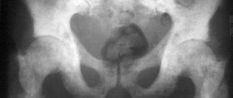
Pathological processes in the bladder often have similar clinical manifestations: pain in the lower abdomen, pain during urine discharge, frequent urges, episodes of incontinence, detection of leukocytes, red blood cells, bacteria, protein in tests, etc. The multifactorial nature of the occurrence of disorders leads to difficulties in diagnosis.
MRI of the bladder is a non-invasive study that shows what contributes to the appearance of the above symptoms: inflammation, tumor, deviation in the anatomical position of the pelvic organs, stone, injury, foreign body or other cause. Carrying out magnetic resonance scanning for malignant neoplasms allows one to obtain important information that is the basis for choosing therapy: the stage of the disease is distinguishable in the images.
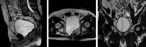
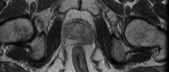
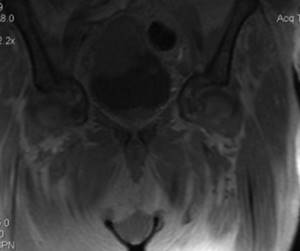
Three-plane visualization of the pelvic organs on MRI
Chelated gadolinium is used to improve imaging capabilities, and contrast-enhanced scanning can be performed in patients with allergies to iodine and seafood for whom enhanced CT is not available.
With MRI of the bladder, images are constructed using magnetic resonance and computer processing of the information obtained. There is no ionizing radiation exposure, changes in cells are completely reversible. This feature makes magnetic resonance imaging the most suitable procedure for dynamic monitoring of the pathological process, including after surgical treatment or conservative therapy. MRI examination can be performed on pregnant women (from the second trimester) and newborns who have reached the age of one month.
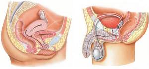
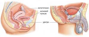
Anatomy of the bladder
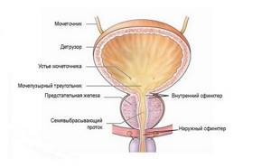
Structure of the bladder
The organ in question is a muscular-membranous sac located in the anterior section of the small pelvis, mainly extraperitoneally - covered with peritoneum on one side. The bladder is separated from the pubic symphysis by loose tissue. When filled, the organ straightens out and occupies a mesoperitoneal position (covered by peritoneum on three sides), rising above the pubis. In case of acute urinary retention, it can reach the navel.
In men, the bladder is separated from the rectum by vesicles and the terminal sections of the vas deferens; in women, by the vagina and uterus. The organ is divided into front, back and side walls, apex and bottom. In Lieto's triangle (formed by the draining orifices of the ureters and the internal opening of the urethra), tumor neoplasms are most often diagnosed. The muscles of the organ form the detrusor and sphincter, which are responsible for expelling and locking fluid.
The size of the bladder depends on the amount of urine contained inside; the capacity is on average 300-350 ml. In pathological processes associated with obstruction of the lower urological tract, it can reach 1.5 liters. Against the background of prolonged artificial diversion of urine, muscle atrophy and a decrease in volume to 30-50 ml are recorded.
Histologically, the bladder consists of the urothelium, vascular plate, muscular and serous layers.
Bladder diseases
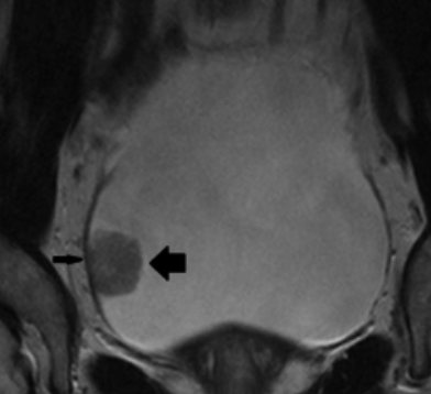
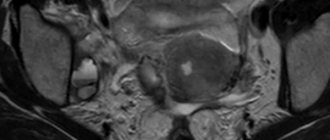
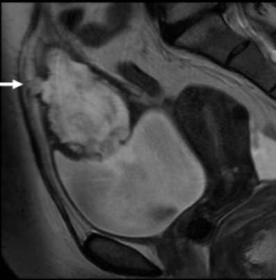
MRI of the bladder, T2-weighted image, axial plane. The thick arrow indicates the tumor, the thin one demonstrates the continuity of the detrusor muscle, which corresponds to the stage of the tumor process T1
Magnetic resonance imaging is used primarily for the diagnosis of malignant neoplasms. More than 90% of carcinomas are transitional cell tumors originating from the urothelium. MRI of the bladder with contrast enhancement shows the exact size and position of the tumor, the degree of invasion (germination) into surrounding tissues, the condition of the pelvic, inguinal, para-aortic lymph nodes, which are targets for the spread of urothelial cancer, and bone structures. The ability to use multiple MR sequences increases the accuracy of the result.
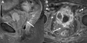
T2-weighted (A) sagittal and T1-post-contrast (B) axial images, white arrow indicates rectal invasion, black arrow indicates abdominal wall involvement, corresponding to cancer stage T4b
If bladder cancer is suspected, cystoscopy is often performed, the advantages of which include the ability to perform a biopsy and determine the histological type of the tumor. The study visualizes parietal changes in the organ, but does not give an idea of the extent of the pathological process into the deep layers. There are certain limitations to endoscopic diagnosis, which is associated with a number of complications (trauma, addition of secondary microflora).
Ultrasound sonography is not always informative for formations smaller than 1 cm. Therefore, patients are often referred for MRI.
Magnetic resonance scanning is performed to determine the extent of the upcoming surgical intervention. Tumors of the rectum, uterus, and prostate can spread to the bladder. MRI helps in differentiating external and mural processes.
Metastatic lesions of the organ are rarely diagnosed. Primary tumors include melanoma, breast and stomach cancer. In the photographs, the dropouts resemble focal formations. Metastasis to the bladder may appear as diffuse wall thickening.
In endometriosis, organ damage is caused by direct implantation of the inner layer of the uterus, which is associated with surgery in the pelvis. The imaging pattern of the pathology is nonspecific, with MRI showing an indentation along the posterior wall of the bladder with obtuse angles (a pattern reminiscent of other parietal processes).
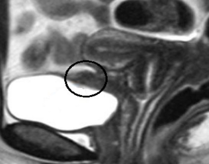
The focus of endometriosis most often occurs on the posterior surface of the bladder (area circled) and is not separable from the adjacent uterus
Areas of hemorrhage and fibrosis, typical of the process, intensively accumulate contrast.
With an acquired (false) pseudodiverticulum of the bladder, a protrusion forms through the muscle wall in the form of a pocket. There is multiple organ damage, characteristic of long-term obstruction of the lower parts of the urogenital tract due to benign prostate hyperplasia/cancer, neurogenic dysfunction, and urethral narrowing.
The arrow on the MR image shows the neck of the protrusion
With a congenital diverticulum, the histological structure is the same as that of the bladder wall. The size of the hernial sac depends on the volume of urine; stones and inflammatory elements may be present inside. A diverticulum on MR scans shows a low signal on T1 WI and a hyperintense signal on T2 WI. There is a risk of urothelial carcinoma developing in the hernial sac.
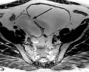
Urinary duct cancer on MRI with invasion of the anterior abdominal wall (arrow), T2 WI, sagittal plane
The urachus is an embryonic appendage that normally closes at birth or in the first years of life. When a limited cavity is formed with fluid accumulation, they speak of a cyst; if there is drainage to the outside, they speak of a vesico-umbilical fistula. Magnetic resonance imaging allows us to clarify the type of embryonic malformation of the urinary duct. The study is carried out if urachal cancer is suspected.
What else will an MRI of the bladder show:
- inflammatory processes;
- foreign bodies;
- iatrogenic damage,
- cystolithiasis (stones);
- genitourinary tuberculosis and other infectious processes;
- ulcerative lesions in interstitial cystitis;
- anomalies in the location of the ureteric orifices, ureterocele;
- cervical sclerosis;
- microcysts (volume reduction);
- incorrect anatomical position (pelvic organ prolapse, prolapse) as a cause of urinary incontinence;
- narrowing of the urethra and the presence of neoplasms.
MRI of the pelvic organs in Izhevsk. Cost, sign up for an MRI in Verum
The pelvic cavity includes the bladder, male and female reproductive organs, lymph nodes, pelvic bones and many other organs.
What organs and systems are examined during an MRI scan of the pelvic organs?
Men and women have different organs located in the pelvis. On MRI of the pelvic organs we examine:
Among women:
body and cervix, vagina, ovaries, bladder, rectum, lymph nodes.
For men:
bladder, prostate gland (prostate), scrotal organs, rectum, lymph nodes.
Advantages of MRI of the pelvic organs compared to other diagnostic methods
An MRI of the pelvis can help check for possible problems found with other diagnostic methods. MRI scans of the pelvis are also used to diagnose unexplained pain, investigate the spread of certain types of cancer, or better understand what is causing certain symptoms.
MRI does not use radiation like X-rays, so it is considered a safer alternative, especially for pregnant women or young children.
The high contrast of tissue images with MRI and the possibility of multiplanar visualization determine diagnostics as the most sensitive and specific method for searching for local prevalence and conducting a complex differential diagnosis of pathological processes of the pelvic organs.
Indications for pelvic MRI in men and women
Common indications for both sexes for pelvic MRI are:
- congenital developmental anomalies;
- pelvic area injuries;
- equivocal radiographic/ultrasound results;
- pain of unknown origin in the pelvic area;
- unexplained difficulty urinating or defecating;
- cancer or suspected cancer of the reproductive organs;
- congenital and acquired fistulas, including anorectal;
- monitoring the local prevalence of rectal and bladder cancer when planning surgical treatment and staging the disease.
Magnetic resonance imaging of the pelvis in women
allows you to assess the condition of the lower intestines, bladder, uterus, fallopian tubes, ovaries, vagina, peri-uterine and peri-vaginal space.
Based on this, the indications for MRI in women are
:
- adenomyosis (internal genital endometriosis) – for identification, differential diagnosis and assessment of changes in dynamics after therapy. It is important that in this case, an MRI examination must be performed on days 20-25 of the menstrual cycle;
- external genital endometriosis – to search for implants throughout the peritoneum along its entire length, assess the depth of invasion of nearby organs and plan surgical treatment tactics;
- space-occupying formations of the ovaries - when conducting a differential series and assessing the condition of the peritoneum throughout;
- inflammatory changes, pelvic pain - in order to exclude abscesses and analyze the local extent of changes before surgical treatment;
- volumetric formations of the body and cervix - for staging cancer of the body and cervix, assessment of pelvic and para-aortic lymph nodes;
- to assess the condition of the scar after cesarean section;
- with abnormal development of the uterus.
A routine examination of the internal genital organs of menstruating women (uterus, fallopian tubes, ovaries) is carried out on days when there is no menstruation.
Indications for pelvic MRI in men are:
- acute inflammatory diseases of the genitourinary system (prostatitis, cystitis) - with contrast to exclude abscesses;
- non-descent of the appendages (cryptorchidism) - for localizing and assessing the condition of the gonads when ultrasound diagnostics are uninformative;
- suspicion and assessment of the local prevalence of prostate cancer, stratification of pathological changes using the Pi-RADS system (study with dynamic contrast) for planning targeted biopsy, determining the stage of the disease.
The administration of a contrast agent during MR examinations of the pelvic organs is carried out for the following diagnoses (or suspicion of these diagnoses):
- volumetric formations of the ovaries (according to the doctor’s decision);
- cancer of the uterus;
- prostate cancer;
- acute prostatitis;
- fistula (fistula);
- abscesses of the pelvic organs;
- exclusion of metastases in the organs and bones of the pelvis.
MRI OF THE BLADDER
If your doctor says a bladder test is needed, a contrast agent will also be given.
This is due to the fact that MRI of the bladder is not a routine examination.
, it is indicated in the following cases:
- to assess the prevalence of the process in confirmed bladder cancer;
- if a malignant tumor is suspected - the presence of impurities of blood, mucus and pus in the urine, a palpable neoplasm, enlarged inguinal lymph nodes, etc.;
- to monitor the treatment of malignant neoplasms with an already established diagnosis;
- to determine optimal treatment tactics, when planning surgical interventions and monitoring after them;
- for bladder injuries and assessment of their consequences (fistulas, strictures);
- if developmental defects, both congenital and acquired, are detected or suspected;
- with prolapse and prolapse of the bladder.
Magnetic resonance imaging of the pelvic organs is an informative and painless diagnostic method for determining the presence of tumors, their location, degree of spread or metastasis. This technique allows the doctor to determine the tactics of patient management, make adjustments to treatment and give a prognosis regarding the results of treatment.
Contraindications to MRI of the pelvic organs
Despite the safety and painlessness of the MRI method, there are a number of contraindications.
Contraindications, for the most part, relate to the specifics of the method.
The tomograph creates a magnetic field; therefore, the following are absolute contraindications:
- cochlear implants;
- metal clips, ferromagnetic stents or staples;
- artificial valves in the heart;
- implanted cardiac and neurostimulators or defibrillators;
- metal objects in the body.
Relative contraindications:
- The study is not recommended for women who are in the first trimester of pregnancy. This is permissible only if there are serious indications and special instructions from a doctor.
- pathological processes in which a person cannot maintain a still state, neurological diseases;
- fear of confined spaces. If you have claustrophobia or excessive anxiety, you can ask your doctor to prescribe mild sedatives for you before your scheduled test. Be sure to warn our Center staff about claustrophobia. We suggest being present with one of your relatives or friends, coming in advance to look at the tomograph and even try to lie down;
- extremely serious condition;
- weight limit (due to technical features of the tomograph).
Preparing for an MRI of the pelvis
During pregnancy, no preparation for the study is required. In other cases:
- 2 days before the test, exclude from the diet coarse fiber (vegetables, fruits), carbonated drinks, brown bread, fermented milk products that cause gas formation, i.e. anything that can cause fermentation in the intestines. You can eat boiled chicken fillet, chicken broth, boiled or steamed low-fat fish, boiled squid, buckwheat, rice, white bread.
- Avoid eating 3-4 hours before the test.
- 2 hours before the start of the study, you should take 3 No-Spa tablets (not No-Spa-Forte).
- An hour before the examination, empty your bladder, then take fluids as desired. As a general rule, the bladder should not be too full or empty.
- If the study is carried out in the morning, then before bedtime you can do a microenema (Microlax), if the study is in the afternoon, then the microenema should be done in the morning.
IMPORTANT! The study is not carried out during menstruation!
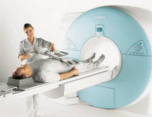
How is the MRI procedure of the pelvic organs performed?
The magnetic field generated by the MRI temporarily aligns the hydrogen proton atoms in your body. Radio waves pick up these aligned particles and create weak signals that the machine records as images.
A magnetic resonance imaging scanner is a large metal cylinder with a movable table. You must follow the instructions and remove all metal objects and elements.
You will lie motionless on your back on a table that is slowly moved to the center of the CT scanner. The operator may place small coils around the pelvic area (depending on the organ of interest) to improve the quality of the scan images.
Survey results
You will receive a disk with the images immediately after undergoing the MRI. The results of the examination (description and conclusion of a specialist) can be given to you within 60-90 minutes after the procedure. If a consultation is necessary, the description can take up to three days. If you wish, you can receive all the necessary information electronically.
Prices and list of abdominal MRI services in Izhevsk:
| № | Magnetic resonance studies: | Prices in rubles |
| 3 | Magnetic resonance imaging studies of the pelvic organs: | |
| 3.1 | Small pelvis | 4 700 |
| 3.2 | Small pelvis with contrast. (10 ml, weight up to 62 kg))* | 9 100 |
| 3.3 | Small pelvis with contrast. (15 ml, weight from 63 to 89 kg))* | 10 000 |
| 3.4 | Small pelvis with contrast. (20 ml, weight from 90 kg))* | 10 900 |
| 3.5 | Pelviometry | 2 400 |
| 3.6 | Placenta | 3 000 |
| 3.7 | Scrotal organs + pelvic and para-aortic lymph nodes | 7 800 |
| 3.8 | Organs of the scrotum + pelvic and para-aortic lymph nodes with contrast. (20 ml))* | 12 200 |
| 3.9 | Scrotal organs and inguinal canals | 4 500 |
Sign up for an MRI
Preparing for an MRI of the bladder
To undergo the diagnostic procedure, choose loose-fitting clothing without metal components
The study is often carried out as planned. Preparing for an MRI helps you obtain better images.
The bladder is adjacent to the intestines, so increased peristalsis of the latter against the background of putrefactive fermentation processes will interfere with visualization and lead to the appearance of artifacts on the films. 2-3 days before the diagnostic procedure, you must follow a special diet. The diet involves limiting carbohydrate intake and avoiding foods containing large amounts of fiber:
- confectionery, chocolate, cocoa;
- black bread, pastries;
- raw vegetables (potatoes, tomatoes, cabbage, legumes) and fruits (apricots, bananas, apples, pineapples, citrus fruits);
- seeds and nuts;
- mushrooms;
- fatty meat, fish;
- butter, sour cream;
- marinades, smoked spices, semi-finished products, etc.
Avoid drinks that increase peristalsis:
- milk, curdled milk, cream;
- fresh juices;
- kvass, jelly;
- lemonade, sparkling water;
- alcohol;
- strong tea and coffee.

On the day before the study, it is preferable to consume products made from chopped chicken/turkey breasts, rabbit meat, steamed vegetables, non-rich broths, dry cookies, unsweetened compotes
Preparing for an MRI of the bladder involves the use of gentle thermal methods of processing foods: deep frying (with a large amount of boiling fat) or over open coals is unacceptable. You must appear for the diagnostic procedure after 4-6 hours of fasting.
For irregular bowel movements and digestive problems, the doctor may prescribe antispasmodics, sorbents, anti-bloating agents, enzymes, laxatives, and cleansing with an enema.
If an MRI with contrast is planned, a light snack is allowed 20-30 minutes before the start of the diagnosis: the measure is aimed at preventing vegetative reactions - nausea, drooling, dizziness, etc. Women during lactation are recommended to stock up on milk for 2-3 feedings.
The bladder is viewed as full, which ensures straightening of the folds and better visualization of changes. To do this, you need to refrain from going to the toilet for 2 hours and drink an additional 350-500 ml of still water within 40 minutes.
Don’t forget to take a referral, passport, results of previous studies related to the disease - ultrasound, cystoscopy, intravenous urography, etc.
Decoding the results
MRI is one of the most highly informative methods for studying the bladder. Interpretation of the received images is possible only by a qualified radiologist. In difficult cases in our clinic, the decision is made by a council of specialists.
Most often, an MRI of the bladder is prescribed to detect tumors. Although the study will not allow us to identify the histological appearance of the tumor, it will make it possible to predict the degree of its malignancy and choose further tactics for managing the patient.
If the diagnosis has already been established, MRI is prescribed to clarify the extent of the malignant process - the scan will allow you to visualize all tissues of the bladder, determine the depth of tumor growth into the walls of the organ, its spread to nearby tissues, as well as metastasis to regional lymph nodes. If greater detail is required, contrasting is performed.
MRI also allows you to identify ultrasound and X-ray-negative stones - stones that are not detected by other methods of radiation diagnostics.
Indications for MRI of the bladder

MRI scans are considered expert diagnostics; sometimes ultrasound, assessment of symptoms and tests are sufficient
A magnetic resonance imaging procedure is justified if:
- Ultrasound and computed tomography data are ambiguous or suspicious for a malignant neoplasm;
- a preoperative assessment of the anatomical features of the bladder and determination of the stage of cancer are required;
- dynamic monitoring of the pathological process is indicated;
- the patient is diagnosed with an allergy to an iodine-based radiocontrast agent;
- changes in tests and symptoms (blood in the urine, pain in the lower abdomen, dysuric disorders, urinary incontinence, etc.) cannot be explained using other methods of examination);
- There are contraindications to cystoscopy.
Magnetic resonance imaging of the urogenital tract is a safe study that does not involve ionizing radiation exposure to the body, therefore it is more suitable for pregnant women and children. Indications for MRI of the bladder are determined by the doctor taking into account the clinical situation. If cystitis is recurrent and the effect of treatment is short-term, you can undergo diagnostics to identify diseases that support inflammation.
Cancer of the urinary duct with infiltration of the surrounding tissue, the bladder wall at the apex on magnetic resonance imaging
How the research is carried out
In our center, MRI of the bladder is performed on an open-type tomograph - Siemens Magnetom S with horizontal loading. This means that during scanning the patient will not lie in the tomograph tube, but inside a C-shaped contour, which is open on the side surface. In this case, a closed space is not formed, and the noise of the tomograph is less felt. During the study, medical staff is in an adjacent room, and communication with them is carried out via a signal bulb. The scanning duration is 30 minutes, and you must remain motionless throughout this time.
The scan is performed with a full bladder. To do this, you need to either not urinate 2-3 hours before the test, or drink a liter of non-carbonated liquid an hour before the procedure.
MRI of the bladder: how it is done for a man
The diagnostic procedure will be performed after appropriate preparation. In addition to the bladder, MR scans evaluate the prostate, seminal vesicles, rectum and urethra.
The study does not require sexual abstinence. The magnetic field does not have a negative effect on spermatogenesis, potency, or libido. Before the procedure, the patient must warn the doctor about a history of brachytherapy (treatment for prostate cancer associated with implantation of radioactive metal grains into the organ), allergies to gadolinium, functioning devices (cardio-, myo-, neurostimulators, insulin pump, fixed auditory prosthesis, etc. .), orthopedic structures, claustrophobia (when using equipment with a closed circuit).
MRI of the bladder: how it is done for a woman
Uninformative results of MR scanning in patients can be caused by:
- Intrauterine device. Modern contraceptives are made primarily of copper and silver, which do not react to magnetic fields, but other examples are also found. To ensure safety, it is enough to know the brand of the installed IUD; information is in the product description.
- Inappropriate day of the cycle. Magnetic resonance imaging is rarely performed solely to examine the bladder. The nonspecificity of symptoms in diseases of the pelvic organs implies a broader assessment, which includes examination of the uterus with the cervix, ovaries, fallopian tubes, and lower intestines. If you suspect a concomitant pathology of the internal genitalia, it is important to choose the optimal time for the study: MRI for endometriosis is performed in the second half of the menstrual cycle, in all other situations - from 6 to 11 days from the beginning.
- Gestation. MRI scanning is not recommended in the first trimester of pregnancy, but there is no evidence in the world literature about the harm of a magnetic field on fetal development. Other contraindications for diagnosis are similar to those listed above.
Carrying out MRI using modern equipment
Before undergoing an MRI of the pelvis, all jewelry and metal objects must be removed. After this, the patient is positioned on a special table, which takes her inside the tomograph. During the MRI, you need to relax and lie still. An MRI does not cause any pain or discomfort. Some women report feeling warmth as the scanner approaches the area being examined. This is a completely natural and predictable reaction of the human body to direct exposure to magnetic fields.
The process of examining the pelvis in women is closely monitored by qualified staff at the medical clinic. The CT scanner operator may issue small commands from time to time. For example, he may ask you to hold your breath for a few seconds. By executing these commands, you can ensure that you receive the highest quality images possible. After the procedure is completed, the patient returns to her usual activities.
CT or MRI of the bladder

In difficult cases, the patient management algorithm is determined collectively
The choice in favor of the study is made by the attending physician, who, based on an analysis of all available data, chooses therapy. CT is used in urgent situations associated with trauma, when quick results are needed. Computed tomography is prescribed for cystolithiasis (shows the density of stones), to evaluate the cyst and ureteral orifices. In oncourology, preference is given to magnetic resonance scanning. Often the diagnosis is established after a combined examination, including radioscintigraphy of skeletal bones, positron emission tomography, morphological examination, etc.
At the Magnit Medical Center in St. Petersburg, MRI of the pelvis is performed on a high-field, expert-class scanner from Siemens (Germany). You can sign up for the clinic by calling +7 (812) 407-32-31. At night the cost of the procedure is cheaper. We serve not only private clients, but also work under the compulsory medical insurance/voluntary medical insurance system, with payment through an insurance company. Don’t delay diagnostics and come - we’ll be happy to help!
Results of MRI of the pelvis in women
Examination of the pelvic organs using MRI allows you to:
- establish with high accuracy the absence or presence of a tumor formation;
- identify the size, contours and internal structure of the neoplasm;
- clarify the anatomical position of the tumor in relation to other organs;
- detect pathological enlargement of lymph nodes.
Based on the MRI results, the attending physician can see even the smallest tumors. Thanks to this, a woman gets a chance to completely cure cancer in the early stages of its development.