Home > CT and MSCT
X-ray computed tomography is a method of studying the internal systems and organs of a patient using x-rays. With the advent of CT, many diagnostic tests are now performed by radiology specialists. Many invasive methods have lost their relevance in diagnosing diseases.
Sensors of special sensitivity register X-rays penetrating through body organs. The software allows you to conduct complex studies, process the resulting images and analyze them.
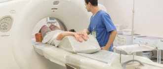
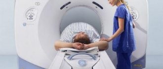
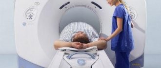
The gantry is an important part of the tomograph - an X-ray unit rotating around a lying person, recording images of the body from different angles. The images are subsequently processed and summarized using complex computer programs.
Multislice computed tomography is a progressive method of computed tomography. MSCT differs from conventional CT in that images of sections of the patient’s body are virtually obtained in the form of a “serpentine” rather than simple “slices” - due to the movement of the device around the body in a spiral. In just a few seconds, the structure of the layers of a certain organ or area is revealed.
Multislice tomography is also called multislice. The difference between MSCT and CT is that, due to continuous improvement, the latest generation multislice tomographs produce images several times faster and more comprehensively than conventional tomographs.
CT, SCT and MSCT - what is the difference?
Computed tomography, depending on the device used for scanning, is divided into traditional step-by-step CT, spiral and multispiral.
With a conventional CT scan, each slice is scanned with the patient moving step by step through the machine. In this case, the minimum thickness of the slice coincides with the minimum step of the table on which the patient lies, which does not exceed 1 cm. Such an examination is significant in terms of time and degree of irradiation, and does not allow assessing pathology with a size smaller than the thickness of the slice.
With the advent of spiral devices, the diagnostic capabilities of CT improved significantly, since the minimum slice thickness was now 5 mm. This was achieved by the fact that the patient smoothly entered the device, and at this time the radiation source rotated around him in a spiral. This made it possible to reduce the examination time, reconstruct images in the planes of interest (and not in one, as with conventional CT), and the images became clearer.
The invention of the multislice scanning principle increased the diagnostic capabilities of CT to the maximum. Unlike a spiral apparatus, in which the radiation passing through the body was perceived by one row of detectors, with MSCT it is perceived by at least two rows (currently the number of rows of detectors is 32, 64, and even 128), which allows scanning the corresponding number of sections in one turn of the tube. At the same time, the slice thickness began to reach only 0.5 mm.
Cost of diagnostics
The cost of research depends on the quality of the device. Experts advise choosing clinics that use a 16-slice and more powerful device. Also, the price may increase due to the administration of contrast or anesthesia.
The cost of diagnostics is:
- head, from 3-4 thousand rubles,
- cervical spine, from 3-3.5 thousand rubles,
- OGK, from 4 thousand rubles,
- OBP, from 4 thousand rubles,
- kidneys and bladder, from 4.5 thousand rubles,
- retroperitoneal space, from 4 thousand rubles,
- lumbosacral spine, from 4 thousand rubles.
Multislice CT is a modern method of organ imaging, which is actively used to quickly identify pathologies.
MSCT or CT - which is better?
Without a doubt, MSCT is the priority type of scanning among all types of computed tomography. Step CT is bad because the slice thickness is at least 10 cm, which is why a large number of pathologies simply do not fall into the scanning area, which is especially important when identifying metastases, strokes, and small focal formations. In addition, long-term scanning with a stepper inevitably led to motion artifacts, especially when scanning the lungs, since it is impossible to hold your breath for a long time.
Due to the short scanning time, the MSCT device allows one to avoid such artifacts, and the lungs are now examined in just one breath-hold. The introduction of ECG synchronization, in which a slice is made only between heart contractions, as well as the use of a contrast agent, made it possible to examine in detail the heart, aorta and other “pulsating” vessels. The main thing is that high spatial resolution has been achieved with the ability to scan in any plane with the construction of three-dimensional images, which allowed the MSCT device to show oncology with an accuracy of 0.5 mm, examine even the smallest metastases and identify the extent of the process.
If we take into account the same number of slices for stepwise CT and MSCT, then with MSCT the radiation dose will be significantly lower. However, MSCT allows one to examine thin sections, and their number during scanning can reach several thousand, which undoubtedly ultimately increases the radiation dose. Therefore, unreasonable multiphase scanning and reduction of slice thickness is unacceptable.
Advantages of conventional CT over MSCT
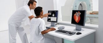
The difference between CT and MSCT is also in the time of interpretation of the results. Processing (image synthesis) and interpretation of MSCT results takes significantly more time than with a simple computed tomograph, since the radiologist needs to process and make descriptions of primary images and reconstructions, the number of which reaches two thousand. Diagnostic image processing is based on mathematical algorithms, so the results of accurate studies reflect the real characteristics of the disease process. Analysis of images obtained by a tomograph occurs by combining a large number of thin-section images into one volume, which are reconstructed into a three-dimensional model.
Indications for MSCT
MSCT is preferable to MRI for:
- suspected stroke;
- traumatic brain and associated injuries;
- the patient's serious condition;
- the need to study the bone apparatus;
- assessment of atherosclerotic plaques and internal walls of blood vessels;
- presence of contraindications to MRI.
For other pathologies, the decision to scan using CT and MRI remains with the patient. It should be noted that MSCT scanning is more accessible.
Indications for computed tomography
CT is widely used in medicine:
As a screening test:
- for headaches and migraines;
- with a head injury that does not lead to loss of consciousness;
- with frequent fainting;
- to exclude lung cancer, it is carried out routinely if computed tomography is used for screening.
For CT diagnostics for emergency indications:
- severe injuries;
- suspicion of cerebral hemorrhage, vascular damage;
- suspicion of other acute injuries of hollow and parenchymal organs;
For routine CT diagnostics:
- Most CT scans are carried out routinely, upon the direction of a doctor, for final confirmation of the diagnosis. Before performing a computed tomography scan, simpler studies are done - x-rays, ultrasound, tests, etc.
- To monitor the results of treatment
- For carrying out therapeutic and diagnostic procedures.
Contraindications to MSCT
Contraindications for CT:
- pregnancy at any stage of gestation;
- not recommended for children under 14 years of age;
- not recommended for existing multiple myeloma and recent ionization examinations;
- excessive weight of the patient;
- inability to take a supine position.
Contraindications for contrast enhancement:
- uncompensated thyrotoxicosis;
- history of severe reactions to an iodine-containing drug;
- renal failure with a significant decrease in clearance;
- with caution in severe bronchial asthma.
Contraindications for cardiac CT:
- an irregular rhythm that cannot be made regular with medication (for example, a permanent form of atrial fibrillation);
- inability to hold your breath for 15 seconds.
Spiral computed tomography
Helical CT has been used in clinical practice since the launch of the first helical CT scanner by Siemens Medical Solutions in 1988.
Spiral scanning consists of performing 2 actions simultaneously:
- continuous rotation of a radiation source —an X-ray tube—around the patient’s body;
- continuous translational movement of the table with the patient along the longitudinal scanning axis through the gantry aperture (the ring-shaped part of the computed tomograph).
The trajectory of the X-ray tube relative to the direction of movement of the table with the patient's body in this case will take the form of a spiral . Unlike sequential CT, the table speed can take values determined by diagnostic purposes. Increasing the table speed increases the length of the scanning area.
Monitor of multislice CT tomograph Toshiba Aquilion 16
Spiral scanning technology allowed:
- significantly reduce the duration of CT examination;
- seriously reduce the radiation dose to the patient.
How is MSCT performed?
Preparation for MSCT with intravenous contrast
The MSCT procedure is carried out on an empty stomach; you cannot eat or drink for at least 4 hours; in addition, when examining the heart, you cannot smoke or drink coffee for 4 hours. Also, due to the fact that when scanning the heart and coronary vessels, the heart rate should not exceed 65 beats/min, take 50 mg of metaprolol or any other beta blocker 40 minutes before the scan. When examining the intestines, the procedure is carried out before fluoroscopy with barium, or after its complete removal from the body; if it is necessary to contrast intestinal loops, drink a liter of oral contrast within an hour before the procedure; if you need to examine the large intestine in detail, then add 0.5 liters the night before. When performing MSCT cholangiography, you must drink at least 1.5-2 liters of still water an hour before the examination.
If there is a history of allergy to contrast, premedication (with prednisone/antihistamines), hydration, and only low-osmolar non-ionic contrast are used.
Carrying out the procedure
Before the procedure, it is necessary to provide the doctor with a direction indicating the purpose of examining a certain area and the conclusion of the examinations already carried out. Next, the patient lies down on the table deck, contrast is injected intravenously with an injector, the table moves through the device and scanning is performed. If necessary, you will need to hold your breath for up to 15 seconds. Depending on the area being examined, MSCT lasts from 10 to 40 seconds. Most of the time is spent deciphering MSCT images, since their number is often measured in thousands.
Preparing for an abdominal CT scan
If you are scheduled for a CT scan of the abdominal organs, refrain from eating solid food the evening before the examination. Before the procedure, you may be asked to drink a contrast agent, and in some cases, take a mild laxative or have a barium enema.
CT and MSCT of the abdominal organs are performed on an empty stomach or no earlier than 3-4 hours after the last meal. It is allowed to take medications (with a small amount of water). Immediately before the examination, the department may ask you to drink about 500-600 ml of water.
MSCT examination is performed after preliminary contrasting of the intestine with a diluted contrast agent. The gastrointestinal tract is filled either on the day of the study or the day before: On the eve of the study, drink 0.5 liters of solution in the evening, in the morning (06:00-07:00) drink the remaining 0.5 liters. The study is carried out with a full bladder. Women must have a sanitary tampon with them.
Attention! After an X-ray examination of the stomach, colon with barium (gastric fluoroscopy, irrigoscopy), CT examination of the abdominal organs is recommended no earlier than 5-7 days (at least 3 days). In cases of emergency - after cleansing enemas.
How many times can MSCT be done per year?
The answer to this question, how often an MSCT examination can be done, depends on the area being examined. For example, with MSC scanning of the head, the radiation dose is on average 1.4 mSv, for the chest and abdominal cavity - 2 times more, pelvis - 2.5 times more. The safe radiation dose for the population is 1 mSv per year (equivalent to two x-rays of the lungs), if necessary, it can be increased to 5 mSv per year. As you can see, the permissible number of studies for the head is 3 per year, for other areas - 1 time per year (all this taking into account the absence of other ionizing procedures). If research is still necessary, it is recommended to use a safe and equally high-precision imaging method - MRI (in the absence of contraindications).
Comparison of surveys
The principle of CT and MSCT is identical. The difference lies in the amount of time spent and the dose the patient receives.
A special feature of the multispectral tomography apparatus is the location of the X-ray emitting sensors. During the examination, the tomograph rotates around a lying person, and the couch moves inside the device in a horizontal plane. The sensors move in a spiral, which is why the study is called spiral CT, and such tomographs are multidetector.
The difference between modern technology and traditional CT:
- reduced radiation exposure, total radiation exposure is 30% less,
- minimum time spent per patient,
- a larger number of images received, because scanning is carried out continuously,
- efficient placement of sensors, which allows obtaining thin sections,
- less interference during scanning,
- thanks to the design features, an effective signal-to-noise ratio of the device has been achieved,
- improved contrast resolution,
- used to detect hemorrhages, hematomas, neoplasms measuring 0.5-1 mm,
- MSCT images show pathologies of the biliary tract, liver, pancreas, and deformation of the aorta.
The diagnostic capabilities of the multislice method are better than conventional CT. MSCT gives the doctor the opportunity to assess the condition of bones, blood vessels and soft tissues.
Indications and contraindications
Multislice, or multislice, CT allows you to clarify the diagnosis through the use of three-dimensional reconstruction of organs or bone structures of the body. It is used to substantiate the diagnosis and confirm the need for surgical treatment.
MSCT is prescribed in the following cases:
- the need for urgent examination of the aortocoronary vessels, abdominal aortic aneurysm,
- diagnosis of internal bleeding of unknown origin,
- TELA,
- suspected cardiac, pulmonary, renal or vascular failure,
- the need to determine the size of tumor formations,
- measurement of bone tissue density in osteoporosis,
- diagnosis of diseases of the maxillofacial region.
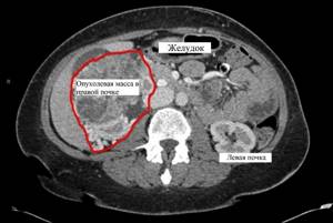
An alternative to MSCT for identifying pathologies of the maxillofacial apparatus is cone-beam CT.
Contraindications to the examination are divided into absolute (permanent), relative (temporary) and individual prohibitions.
Absolute contraindications:
- severe allergy to substances used as contrast,
- the patient’s body weight exceeds the load value established by the tomograph manufacturer,
- pregnancy,
- severe form of renal or liver failure, bronchial asthma.
Relative indicators:
- the presence of metal implants or alloys in the body,
- mental disorders, inability to self-control,
- childhood,
- mild or moderate allergy to contrast media,
- late pregnancy,
- bronchial asthma that can be controlled,
- serious condition of the patient.
Individual contraindications include:
- Parkinson's disease,
- eating before abdominal diagnostics,
- tachycardia or dysfunction of the heart muscle,
- calcification of arteries and vasoconstriction,
- inability of the patient to hold his breath during examination of the OGK.
How to prepare
Preparation for tomography depends on the organ being diagnosed and the need to use a contrast agent.
To conduct MSCT of the OGK, soft tissues, cervical spine, brain without the use of contrast, a person is allowed to wear loose clothing and must take the necessary documents. No specific preparation is required.
If an examination of the peritoneal organs is prescribed, the following rules must be followed:
- Check for an allergic reaction to iodine.
- The day before the test, do not eat solid foods or foods that cause gas. If you have such problems, you should take carminative medications.
- The last meal should be no later than 5 hours before MSCT.
- 3 hours before the procedure, you should drink a radiopaque contrast agent dissolved in the prescribed proportion. Then perform an enema.
If an examination with contrast is prescribed, the person undergoes blood and urine tests 3 days in advance. If an increase in urea levels or the presence of creatinine is detected, the study is postponed.
The patient must remain motionless during the scan. The scanning is carried out within 5-30 minutes and depends on the technical capabilities of the device.
What is the radiation dose and how often can it be done?
MSCT has become more widespread due to reduced examination time and less radiation exposure to the patient.
The radiation dosage depends on the organ being examined:
- spine, 6 mSv,
- limbs, 1-2 mSv,
- head, 2 mSv,
- OGK, 11 mSv,
- hip area, 9 mSv,
- internal organs, 10 mSv,
- Gastrointestinal tract, 14 mSv,
- paranasal sinuses, 0.6 mSv,
Doctors recommend examinations no more than once a year, unless medical indications otherwise require. The received dose level of 150 mSv per year is considered critical.
Is it harmful to the baby during pregnancy?
Children are more sensitive to radiation exposure. Their risk of developing cancer is higher due to the fact that the body's cells are constantly growing and are predisposed to DNA changes.
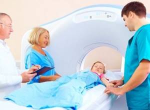
Without medical prescription, MSCT is contraindicated for children under 14 years of age, i.e. until the end of active growth.
Pregnant women are not recommended to undergo MSCT. The examination is prescribed if there is an existing risk to life or if it is impossible to perform an MRI. Since the fetus is in constant motion, the examination is carried out under general anesthesia.
Is MSCT harmful to a child and is MSCT dangerous during pregnancy?
Children are especially susceptible to radiation exposure (and with MSCT, the radiation dose can reach up to 12 mSv versus 0.5 mSv with conventional radiography), they have a greater risk of developing cancer. This happens because the tissues of a child’s (and even more so the unborn) body are in a phase of continuous growth, and, therefore, are more susceptible to DNA changes. Also, the chance of developing a tumor after MSCT is much higher in a child than in an adult, because there is actually more time for a tumor to develop in children than in adults and the elderly. To reduce the risk of consequences, it is highly not recommended to perform MSCT on children until the process of active tissue growth has completed (up to 14 years). During pregnancy, MSCT is an absolute contraindication and can be performed only if there is an absolute risk to life and it is impossible to urgently perform an alternative non-ionizing scan (MR imaging).
Advantages and disadvantages of MSCT
Advantages of MSCT over CT:
- the images better display soft tissues, arteries, veins;
- research time is reduced;
- the radiation dose is reduced by 30%;
- the amount of contrast agent decreases, the load on the kidneys decreases;
- results are less dependent on random movements of the patient;
- formations are identified that are smaller in size than the size of the cut;
- When processing the data, a full three-dimensional image is formed.
The disadvantage of MSCT is its high cost. In remote regions, the procedure is not always available due to lack of equipment.
Areas of research and capabilities of CT (what does it show?):
- MSCT of the brain: acute cerebrovascular accidents;
- moderate to severe traumatic brain injuries;
- brain tumors with contrast;
- MSCT of the spine: traumatic injuries;
- tumor and inflammatory diseases;
- degenerative changes;
- detection of herniated intervertebral discs;
- postoperative changes;
- MSCT of the paranasal sinuses: sinusitis;
- sinus cysts;
- before dental implantation;
- MSCT of the orbits: tumor and inflammatory diseases;
- traumatic injuries;
- MSCT of the temporomandibular joints;
- MSCT of soft tissues and organs of the neck: tumor and inflammatory diseases;
- traumatic injuries;
- CT scan of the chest (organs and bone structures): diseases of the lungs, mediastinum, ribs, sternum, shoulder blades;
- CT scan of the abdominal cavity and retroperitoneum: diseases of the liver, kidneys, adrenal glands, pancreas, spleen;
- damage to the lymph nodes and blood vessels of the abdominal cavity;
- MSCT of the pelvis: tumor, inflammatory, vascular diseases;
- traumatic injuries;
- developmental anomalies of the pelvic organs;
- MSCT of the vessels of the head and neck, vessels of the upper and lower extremities, MSCT of the thoracic and abdominal aorta: detection of aneurysms, thrombosis, vascular malformations;
- MSCT of joints and bone structures: tumor, inflammatory diseases;
- traumatic injuries.
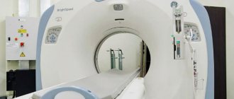
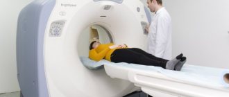
Multislice computed tomograph Toshiba Aquilion 16
At the North-Western Medical Center of St. Petersburg on the street. Borovaya, 55, when diagnosing internal organs, a Toshiba Aquilion 16 multislice computed tomograph (Japan) is used.
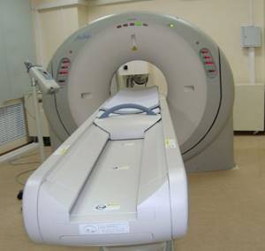
Multislice computed tomograph Toshiba Aquilion 16
MSCT Toshiba Aquilion 16 allows you to perform computed tomography:
- in a short time;
- with minimal radiation exposure to the patient.
CT machine Toshiba Aquilion 16 consists of:
- ring-shaped part (gantry) , containing: an x-ray tube;
- radiation detectors;
The X-ray emitter rotates around the patient's body , generating a thin fan-shaped beam of X-rays. This beam passes through the human body and is recorded by detectors located opposite the X-ray tube.
Information is read from radiation detectors and transmitted to a computer complex, which reconstructs the image of the area under study. An image of the area is displayed on the Toshiba Aquilion 16 CT screen and can be examined and assessed by a radiologist.
Scanning speed of the Toshiba Aquilion 16 in 16-slice computed tomograph:
- 24 times higher than 1 slice;
- 4-5 times higher than 4 slice systems.
This reduces the time of CT examination by more than 30 times , eliminating motion artifacts and significantly reducing radiation exposure by reducing X-ray exposure. The Toshiba Aquilion 16 16-slice multislice computed tomograph contains application programs that allow you to conduct all types of CT studies for all organs and systems of the body.
Material X-ray computed tomography in St. Petersburg (CT) was viewed 65570 times
Comments:
- In contact with
- JComments
Add a comment
JComments
Download SocComments v1.3
| < Computed tomography of the lungs in St. Petersburg |
