Heel spur (or plantar fasciopathy)
is a chronic disease of the musculoskeletal system associated with constant trauma to soft tissues due to a growth on the heel tubercle.
As the name suggests, the cause of these injuries is the so-called. spur
is a sharp bone growth that disrupts the physiological position of the musculo-ligamentous apparatus of the foot, causing compression when walking, pain and inflammation.
Heel spur is a disease of the musculoskeletal system in which soft tissues are injured due to a growth on the heel tubercle
Heel spurs usually result in difficulty maintaining daily activities and physical inactivity. Without treatment, the pathology leads to progressive degenerative changes, persistent disruption of tissue trophism and fibrosis (formation of obstructive compactions). Calcium salts can also deposit on the spur, which increase its size. In this case, the foot loses normal mobility, becomes stiff, and does not bend when walking. Injuries caused by a sudden step on the heel can take a very long time to heal and can be complicated by infection.
,
nerve conduction disorders
(including compression of the tibial nerve),
suppuration in the periosteum
.
A heel spur can have other serious complications - for example, destruction of the connective tissue membranes of the muscles
,
nerves and blood vessels or tissue necrosis
(with advanced plantar fasciitis). If the growth continues to grow, if it is more than 20 mm in size, it can interfere with walking - in this case, patients require auxiliary aids (walking sticks, crutches, walkers).
According to orthopedists and traumatologists, from 15 to 30% of people over 40 years of age suffer from heel spurs. However, the disease does not cause serious discomfort to everyone.
- some do not even suspect its presence, attributing fatigue and discomfort in the legs to corns.
According to statistics, a more severe course of the disease is observed in women - they make up up to 70% of patients who seek help.
Causes of heel spurs
The direct cause of heel spurs is plantar fasciitis.
. This fasciopathy is a multifactorial pathology. The onset of the disease and its further progression depends on many factors. Causes of heel spurs include:
- congenital or acquired flat feet;
- chronic injuries and diseases of the joints of the lower extremities and spine, due to which the normal distribution of load over the entire area of the foot is disrupted;
- intensive physical training (both classes with heavy weights and ballet and dance classes);
- overweight;
- age-related changes (hormonal levels, metabolism, as well as the quality of blood supply in the lower extremities);
- pathologies of the musculoskeletal system, internal organs and metabolism (rheumatism, gout, diabetes mellitus and others), as well as autoimmune diseases (rheumatoid arthritis, psoriasis);
- vascular diseases (varicose veins, atherosclerosis of the vessels of the lower extremities and others);
- incorrect gait, uncomfortable shoes (too narrow or high heels);
- professional activities associated with long periods of standing (standing work, courier profession, teachers, etc.);
- factors associated with pregnancy, especially multiple or complex pregnancies;
- prolonged depression, stress and other psycho-emotional causes of heel spurs;
- posture disorders;
- the presence of foci of chronic infection in the body, as well as past infectious diseases;
- long-term use of hormonal drugs;
- the presence of local inflammatory processes in the foot and lower leg;
- hereditary predisposition;
- bad habits;
- sedentary lifestyle;
- individual constitutional characteristics;
- unfavorable environmental situation
The rate of increase in symptoms and growth of the spine is individual in each case and depends on the causes of the heel spur. In the initial stages, there is practically no pain - up to 3-4 years can pass from the onset of the disease to the appearance of pronounced symptoms
. Pain syndrome with heel spurs is formed under the influence of several factors:
- the appearance of microcracks on the heel spur;
- compression of blood vessels and disruption of tissue trophism;
- the presence of inflammation in places where the spur injures the tissue.
The risk group for the disease includes:
- women over 40 years of age and people of retirement age;
- professional and amateur athletes;
- overweight persons;
- patients with a history of injuries and their complications.
Causes
The causes of heel spurs include the following:
- Injuries. Basically, such growths begin to form after jumping from a great height, when upon landing the heel and surrounding tissues are damaged, and therefore inflammation begins.
- Flat feet. This reason is due to the fact that the load on the foot is distributed incorrectly. As a result, the heel experiences more stress than in the correct position.
- Various joint diseases (for example, rheumatoid arthritis), as well as sprains.
- Overload of the foot area associated with external influences. A very common situation is the wrong choice of shoes. Moreover, the problem is not only in heels - for example, narrow ballet flats and shoes with very thin soles are also undesirable for the heel. Overload of the feet is often associated with work, if a person stands or walks all day.
- Gout. With this disease, excessive salt deposition occurs, which affects all joints and bones.
- Diseases associated with poor circulation, such as diabetes.
- STI. Chlamydia, gonorrhea and other infections affect the condition of the joints, and therefore, over time, a heel spur may develop.
- Natural changes in the body that are associated with age. Again, as the body ages, tissues become less healthy and perform their natural tasks less well, so the appearance of bone growths is quite common.
The causes of the problem cannot always be determined accurately - they are often identified during other examinations or are not relevant right now for the treatment of the disease.
Symptoms of heel spurs
Symptoms of heel spurs
depend on the course of the disease, the availability of treatment and other limiting factors, the size and location of the growth on the heel tubercle. The determining role is played by the location of the spur in relation to the nerve fibers and endings - the closer it is, the more intense the signs of the disease appear. In rare cases, the thorn may be so “successfully” located that it can only be identified by the erased symptoms of inflammation and during an X-ray examination.
The most noticeable symptom of a heel spur for a patient is pain. In the early stages of the disease, they can be muffled, expressed in discomfort after prolonged standing or walking. The nature of the pain is usually pressing, as if a small pebble had hit the shoe. Sometimes patients notice increased fatigue and a general decrease in leg endurance.
Soon complaints of soft tissue injury appear when abruptly rising from a bed or from a chair (“got off on the wrong foot” - this is about a heel spur!), going up and down the stairs, jumping or running, or striding.
Subsequent stages of the disease are characterized by severe inflammatory symptoms of heel spurs:
- local increase in temperature, feeling of heat and tension in the heel area;
- swelling that causes discomfort when trying to take off or put on shoes (especially those that were previously comfortable for the patient);
- redness of the skin over the heel tubercle;
- a feeling of numbness in the foot, which can even rise above the ankle;
- the appearance of foci of coarsening - thickening of the stratum corneum on the skin of the heel;
- increased pain when walking and other activities associated with pressure on the heel (for example, sitting cross-legged);
- a noticeable protrusion under the skin;
- formation of painful calluses;
- tingling and burning sensation at the end of the day, while resting.
As the pain becomes continuous, the patient's gait begins to change - lameness appears
, an attempt to transfer weight to the toe or turn out the leg.
In advanced cases, a characteristic symptom of a heel spur is acquired transverse flatfoot
, which only complicates the situation and contributes to the growth of the heel spur.
With flat feet, the patient's foot lengthens, and the muscles of the lower leg of the affected leg grow unnaturally. The pain can radiate not only to the knee or hip, but also to the back. Complications can be expressed in postural curvatures
,
intervertebral hernias
.
In the later stages, patients complain as if there is a sharp thorn or nail stuck in the heel - during the exacerbation period, every step is like stepping on broken glass. The leg begins to hurt even without any load, the symptoms of heel spurs do not subside even at night. Early morning pain (sharp and throbbing) becomes an excruciating routine. Pain in the Achilles tendon area may also develop. In the later stages, a compensatory growth of dense connective tissue is also observed, in which calcium salts are deposited. They cause a strong reaction from the nerve endings and cause a complete loss of flexibility in the foot.
- to walk, patients have to actively use the hip joint.
Diagnostic process
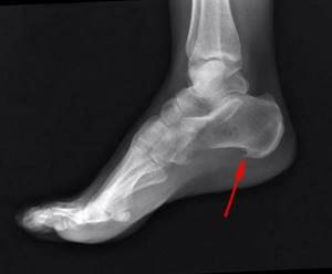
It is easy for a doctor to diagnose heel spurs. Based on the patient's complaints about heel pain, the diagnosis is made immediately in 95% of cases. The doctor will examine the limb visually and, by pressing on certain points on the heel, accurately diagnose the disease. To confirm, the patient is given an x-ray, from which the doctor will determine the size and shape of the spur. Prescribe the correct treatment and assess the level of disease progression.
Over time, the growth can reach a size of more than 1 cm. It grows on the heel tubercle towards the toes. May be diagnosed by x-raying another area of the legs and seen by a specialist. In this case, one patient may have limited movement due to severe pain, while another does not experience any sensations.
Diagnosis of heel spurs
To diagnose a heel spur, at the initial appointment, an orthopedist or rheumatologist takes a medical history and palpates the affected area, checking the patient for inflammation, tenderness, palpable bone deformities, and osteophytes.
Making an accurate diagnosis requires additional research:
- X-ray
(helps to establish the presence of a heel spur, its location and size); - Ultrasound
(shows the condition of soft tissues and tendons, determines the outline of the inflammatory focus).
To exclude ankylosing spondylitis
,
rheumatoid arthritis
, and
other autoimmune diseases
, as well as
gout
, specialists usually prescribe a general biochemical blood test (to detect inflammation, uric acid levels and a specific marker - rheumatoid factor).
In the presence of concomitant diseases and complications of the vessels, Dopplerography of the vessels of the lower extremities and nerve conduction studies may be prescribed. For complaints indicating serious nerve conduction disturbances, patients may be advised to undergo an MRI.
The combination of these diagnostic techniques makes it possible to reliably differentiate a heel spur from gout, osteomyelitis, bone tuberculosis, old injuries and other pathologies with similar symptoms.
Indications and progress of the operation
When the first signs of a heel spur appear, do not self-medicate and seek medical help immediately. Surgery is rarely performed, but sometimes it becomes simply necessary. The effectiveness of removing a bone growth through an incision in the skin brings results in 70% of cases, otherwise the pain returns.
Indications
1. No improvement in the patient’s condition within six months.
2. Inability to wait and carry out drug treatment.
3. Decreased performance and restrictions on movement.
Heel spur treatment
Treatment of heel spurs is determined by the severity of symptoms, location and length of the osteophyte. In some cases, the disease can be kept under control only with the help of exercise therapy, periodic courses of physiotherapy and, if necessary, the use of local anti-inflammatory drugs. However, heel spur therapy is often carried out in several directions:
- taking systemic and local anti-inflammatory drugs and microcirculation correctors;
- taking chondroprotectors to relieve inflammation, slow down the growth rate of osteophytes and improve the shock-absorbing characteristics of the foot joints;
- physiotherapeutic measures (10-24 sessions, depending on the chosen technique);
- performing special gymnastic warm-ups for the feet, lower extremities and, if necessary, the spine.
In addition, the doctor gives patients recommendations on lifestyle changes and maintaining the correct orthopedic regimen.
Correctly selected treatment for a heel spur can slow down its growth, and sometimes even stop it completely.
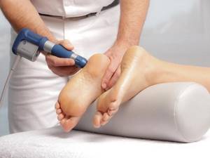
Heel spurs can be treated using several methods Alt: Heel spur treatment
Physiotherapeutic complex
All traumatologists agree on the question of how to treat heel spurs: a set of physiotherapeutic procedures in the early stages of the disease allows you to completely solve the problem
. But even subsequently, physical therapy can significantly alleviate symptoms. Typically, patients are prescribed one or more techniques from this list.
- magnetic therapy
(eliminates pain, inflammation, restores vascular function); - ultrasound therapy with medications
(improves local metabolism, relieves inflammation); - laser therapy
(provides a biostimulating effect, promotes the healing of injuries and relieves swelling); - massage
(in the absence of acute inflammation and unhealed injuries).
Cryotherapy, ultra-high-frequency therapy, paraffin baths, warm and mineral baths, and mud therapy can be used less frequently, but they usually give a temporary effect. X-ray therapy is only used to treat and relieve pain of heel spurs in young people (under 40 years of age).
Treatment of heel spurs with ultrasound
Shock wave therapy
is one of the types of ultrasound therapy, in which the device generates shock waves of a certain frequency (penetrate to a depth of 8 cm). These waves improve metabolism, promote tissue regeneration and speedy healing of injuries caused by spurs. A course of UVT helps remove calcium deposits in the tissues around the heel tubercle, prevent calcification of the tendons, and improve blood supply in small capillaries. After the first session, 4 out of 5 patients note noticeable relief, a decrease in pain and a decrease in fatigue.
Shock wave therapy has a beneficial effect on nerve endings - it helps relieve compression, blocks pain receptors and eliminates overstimulation.
In most cases, with a heel spur, a course of 5-7 procedures with an interval of 3-7 days is indicated (the pain can stop either after a week or after a couple of months - everything is individual. Sessions are not time-consuming and last from 10 to 20 minutes The intensity of the impact with a circular applicator is selected individually, based on the patient’s sensations - shock wave therapy should not cause pain, however, discomfort and mild pain for the first procedure are considered normal.
In the early stages of the disease, when the spur is still small, shock wave therapy can completely destroy it
.
Surgical treatment of heel spurs
Surgical removal of heel spurs
is prescribed as a last resort when shock wave therapy, massage, exercise therapy and medications are powerless in relieving pain. Typically, surgery is performed in cases where the osteophyte reaches an impressive size and creates severe compression.
Previously, surgery to remove heel spurs helped eliminate pain, but often served as a reason for assigning disability to the patient. Modern medicine follows the principle of minimally invasive intervention: the spur is removed through small incisions, and in the absence of severe pain, the patient can step on the operated leg immediately after it.
Long-term rehabilitation after surgery is not required, although in the first 2-3 weeks after surgery, the doctor may recommend a walking stick to protect the foot.
Medicines to treat heel spurs
How to treat heel spurs with medications? For this purpose, several groups of drugs are used that have a complex effect not only on the site of inflammation, but also on adjacent tissues and associated joints (for example, the knee, intervertebral joints and others that may suffer from this disease).
- Non-steroidal anti-inflammatory drugs
- taking them in courses of 10-12 days can reduce pain and relieve swelling, which helps restore normal metabolism. However, long-term oral administration of drugs in this category is not possible due to their negative effect on the gastrointestinal mucosa. For heel spurs, they are mainly used topically. - Corticosteroid drugs
- used in the form of fascial injections and blockades. They allow you to quickly stop the inflammatory process. These powerful drugs are usually used no more than 2-3 times a year due to the risks to the patient's health. - Chondroprotectors (Artracam)
- are used to contain the disease and eliminate pain, as well as normalize metabolism. They are especially useful in the formation of microcracks in the heel aponeurosis, which cause severe pain in many patients with heel spurs. Bone and cartilage metabolism correctors help protect all joints that suffer due to changes in gait and posture due to heel spurs, therefore they are an indispensable aid.
Narrow-profile drugs can also be prescribed for the treatment of underlying diseases, for example: antibiotics - for purulent inflammation, anti-gout drugs - for gout, blood microcirculation correctors and vascular strengthening drugs - for varicose veins, atherosclerosis of the vessels of the lower extremities. Blockades are selectively used - injection of an anesthetic and HA into the heel, which can be repeated after a month (if necessary). But this method is not very effective, because... does not eliminate the root cause of pain and loses its effectiveness over time. It is often used during complex treatment or in preparation for surgery.
Medicines for heel spurs are used both in the form of tablets and injections (systemically), and locally - in the form of gels, creams, ointments, balms, compresses and patches.
Therapeutic gymnastics and massage
Exercise therapy is one of the most effective areas in the conservative treatment of heel spurs
. Provided daily exercise, it triggers tissue regeneration processes, improves the shock-absorbing functions of the foot by strengthening muscles, and promotes the removal of calcium from soft tissues.
For heel spurs, the following gymnastic exercises are recommended:
- rolling a bottle of water for 5 minutes (your back is pressed against the back of the chair, in the starting position your shins are perpendicular to the floor);
- picking up small objects (for example, beans, pencils) from the floor with your toes - can be done both standing and sitting (for severe pain);
- sitting on a chair, the patient places the foot of the right leg on the knee of the left and pulls its toes towards himself so that the sole curves outward (repeated on both legs);
- Having passed the towel under the arch of the foot, the patient pushes the leg away from himself and pulls the towel towards himself.
Exercise therapy exercises can be combined with massage techniques (it is advisable to perform a massage before, but it is also possible after exercises). First, you need to rub your feet thoroughly until a feeling of warmth appears. After this, you can perform longitudinal pinching, circular rubbing, intense kneading, and light tapping with the edge of your palm. If pain occurs, massage should be stopped! When performing a massage, you can use painkillers and warming gels, massage mittens, and electric massagers.
Prevention of heel spurs
Prevention of heel spurs can be started at any age. For example, patients may require treatment for flat feet as early as 19-20 years of age. Patients over 40 years of age are required to pay special attention to their joints.
. They are recommended:
- timely treatment of diseases of the musculoskeletal system, infections, inflammatory vascular diseases;
- posture correction;
- wearing comfortable shoes with orthopedic insoles - they should be weather-appropriate and have a heel about 2-4 cm high;
- refusal of increased physical activity, convenient organization of the workplace (for example, when standing, it is recommended to place a low stool at the feet and alternately place the legs on it so that they can rest and restore normal tissue trophism);
- maintaining muscle tone and ligaments through moderate physical activity and self-massage;
- walking barefoot (for example, on grass, sand, pebbles), rolling chestnuts or other small objects with your feet;
- adjust your diet in such a way that the diet contains enough vitamins, minerals, clean water, a minimum of salts and carbohydrates - long breaks between meals and dehydration are unacceptable;
- weight loss.
Prevention of heel spurs involves avoiding smoked, salty foods, soda, flavored foods, strong broths, processed foods, confectionery, chocolate bars and baked goods.
Prevention
Preventive measures in this case include the following:
- Timely treatment of all orthopedic diseases: diseases of the spinal column, flat feet, etc.
- Increased attention to the condition of the joints, preventing their diseases.
- Choosing the right shoes - without high heels, with a stable last, but not too flat.
- Supplementing shoes with special insoles, which are selected with the participation of an orthopedist.
- Moderate physical activity for the harmonious development of muscles, ligaments and joints.
- Elimination of foot overloads and injuries of various types (avoidance of dangerous situations).
- Regular foot exercises.
- Weight control. Excess weight puts significant stress on the heel area, which can contribute to the development of spurs.
- Proper nutrition with enough nutrients.
It is worth adhering to these recommendations at a young age in order to eliminate the risks of developing the disease in the future or to delay the first signs of a heel spur much further in time.

