Chest X-ray is a projection hardware diagnostic of the chest area, one of the most frequently performed.
Dear patients! The result of the x-ray examination is recorded on disk or film. The service is free.
The examination makes it possible to assess the condition of organs and identify pathologies not only of anatomical structures in the chest cavity, but also nearby ones.
Organs and tissues of the body transmit X-rays differently. The densest objects are the lightest in the photo. When examining the chest, it is possible to visualize bones, soft tissues, joints, and evaluate the structure of the mediastinum, lungs, and pleura.
Indications for plain radiography of the chest organs
Plain radiography of the chest organs is prescribed:
- for pain in the chest area;
- when you feel short of air;
- with a decrease in appetite and body weight (if gastrointestinal diseases and endocrine disorders are refuted);
- with frequent increases in pressure and swelling of tissues;
- with painful breathing;
- whistling or wheezing when breathing;
- in case of injury to the sternum area;
- as a preventive examination during medical examination or during a routine examination.
Diagnosis is often carried out when pneumonia, abscess, or silicosis is suspected. The study is informative in cases where foreign objects enter the esophagus or respiratory tract.
Examination of a child's chest using x-rays - MEDSI
Table of contents
- What is the harm of x-rays?
- When can the procedure be scheduled?
- Carrying out X-ray diagnostics
- Types of X-ray examinations
- How often can an x-ray be taken?
- Advantages of the procedure at MEDSI
Radiography is an informative and cheap method of radiation research.
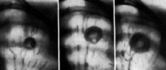
Having passed ionizing rays through the area under study, the device produces a black and white image in which tissues of different densities receive different shades: the denser the tissue, the more rays it intercepts and reflects, the lighter the shadow in the image (example: the bone structures of the chest are visible on an x-ray like white, lung tissue is dark). In this way, compactions or gaps can be detected in uncharacteristic places, which allows one to suspect the presence of a neoplasm or a violation of the integrity of the organ. Advantages: non-invasiveness, accessibility (X-rays are available in all large clinics), speed, obtaining an image that can be presented to different doctors upon request.
What is the harm of x-rays?
One of the few disadvantages of the method is its radioactivity. Large doses of radiation can provoke changes in the structure of cells and serve as an impetus for the development of tumors and the malignancy of hyperplasias. Therefore, irradiation is strictly dosed - the study is rarely carried out more than 3 times a year.
This is why, unlike adults, children do not undergo fluorography: increased cell division in childhood increases the risk of developing cancer pathologies.
Only after carefully assessing the balance of harm and benefit can a doctor prescribe a chest x-ray for a child.
When can the procedure be scheduled?
- If serious diseases of the lungs and bronchi are suspected: pneumonia, obstructive bronchitis, asthma, tuberculosis, abscess, pleurisy, tumors
- To assess the condition of the thymus (thymus gland) if a tumor is suspected or there are problems with the immune system
- After injury with a high probability of dislocations, fractures, pneumothorax, hemothorax, the presence of traumatic foreign bodies
- For symptoms of asphyxia (suffocation) to detect the cause of obstruction (clogging) of the trachea, examining blood vessels for damage or the presence of blood clots
- When planning surgery for a child with cardiac pathologies
Carrying out X-ray diagnostics
X-rays of a child’s lungs should not be performed using adult equipment, as it requires a reduction in the radiation dose. Modern digital devices for pediatric use make it possible to minimize radiation exposure, and in addition, they are adjusted to children's dimensions and equipped with special non-traumatic clamps.
The procedure is quick:
Small children are restrained upright or examined lying down with soft straps on a couch. Parts of the body not involved in the study are covered with a lead apron of the appropriate size.
- The baby can be held by the mother, who is also given an apron for protection
- Adult children who are able to remain still for the required time are examined while standing.
The procedure takes no more than a few seconds. It is important to remain completely still in a given position during this time in order to obtain a clear image.
Types of X-ray examinations
In addition to static radiography, there are other methods of radiation examination.
- Fluorography is a photograph taken from a fluorescent screen, capturing the organ under study in a reduced form.
- X-ray (X-ray television) - demonstrates the organ on the screen in real time. Previously, fluorescent screens were used to display images of the organ. With the development of digital technologies, images began to be broadcast on a monitor and also saved on digital media. The radiation dose during fluoroscopy is higher than during radiography, but the method is indispensable for some manipulations, as it allows you to observe instant changes in the organ (during bronchoscopy, some operations)
- Computed tomography allows you to examine the structures of an organ in detail, section by section. Some operations are also performed under CT guidance. However, up to 7 years of age, the examination is carried out under anesthesia, since the patient is required to lie still for 15–20 minutes
These chest X-ray methods are performed strictly according to indications (for example, in cardiac surgery).
How often can an x-ray be taken?
Unlike radioactive substances, rays do not accumulate in the body; the effects of radiation stop along with the procedure. Therefore, when performing an X-ray of a child’s lungs, the single dose of radiation, duration and frequency of exposure will be important.
Radiation exposure during radiography is measured in Sieverts and averages from 0.1 to 0.42 millisieverts for one image (for a chest CT scan it is about 7 mSv). Digital devices allow you to further reduce the dose.
At the same time, according to the recommendations of the Ministry of Health of the Russian Federation, the maximum annual radiation dose should not exceed 1 mSv per year on average (over the next 5 years) and a maximum of 5 mSv per year.
Thus, radiological diagnostics of the chest can be carried out from 3 to 10 times a year without harm to health (depending on the settings of the device, the age and state of health of the child).
Advantages of the procedure at MEDSI
- Availability of the latest generation of children's digital devices with comfortable fixation devices - safe research in a relaxed environment
- Visit at a time convenient for you
- Image interpretation by experienced diagnosticians
- Possibility of carrying out the procedure and visiting a pulmonologist, phthisiatrician or pediatrician with examination results in the same place
To make an appointment, call 24/7 8 (495) 7-800-500.
How is a plain chest x-ray performed?
Before the examination, the patient removes metal jewelry and frees the sternum area from clothing. After this, the doctor protects certain areas of the patient’s body with a lead apron, fixes the position of the patient and leaves the diagnostic room, maintaining contact with him. On command, you need to hold your breath for a couple of seconds. The scan is completed in a matter of seconds, without causing any pain or discomfort.
After the survey x-ray, you can return to your usual activities. No recovery required.
Acquired chest deformities
Acquired chest deformity in a child develops against the background of diseases of the lungs and ribs (including tumor-like formations). This pathology can lead to other disorders of the body, for example, improper functioning of the respiratory system or psychological problems.
The acquired deformity is characterized by weakened immunity; the child often suffers from acute respiratory viral infections.
Physiological development is inhibited, fatigue appears after weak physical exertion. There are sharp changes in blood pressure.
Acquired curvature of the chest in a child can develop after suffering musculoskeletal diseases:
- Tuberculosis;
- Rickets;
- Scoliosis (Taking into account the fact that the spine and the sternocostal complex are an interconnected system, with severe deformities of the spine, a pronounced deformation of the chest is sometimes observed. More often, the posterior chest wall is deformed in the form of a rib hump, but there are also accompanying deformities of the anterior chest wall);
- Costal osteomyelitis;
- Rib tumors.
Pathology can be provoked by purulent-inflammatory processes in the soft tissues of the chest walls and pleura, injuries and burns of the chest. In some cases, the deformity is a consequence of cardiac surgery after median sternotomy, which can change the growth of the sternum in a child.
Advantages of performing a plain chest x-ray at the Mother and Child Clinic
The group is an undisputed authority in the provision of medical services. We have created comfortable conditions for conducting survey radiography, we have taken care of your safety and offer convenient diagnostic times.
Our advantages:
- plain radiography of the chest organs is carried out using modern equipment;
- strict adherence to study protocols;
- high accuracy of radiography;
- reasonable cost of plain chest x-ray;
- the opportunity to consult a pulmonologist or therapist immediately after receiving the X-ray results.
It is important to get diagnosed on time! Contact the group if you need a high-tech lung examination and competent medical advice!
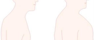
Congenital chest deformities in children
Congenital deformity of the chest in children may be associated with genetic characteristics and changes in the formation of the sternocostal complex, which can lead to a gradual increase in deformity until the growth of the child’s or adolescent’s skeleton is completed.
Congenital pathology is associated with improper development of the skeleton (vertebral column, ribs) due to an imbalance of mineral and endocrine metabolism. The consequence may be a specific development of the body:
- painful thinness;
- narrow shoulders;
- high growth;
- protrusion of the shoulder blades and collarbone;
- sunken chest when inhaling;
- long limbs;
- curvature of the spine (scoliosis or kyphosis).
Hereditary deformity is determined in 20-65% of cases of chest deformity. There are diseases and specific syndromes where this type of deformation is one of the symptoms. For example, pathology often develops against the background of Marfan syndrome.
Marfan syndrome
This disease is characterized by funnel-shaped and keeled deformities of the chest.
Marfan syndrome has the following symptoms:
- asthenic physique;
- arachnodactyly;
- dissecting aortic aneurysm;
- dislocation or subluxation of the lenses of the eyes (or other vision pathology);
- biochemical changes in the metabolism of collagen and glycosaminoglycans.
The development of chest deformity can be facilitated by dysplasia of connective and cartilaginous tissues, which is caused by enzymatic disorders.
Sporadic (non-hereditary) forms of deformity
Non-hereditary deformity of the chest develops due to teratogenic factors that affect the fetus during its development. Most often, abnormal development is caused by asynchronous, inharmonious growth of the sternum and costal cartilages.
Types of chest deformation
Ryzhikov Dmitry Vladimirovich
Ryzhikov Dmitry Vladimirovich (Head of the Department of General Bone Pathology of the Federal State Budgetary Institution "National Medical Research Center for Pediatric Traumatology and Orthopedics named after G.I. Turner, Candidate of Medical Sciences, doctor of the highest qualification category, traumatologist-orthopedist)By type, we most often see the corpo-costal type, this is a deformation of the sternum in the lower part with the involvement of the ribs.
The manubrial type (manubrio-costal) is much less common, this type includes deformation of the manubrium of the sternum (this is the upper part of this bone).
Orthopedists also differentiate between asymmetric forms of deformation and its elasticity.
At what age and by what symptoms can chest deformation be detected in a child?
Among the patients of our Center there are children of any age. Most patients are admitted with a congenital form of the pathology. Sometimes a child is born with an already noticeable chest deformity, but most often we see situations where the deformation becomes noticeable for the first time at the age of 6-8 years and progresses significantly at 10-13 years. Chest deformities can increase while there is potential for skeletal growth, that is, on average until 15-17 years of age. And the higher the height of the parents and the more active the growth of the children, the higher the risk of developing a very pronounced deformity. In contrast to limb deformities, chest deformities often include in their list of symptoms and disturbances in the functioning of the chest organs.