From this article you will learn:
- what is periodontitis - photo, diagram,
- what does it look like on an x-ray,
- symptoms and treatment of periodontitis.
Dental periodontitis is a disease characterized by the occurrence of a focus of acute or chronic inflammation at the apex of the tooth root. In relation to periodontitis, dentists often use the term “apical” - this implies the localization of inflammation precisely at the apexes of the roots of the teeth (from the Latin word “apex” - apex).
Apical periodontitis most often occurs - 1) in the absence of timely treatment of pulpitis, 2) as a consequence of poor-quality root canal filling in the past. With periodontitis, a so-called “periodontal abscess” is formed at the apex of the tooth root, which at first can only be a focus of infiltration of bone tissue around the apex of the tooth root - pus (Fig. 1). At this stage, destruction of the integrity of the bone has not yet occurred, but all this is accompanied by severe pain - especially when biting on a tooth.
But if left untreated, acute purulent periodontitis can turn into a chronic form, in which a focus of chronic inflammation forms at the apex of the tooth root - in the form of so-called “purulent sacs” (Fig. 2-3). In such foci of inflammation, bone tissue is destroyed, as well as periodontal fibers that provide attachment of the tooth to the bone. The chronic form of periodontitis can be virtually asymptomatic for years (patients sometimes report only periodic discomfort that occurs when biting on a tooth).
Apical periodontitis of the tooth: what is it?
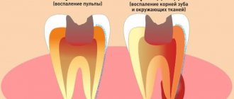
Comparison of pulpitis and periodontitis (Scheme 1) - with pulpitis, the neurovascular bundle (pulp) located inside the tooth is infected, but it still retains its vitality. With periodontitis, the pulp is completely necrotic and is a source of infection for the tissues surrounding the tooth. The infection enters the surrounding tissues through root canals, which open through holes in the area of the apex of the tooth roots. It is around the latter that foci of inflammation form during periodontitis. This is what the main differences between pulpitis and periodontitis look like.
Symptoms of acute periodontitis -
This form always occurs with severe symptoms: pain, swelling of the gums, sometimes even swelling of the gums/cheeks. The following symptoms are characteristic of acute periodontitis:
- aching or sharp pain in the tooth,
- tapping or biting on a tooth causes increased pain,
- in the absence of treatment, the aching pain gradually turns into throbbing, tearing, with very rare pain-free intervals,
- weakness, fever, sleep disturbance,
- There may be a sensation that the tooth has moved out of the jaw.
On an x-ray, the acute form is understood as primary periodontitis with acute symptoms, in which only bone infiltration with pus occurs in the area of the apex of the tooth roots, but there is no actual destruction of the bone tissue.
Therefore, on an x-ray, it will be impossible to see any significant changes other than a slight expansion of the periodontal fissure. A visual examination can reveal - on a diseased tooth you can always find either a carious defect, a filling or a crown. The gums in the projection of the root of the diseased tooth are usually red, swollen, and painful when touched. You will often find that the tooth is slightly loose. In the projection of the root of the diseased tooth, swelling of the gums may also appear (Fig. 4-6) and even swelling of the soft tissues of the face.
Apical periodontitis: photo
Symptoms of chronic periodontitis –
This form of periodontitis very often occurs asymptomatically or with minimal symptoms. In some cases, biting on a tooth or tapping on it can be painful. But the pain in this case is moderate, not severe. Sometimes the tooth may react to heat, which may cause mild pain.
During a visual examination, you can find - on the diseased tooth, you can again find either a carious defect, or a filling or crown. From time to time, a fistula opening may form on the gum in the projection of the apex of the root of a diseased tooth, from which a scant purulent discharge will be released (Fig. 6-7).
Due to such sparse symptoms, the main diagnosis is carried out using an x-ray, because with long-term chronic inflammation, bone destruction always occurs at the root apex (it is clearly visible on x-rays). Moreover, depending on the X-ray picture, chronic periodontitis is usually divided into the following 3 forms:
- fibrous form,
- granulating form,
- granulomatous form.
Diagnostics periodontitis by x-ray –
Understanding the form of periodontitis is very important for the doctor, because... The tactics of the treatment will depend on this.
- Fibrous form of chronicle. periodontitis (Fig. 10) –

with this form of inflammation, fibrous tissue grows in the periodontium. In this case, an X-ray will show a pronounced expansion of the periodontal fissure (24stoma.ru). This form of periodontitis is very easily treated in 1-2 visits: for this you only need to properly fill the root canals.
- Granulating form (Fig. 11) –
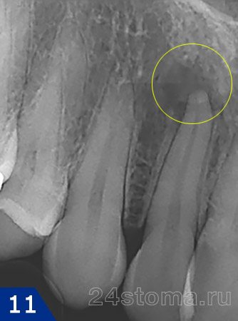
is the most aggressive form, characterized by rapid destruction of bone tissue around the apex of the tooth root. On an x-ray, this form of periodontitis will look like a candle flame without clear contours. The absence of clear contours indicates the absence of a membrane around the source of inflammation.
- Granulomatous form (Fig. 12) –
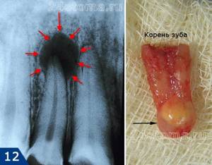
with this form of apical periodontitis, the focus of inflammation on an x-ray will appear as an intense darkening with clear rounded contours.
Moreover, depending on the size of the inflammation, the granulomatous form of periodontitis is divided into 3 more forms: → granuloma (sizes up to 5 mm), → cystogranuloma (sizes from 5 to 10 mm), → radicular cyst (sizes more than 1 cm).All these 3 formations are tightly attached to the apex of the tooth root. On the outside they have a dense shell, but on the inside they are hollow and filled with pus. If the causative tooth with a granulomatous form of periodontitis is subject to removal, then most often when the tooth is removed from the socket, we will see a rounded formation (“purulent sac”) at the apex of its root. You can see what a granuloma or cystogranuloma looks like at the apex of the root of an extracted tooth in the video below.
Appearance of granuloma/cystogranuloma –
Symptoms of exacerbation of chronic periodontitis -
The chronic form of periodontitis is characterized by a wave-like course with periods of periodic exacerbation, during which the symptoms become characteristic of the acute form of periodontitis, i.e. severe pain, possibly swelling and swelling of the gums. Typically, exacerbation of a chronic inflammatory process is associated with hypothermia or other causes of decreased immunity.
If, against the background of exacerbation of chronic inflammation, a fistula appears on the gum (which allows the outflow of purulent discharge from the source of inflammation), acute symptoms may decrease again and the process gradually becomes chronic again. And so on until a new aggravation...
Causes of dental periodontitis
Various reasons can lead to the appearance and development of dental periodontitis, but in the vast majority of cases the disease appears:
- Against the background of untreated dental caries and pulpitis;
- Due to poor quality treatment of tooth canals, or more precisely, mistakes made when filling the canals.
Less commonly, periodontitis occurs after injuries, incorrectly installed dental fillings, or violation of the dosage of dental medications.
Whatever the cause of periodontitis, its treatment cannot be delayed due to fear of dentists. It is important to understand that you will not stop the inflammation on your own, with pills or folk remedies, and the more it progresses, the less chance you have of saving the tooth. In addition, periodontitis can also affect neighboring healthy teeth.
How to identify periodontitis and not confuse it with caries or pulpitis? We already talked about the most striking symptom of the disease at the very beginning of the article, but the signs of periodontitis may vary depending on the form of the disease. Therefore, below we will consider the main types of dental periodontitis, as well as talk about their characteristic symptoms.
Take a short test and calculate the cost of treatment!
Take a short test
- Which teeth have caries?
- Visual assessment
- Reaction to stimuli
- Cost calculation
×

Manukyan Artavazd Genrikovich
Chief physician of the clinic
How is periodontitis treated?
Regardless of the form of apical periodontitis, treatment will begin with an analysis of your complaints and an x-ray. Based on this, the doctor will draw up a treatment plan. An x-ray and examination will show whether it is possible to cure this tooth or whether it needs to be removed.
Emergency care for acute periodontitis (exacerbation of chronic) –
The doctor’s main task is to open the tooth and leave the root canals open for several days. This is necessary to drain the pus and relieve acute pain. If this requires removing a crown, filling, or unsealing previously poorly filled root canals, the doctor will definitely do this on the first visit. In addition, if you have gumboil on your gums, then it will be necessary to open the purulent abscess (by making a small incision).
Emergency care - in video 1 - opening a tooth to create an outflow of pus through the root canals, in video 2 - making an incision to open an abscess on the gum.
Open root canals will allow the pus to escape, and this in itself will significantly reduce pain. During this period, you will be prescribed rinses and antibiotics. You will be scheduled for a second visit (in 3-4 days), and when the doctor sees that the pus is no longer draining from the canals, a special antiseptic will be placed in the canals for several days.
Further treatment will depend on the size of the inflammation at the apex of the tooth root, and the larger it is, the longer the treatment will be. The treatment methods that will be further used will be fully consistent with the treatment of chronic periodontitis.
2) Treatment of chronic forms of the disease –
A separate article is devoted to the treatment of chronic forms of periodontitis (see link), because This is a very complex and voluminous topic. But in short, here only the treatment of the fibrous form of periodontitis is quite simple, and usually requires only 2 visits within 1 week. This is due to the fact that with fibrous periodontitis there are no significant inflammatory changes at the root tips, which means that long-term treatment with temporary filling materials based on calcium hydroxide is not required.
But for granulating and granulomatous forms, treatment can take several months. A special anti-inflammatory material based on calcium hydroxide is introduced into the root canals of such teeth, which will reduce foci of inflammation at the apexes of the roots and cause restoration of bone tissue. The action of the materials is slow, which is what causes the duration of treatment.
In some cases, it is simply impossible to cure periodontitis with conservative methods. This happens when very large cysts are discovered: from 1.5 to 4-5 cm. Then, after preparing the tooth (root canal filling), a tooth root resection operation is performed, during which the doctor, through a small incision, cuts off the apex of the root from the tooth together with a cyst, and remove them. We hope that our article on the topic: Periodontitis symptoms and treatment was useful to you!
Sources:
1. Dental education of the author of the article, 2. Based on personal experience as a dentist, 3. National Library of Medicine (USA), 4. “Therapeutic dentistry: Textbook” (Borovsky E.), 5. “Practical therapeutic dentistry” ( Nikolaev A.).
What to do if you have periodontitis
If the diagnosis is confirmed, the disease must be treated. How dental periodontitis is treated depends on the form and stage of inflammation. The correction program may include therapeutic and surgical techniques.
Conservative therapy
Conservative methods are aimed at maximizing the preservation of the structure of the unit and restoring its functionality. The program includes elimination of the source of inflammation and endodontic treatment. All manipulations are performed under local anesthesia. Treatment regimen:
- Conducting anesthesia.
- Opening the crown, providing access to the root canals.
- Expansion of the cavity.
- Pulpectomy (if the segment has not been previously treated) or unfilling of the canals.
- Antiseptic, medicinal treatment of cavities.
- Temporary filling (for the period of anti-inflammatory treatment).
- Removal of temporary filling, filling of root canal.
- Restoration of the coronal part of the unit.
- Prescribing a course of antibiotic therapy.
Surgical intervention
Invasive methods are used when other methods are ineffective. The operation can be carried out using different protocols:
- Resection of the apex of the tooth root followed by filling the cavity with special bone-forming materials.
- Extraction of the diseased segment.
If periodontitis is not treated, complications are possible: odontogenic periostitis (inflammation of the periosteum), abscess, phlegmon, etc. The infectious process spreads to nearby lymph nodes, and lymphadenitis occurs. In severe cases, possible: tooth loss, osteomyelitis, sepsis.