If a doctor, during an ultrasound examination, tells a woman that she sees a fertilized egg in the uterine cavity, then you can congratulate this patient, because in nine months she will become a mother.
The presence and size of the fertilized egg can be determined by ultrasound as early as the seventh to ninth day of a missed period.
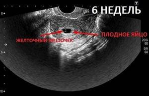
If the fertilized egg is located in the uterus, therefore, the pregnancy is normal. The specialist immediately determines the size of the fetal egg by timing, its shape and location. In addition, he will pay special attention to whether there are detachments or other pathological conditions.
What does a fertilized egg look like?
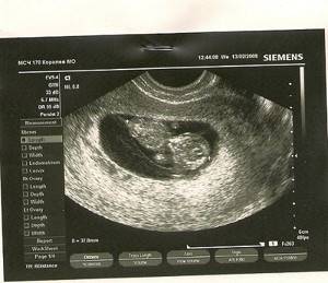
During the sixth week, the fertilized egg can be seen for the first time using ultrasound. At this time, its parameters are about 1 cm. But, if you follow the standards and recommendations of the World Health Organization, such a diagnosis in order to view the ovum is recommended to be carried out no earlier than the 10th week. But, as practice shows, rarely do any future parents have such endurance. Most of them are eager to make sure that the pregnancy is intrauterine and the fetus is developing without pathologies.
Therefore, doctors recommend immediately, after establishing the fact of pregnancy, to contact an antenatal clinic and register with a special register. Doctors will monitor the pregnancy throughout the entire period, monitor the condition and health of the patient and the unborn baby, and also prevent possible risks or pathologies.
Ultrasound examination is the most important diagnostic test that allows you to diagnose pregnancy, monitor the course of pregnancy, and determine possible deviations from the norm of the embryo. Thanks to this research, more than one pregnancy has been saved, and the health of the mother and unborn child has also been saved.
Fertilization process - stages
Many of us know how fertilization occurs in the traditional way. The male sperm merges with the female egg. This requires different amounts of time - from a few minutes to 5 days. It all depends on individual indicators.
The fusion of two cells, female and male, is a complex process. It is preceded by many processes: ejaculation in men, ovulation, penetration into the oocyte shell in women. In order for the egg to form into an embryo, it is important for a woman to monitor her health and plan pregnancy long before it occurs.
Let's consider the main stages of fertilization:
- Ovulation
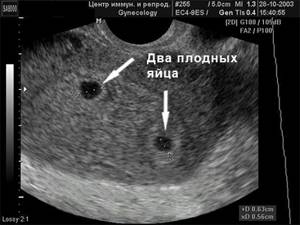
Conceiving a child is impossible without ovulation. It falls in the middle of the cycle, represents the release of the egg from the follicle, and is regulated by hormones. Every month a woman releases one egg, sometimes several, which leads to the development of twins. For conception to occur, the female cell and the male sex gametes must meet in the fallopian tube. If this does not happen, it dies and comes out during menstruation.
- Fertilization
Future parents are interested to know how fertilization is progressing. Often the process takes place in the fallopian tube. But there are exceptions when this happens in the uterus.
- Sperm migration
Everyone knows in general terms how fertilization occurs in humans. This process is impossible without sperm. The most favorable process for pregnancy is 24 hours from the moment of ovulation. Male gametes are active for 2-5 days. If they managed to get to the fallopian tubes before the egg is released, they wait for it.
During sexual intercourse, about 3 million sperm enter the vagina. The more there are, the faster conception occurs. Many gametes die due to the acidic environment of the vagina and the mucus of the cervical canal. The most viable ones make their way to the fallopian tubes and uterus.
- Fertilization
The female reproductive cell is surrounded by a membrane that needs to be dissolved. The acrosome located on the head of the sperm is responsible for this.
Types of ultrasound during pregnancy
Transvaginal and transabdominal diagnostics are considered the most effective in the initial stages of pregnancy. These types of ultrasound make it possible to clearly see the location of the concentration of the fertilized egg in the uterine cavity. In addition, the procedure makes it possible to determine the presence of an ectopic pregnancy.
But, there are cases when it is not possible to examine the egg shell even within the planned time frame. However, an experienced diagnostician can only determine the thickening of the uterine walls characteristic of pregnancy. What to do in such a situation and when can you see the fertilized egg on an ultrasound for sure?
A repeat ultrasound should be performed after a couple of weeks, having previously agreed on this issue with the leading specialist.
Placental abruption in early pregnancy
Detachment of the ovum represents an incipient abortion. In such a case, premature rejection of the fertilized egg from the walls of the uterus is noted. It should be noted that when a spontaneous miscarriage begins, great importance is given to timely assistance, since, in most situations, the pregnancy can be saved. It is very important to do everything correctly and quickly. The detachment is accompanied by nagging pain in the lower abdomen, pain in the lumbar region, and dark red and sometimes brown discharge. The reasons that cause detachment of the ovum include ovarian dysfunction, various diseases of the patient (inflammatory processes, tumors, infectious diseases), underdevelopment of the woman’s genital organs, severe toxicosis, severe physical activity, and constant stress. However, the most obvious cause of detachment of the ovum is a lack of progesterone, which is usually called the pregnancy hormone.
When a pregnant woman shows signs of detachment of the ovum, she (or her relatives) urgently need to call an ambulance, and also call a gynecologist-obstetrician to inform him about what happened. Until the ambulance team arrives, the woman needs to lie down and also raise her legs up. You can also rest them against the wall or place them on the back of the sofa.
Detachment of the ovum is dangerous because it can cause abortion or frozen pregnancy. Therefore, if there is minimal suspicion of detachment, it is necessary to seek help from a doctor.
Additional diagnostics
In addition, you can obtain meaningful information regarding the course of pregnancy using the following modern types of ultrasound diagnostics:
- 3D and 4D studies;
- Dopplerography;
- cardiotocography.
The procedure, which uses three-dimensional imaging (3D and 4D examination), makes it possible to see the child, his limbs, face, as well as the part of the body that the baby is facing at the time of the procedure. This technique is used in later stages of pregnancy, when the body of the unborn child is relatively mature. The image projected onto the screen is in color, usually the fruit “appears” in a golden hue.
With the help of Doppler, a specialist can examine the circulatory system of the unborn child, as well as examine the uterus and placenta. This procedure is recommended for use at any stage of pregnancy. Diagnostics helps to establish the presence of possible pathologies in the cardiac system and makes it possible to establish fetal fading. At a later date, using this technique, oxygen deficiency can be determined.
By performing cardiotocography, the fetal heart rate is established, and possible facts of oxygen starvation are recorded. A referral for the procedure is issued exclusively by a doctor if there are existing indications.
Where does the fetus grow and develop?
The fetus grows and develops in the uterus. Changes are observed every day. Over time, his limbs, organs, and facial features form. From 37 to 42 weeks, labor can begin and the baby is considered full-term. During pregnancy, a woman needs to lead a healthy lifestyle, take care of herself, get more rest, and visit the doctor on time. It is important to take care of yourself, because some infections and diseases pose a threat to the fetus.
Bearing and giving birth to a child is an incredibly complex process, so every woman needs to know how to conceive a child and how to do everything possible to ensure that he is born healthy.
Why is the fertilized egg not visible?
Despite the fact that it is recommended to perform an ultrasound to view the ovum already in the sixth week, it is not uncommon, even at later stages, that it cannot always be examined. Moreover, at week 12, in some cases, even a heartbeat cannot be heard. Doctors say there is no need to panic. Even such a modern and reliable study as ultrasound does not always provide one hundred percent comprehensive information. The lack of proper and expected results can be affected by many nuances, both technical and related to the human factor. Often, in order to get a complete picture of the course of pregnancy, specialists need to study additional results of studies other than ultrasound. These include laboratory tests and examinations by a gynecologist.
If during an ultrasound the embryo in the ovum is not visible, the doctor leading the pregnancy writes out a referral for a blood test for hCG (human chorionic gonadotropin). During normal pregnancy, the level of this hormone gradually increases in accordance with the weeks of gestation.
At the same time, if the fetal egg is not viewed during an ultrasound, the possibility that the period was calculated incorrectly cannot be ruled out. It is quite possible that it is quite small for the fetus to be visible on the monitor screen. Doctors calculate the period in weeks, which are called “obstetric”. They are counted from the first day of absence of menstruation. Therefore, errors in calculations are quite common. To establish the exact date, you should re-take tests and undergo an ultrasound again. To do this, you should contact another medical institution.
Doctors note that the exact period when the embryo can be viewed by ultrasound, and not just the separate fertilized egg surrounding it, directly depends on the personal characteristics of the expectant mother’s body, as well as the location of the fixation of the egg in the uterine cavity. An important role is played by modern equipment with which ultrasound is performed. For this reason, it is recommended to perform ultrasound only in competent clinics that have modern equipment and experienced specialists.
Where fertilization and embryo formation takes place
how a woman is fertilized day by day .
- 1st day. The seminal fluid merges with the egg. Sperm attack it and “pierce” the membrane. The most active one gets inside.
- On the 2nd and 3rd days, the zygote is formed. Cells are actively dividing, and their number doubles every 12 hours.
- On the 4th day, the number of cells is 16. The zygote emerges from the fallopian tube.
- 5th day - the fertilized egg moves along the mucous membrane, looking for a suitable place for implantation.
After the 12th day, cell division stops and the process of implantation of the embryo is completed.
Approximately 2 weeks after conception, “pregnancy hormones” are actively produced.
Now you have learned in more detail how quickly fertilization occurs. After 2-3 weeks from the moment of conception, the test may show a positive result.
Fertilization of an egg - embryo
To understand whether fertilization has occurred, and what is the likelihood that the pregnancy may be terminated in the near future, a woman needs to undergo several studies.

based on the results of which specialists will determine all the necessary indicators. Usually, if the pregnancy has occurred successfully, the woman will know this - either by the results of a pregnancy test, or after taking a blood or urine test for the content of human chorionic gonadotropin (the level will be higher than the established norm), or after consulting a doctor, she undergoes an ultrasound. But if the examination reveals a low level of a hormone such as progesterone, the woman is at risk of miscarriage. In this case, doctors immediately begin complex therapy aimed at replenishing the level of progesterone in the woman’s blood and improving conditions for the further development of the fetus.
What time does the embryo appear in the fertilized egg?
Also, thanks to modern developments of leading foreign laboratories and clinics, today most pregnant women undergo special studies for 9 months, such as pregnancy screening. This analysis includes the results of an ultrasound examination of each trimester of pregnancy, the results of a woman’s blood test, which together give a picture of the possible development of congenital anomalies or genetic diseases of the fetus. One of the latest developments by scientists in the screening structure is a special program that calculates the percentage of damage to a woman’s body over the entire period of bearing a child, the likelihood of developing a somatic disease during pregnancy, etc.