A biopsy and histological examination of a tissue sample obtained with its help is one of the most accurate methods for diagnosing tumors in the human body. In diagnostics, to monitor the condition and identify pathology of the uterine mucosa, gentle aspiration technology is used - endometrial biopsy pipe. Its results, obtained by examining tissue under a microscope, are necessary to identify the causes:
- hyperplastic growth and thickening of the mucous membrane;
- endometrial carcinomas;
- pathological uterine bleeding;
- miscarriage;
- infertility caused by hormonal and hyperplastic disruptions;
- oncological diseases.
Pipel endometrial biopsy is performed before IVF, during bacteriological examination, including before planned conception, as well as to monitor the results of hormone therapy and its effect on the female reproductive system.
Unlike a conventional biopsy, a painful procedure that requires dilation of the cervical canal and produces a high percentage of complications, this technology is painless and non-traumatic
.
Advantages of the method
Pipelle biopsy is a more modern alternative to surgical endometrial biopsy.
Due to its low invasiveness, the procedure can be performed on an outpatient basis. The method by which it is performed does not require painful dilation of the cervical canal. Tissue aspiration is performed with a soft silicone probe with a diameter of about 3 mm. The probe has a hole at one end and a vacuum pump at the other. Despite the fact that the manipulation is a minor surgical procedure, the patient can leave the clinic within two hours after it. The main advantages of pipell biopsy of endometrial tissue include:
- possibility of carrying out on an outpatient basis;
- no need to dilate the cervix;
- minimum of discomfort;
- disposable probe, eliminating the risk of infection;
- low risk of complications after the procedure;
- short recovery period.
What can and cannot be done after the procedure?
After the procedure, the woman can travel home or to work on her own. No hospital or bed rest required
. There are no complications; mild discomfort or pain in the lower abdomen goes away on the second day. Infection is excluded because a sterile, disposable instrument is used.
Are you looking for a clinic in Moscow in the Konkovo area to quickly and inexpensively undergo a pipel endometrial biopsy? Residents of the Yasenevo, Belyaevo, Mosrentgen, and Tikhy Stan districts often contact us.
What is the procedure
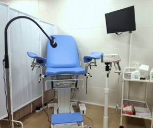
Pipelle endometrial biopsy can be performed on an outpatient basis at any of the clinics in the Medoc network. On the eve of the study, the patient undergoes all the necessary tests, including a gynecological smear for infections and a series of blood tests. The day of manipulation is determined by the obstetrician-gynecologist based on the preliminary diagnosis and symptoms. The procedure is performed on a gynecological chair. To ensure minimal discomfort during the intervention, local anesthesia of the cervix is used. After the anesthesia has taken effect, a special pipell biopsy instrument is inserted into the organ cavity to aspirate endometrial samples. Using a probe, the doctor will suction out a small area of tissue for further examination. After one to two hours (if the condition is satisfactory), the patient can go home.
How is a pipell endometrial biopsy performed?
Benefits of the procedure:
- performed on an outpatient basis;
- does not require anesthesia;
- duration - 10 minutes;
- a disposable pipel is used - a flexible plastic tube with a diameter of 2 to 4.5 mm. The instruments are protected by sterile packaging, so infection of the genitals during the procedure is excluded.
Indications for biopsy
Diagnostics may be recommended for the following diseases and conditions:
- too much menstrual flow;
- spotting outside of menstruation;
- cycle disorders;
- any endometrial neoplasms;
- infertility;
- miscarriage;
- adverse effects of hormonal therapy;
- bleeding during menopause;
- unsatisfactory condition after artificial termination of pregnancy (bleeding, pain, etc.);
- chronic endometritis;
- atrophic changes in the endometrium.
Cost of biopsy by aspiration and Paypel method and features of the procedure
Doctors take material for analysis in two ways:
- Aspiration. In this case, a syringe or an electric vacuum device is inserted into the cavity and the mucous membrane is sucked under the influence of negative pressure.
- Paypel method. A soft, thin tube (3 mm in diameter) with a piston, like a syringe, is inserted, and when the latter is pulled back, tissue samples are sucked in for analysis.
These are modern and safe methods of collecting material. Unlike traditional diagnostic curettage, in which the canal is widened and the mucous membrane is scraped out with a curette - a diagnostic spoon, they do not cause pain and do not require anesthesia. For the current price of biopsy, pipel and aspiration, see the price list below on the page. In both cases, the procedures take about 15 minutes.
Make an appointment for an endometrial biopsy
A biopsy is one of the most informative studies of cervical diseases, such as cancer, polyps, fibroids, etc. The procedure is performed by a gynecologist and requires little preparation. To take part, you must pre-register by phone or through the form on our website.
Leave your phone number. The clinic administrator will call you back.
Make an appointment
Nuances of preparation
Before a pipel biopsy of endometrial tissue, the gynecologist must make sure that there is no acute inflammatory process in the patient’s genitals. This requires taking gynecological smears for infections. You should also consult your doctor about taking blood thinning medications before the procedure. The full list of preparatory measures includes:
- consultation and examination by an obstetrician-gynecologist;
- Ultrasound of the pelvic organs;
- blood test for human immunodeficiency virus and syphilis;
- blood test for hepatitis B and C;
- clinical blood test;
- smear for genital infections;
- smear for cytology.
Sexual rest is required two to three days before the operation.
Methodology
Medicine does not stand still, and today a new approach is being used to study the mucous membrane of the female organ, which allows us to solve many problems that are characteristic of the traditional approach.
Conducting a traditional study is impossible without expanding the cervical canal of the organ. The procedure is performed under hysteroscopic control using intravenous anesthesia. This is not only painful, but can also lead to a number of unpleasant consequences. This is why contraindications arise.
Using a new technique, the doctor gets new opportunities. The procedure itself does not require intravenous anesthesia, and dilation of the cervical canal is also not required.

PET/CT examination
Since the instrument is disposable, it is kept in sterile conditions (packaging) before use, so the issue of possible infection is eliminated.
And another important point: the procedure is more economical than the traditional one.
How is the procedure performed?
An hour before the procedure, doctors advise taking any painkiller in tablet form. In addition, at the request of the patient, the specialist can resort to local anesthesia before the intervention itself. In some cases, he may recommend taking sedatives. But when using them, you need to take precautions, as they can cause drowsiness.
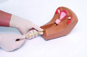
Classic biopsy sampling is performed using a curette, which can cause damage to the walls of the uterus. Using a special probe can reduce this risk to a minimum. To carry out the procedure, a special flexible plastic tube is used - pipel. Its diameter is only 3 millimeters, there is a piston inside, and a special hole on the side.
On the agreed day, the patient must come to the doctor's office with a referral. The procedure itself is carried out as follows:
- The woman is asked to lie down on a chair.
- The external genitalia are treated with an antiseptic solution.
- The doctor inserts a special speculum into the vagina.
- A probe is inserted into the uterine cavity.
- The specialist, by pulling back the piston, creates a kind of vacuum so that part of the endometrium falls into a special hole on the tube.
- The material is collected from different areas (at least 3).
- After receiving the required amount of material, the tube is removed.
The most significant pain is observed during the period of material collection, when the uterus may react with spasm. Patients note that these sensations are similar to those that appear shortly before the critical days. Sometimes stomach pain and dizziness occur. This reaction is called vasovagal.
The duration of the biopsy is from 1 to 3 minutes. Upon completion, all samples received are sent to the laboratory for further research. Immediately after taking the material, the woman can leave the clinic, as she remains fully functional and does not need to stay in a hospital.
How to properly prepare for the procedure?

Although this biopsy method is minimally invasive, it still involves the introduction of certain instruments into the uterine cavity that violate its integrity. Therefore, doctors pay special attention to preparing for taking a biopsy.
Experts recommend:
- During the period of the cycle in which the study is planned, stop taking hormonal medications. This is because such medications can affect the accuracy of the analysis.
- Use contraception before the procedure or refrain from sexual activity. This is necessary in order to avoid pregnancy, because performing a diagnostic procedure can lead to its termination.
- Do not use vaginal suppositories, ointments or creams.
- Do not consume foods that cause increased gas formation.
- Perform a cleansing enema the day before the procedure.
When choosing this research method, the doctor will ask the woman for information about all the medications she is taking. Be especially careful when taking blood thinners. In some cases, the specialist can adjust the time and dosage several days before the date of the planned intervention.
Particular attention is paid to the date of the biopsy. If a woman is not in menopause, then the procedure is carried out on strictly defined days of the cycle. If the patient no longer has menstruation, the material is taken taking into account pathological bleeding.
The most suitable days to perform the procedure are:
- 18-24 – to determine the phase of the cycle.
- 5-10 – with polymenorrhea. Once every 7 days if it is impossible to conceive a child or there is no menstrual flow.
- 17-25 – to monitor the effectiveness of treatment using products containing hormones.
Samples are taken on the first day of bleeding to identify its cause. Also, on the first or last day of the cycle, diagnostics can be prescribed if necessary to confirm infertility. A biopsy is performed on any day of the cycle if the doctor has suspicions about malignant degeneration of the formation.
About the organ
The uterus is a hollow inside, female unpaired organ consisting of muscle tissue, located in the middle part of the pelvic cavity.
The organ is capable of significantly increasing in size during fetal development, and after childbirth it can return to its previous size within a couple of weeks.
Like other organs, the uterus can be subject to various pathologies. They can also cause dangerous complications. Therefore, it is important to regularly visit a gynecologist for preventive purposes.
It's sad, but statistics show that improving medical technology does not reduce the number of patients. Doctors at the Onco.Rehab integrative oncology clinic often encounter cases of advanced forms of cancer due to late detection of the disease, but treating the disease in its initial stages is a significant guarantee of successfully getting rid of the problem.
When to carry out
The doctor determines the time of the uterine biopsy, guided by the most favorable days of the menstrual cycle. They differ for different diseases, for example:
- Infertility – at the very beginning of menstruation or before it;
- Heavy menstrual bleeding – on days 5–10 of the cycle;
- Absence of menstruation and absence of pregnancy - repeated procedures are carried out for 3 - 4 weeks with breaks of 1 week;
- Acyclic bleeding - after the onset of blood smearing, bleeding;
- Suspicion of cancer of the mucosal layer - any day of the cycle.
Research results
If no pathology is detected, then the results of the study will indicate that the mucous layer corresponds to the phase of the female cycle and age, and no signs of atypia were found.
If we talk about pathological conditions, then diagnostics allows us to identify:
- Endometritis.
- Underdevelopment of the endometrium or its atrophy.
- Discrepancy between the thickness of the mucous layer of the uterus and the day of the cycle.
- Atypical endometrial hyperplasia.
- Hyperplasia (glandular-cystic or simple diffuse).
- Adenomatosis (precancerous condition).
- Degeneration of the endometrium into a malignant tumor.
Additional examinations are possible if any disease from the list above is detected. The doctor will talk about this in detail during the consultation. If the biopsy provides enough data to establish an accurate diagnosis, then treatment will be prescribed.
Aspiration biopsy with histological examination
During an aspiration biopsy of the endometrium with histological examination, the gynecologist receives a small amount of material, which is sent to histologists for study. The Healthy Family clinic employs a large staff of various specialists, which allows all studies to be carried out on the basis of the institution.
The histological examination itself is a method of studying affected tissues using a microscope. This method is used to study tumors of various types. With its help, it is determined:
- type (benign or malignant);
- the rate at which cells grow;
- effectiveness of the chosen treatment method.
To conduct research in the clinic, the resulting biopsy is treated with a special substance with fixing properties, which allows it to remain intact, and is also compacted by pouring paraffin. After this, the material is cut into small slices, stained and examined under a microscope.
The resulting samples can be informative or non-informative. An adequate sample contains a sufficient number of cells for analysis. An indication that the sample is inadequate indicates that there are not enough endometrial cells. Instead, there are blood cells and squamous stratified epithelium. This situation is possible if the material was collected in violation of the technology.
The specialist describes each type of tissue found in the sample and evaluates them according to several criteria. The results of the study are ready in 7-10 days. Only the attending physician can decipher the received conclusion. If deviations are not diagnosed, then it will be indicated that the condition of the endometrium corresponds to the age category.
If atypical cells or cancerous processes are detected, the gynecologist may raise the question of using other diagnostic measures, the need for additional examinations or surgical intervention. If the results indicate the presence of inflammatory processes, the doctor prescribes antibiotics or anti-inflammatory drugs.
If the study reveals signs of hyperplasia, the doctor prescribes additional examinations to identify possible endocrine disorders. If the assumptions are confirmed, the woman is prescribed hormone therapy to improve the condition of the endometrium and restore reproductive function.
Indications and contraindications
Endometrial aspiration biopsy is performed only as prescribed by a doctor. Such diagnostics can identify many different diseases. Most often it is prescribed for:
- Endometriosis.
- Infertility.
- Detection of polyps in the uterus.
- Endometrial hyperplasia (thickening of the mucous membrane) of the uterus.
- Unstable menstruation.
- Continuous bleeding after childbirth or abortion.
- Chronic endometritis.
- Miscarriage.
- Pathologies during menopause.
Pipelle biopsy will be indicated in preparation for in vitro fertilization (IVF). The results of the analysis will allow you to find out the condition of the endometrium before starting the use of hormones.
Contraindications to aspiration biopsy are: pregnancy at any stage, acute infectious and inflammatory diseases of the uterus and appendages, poor blood clotting (for example, hemophilia), severe cervical stenosis.