What is the bacterium Helicobacter pylori, Helicobacter?
Helicobacter pylori (Helicobacter pylori bacteria, HP) is currently considered the leading etiological factor in chronic gastritis, gastric ulcers, and gastric cancer. Helicobacter pylori causes 85% of chronic gastritis to be Helicobacter pylori, or HP-associated gastritis (causing serious illness). In the etiology of non-atrophic gastritis, nutritional disorders (improper diet, abuse of spicy, hot or cold foods), disorders of neurohumoral regulation, smoking, strong alcoholic drinks (alcohol, vodka, cognac, whiskey, tequila, moonshine) are of great importance.
How does the procedure work?
Before fibrogastroduodenoscopy followed by an HP test, special preparation is required. A few 2-3 days before the procedure, it is forbidden to consume alcoholic beverages, spicy and highly salty foods, the patient must adhere to a light diet, 12-18 hours before the examination it is necessary to stop eating, and 4-5 hours before the examination, stop drinking water.
FGDS is performed with the patient lying on his left side. In order to eliminate discomfort, the oral cavity is treated with an anesthetic solution (lidocaine). The endoscopic probe is placed on the root of the tongue and, together with the patient’s swallowing movements, it is moved deeper. In general, the procedure is carried out like a standard fibrogastroduodenoscopy, the only difference is that during the examination, a special probe is inserted into the area under study through a probe, with the help of which pieces of tissue are taken for examination.
The obtained biological material can be examined using two methods. The first is the urease test (a piece of tissue is placed on a special chemical medium; if Helicobacter Pylori is present there, the medium will change its color). The second is histological, which consists of examining a piece of tissue under a microscope.
Gastritis and Helicobacter pylori bacteria, Helicobacter pylori, HP
Helicobacter pylori (HP) is the cause of the development of Helicobacter pylori chronic gastritis, the most important factor in the pathogenesis of duodenal ulcer and gastric ulcer, low-grade gastric lymphoma (malt-lymphoma, from MALT - mucosal-associated lymphoid tissue), as well as gastric cancer. In 1994, experts from the International Agency for Research on Cancer (IARC) under the WHO (World Health Organization) classified Helicobacter pylori as a class 1 carcinogen, which means that HP infection is unconditionally linked to the occurrence of stomach cancer. In 1983, Barry J. Marshall and Robin Warren identified the presence of a microorganism in the stomach of patients with peptic ulcers, which was initially called Campylobacter pylori, and since 1989 it has been called Helicobacter pylori (HP), as it belongs to to a separate genus.
IgG antibodies to Helicobacter pylori
Detection of immunoglobulins to Helicobacter pylori is carried out using an enzyme-linked immunosorbent assay, or “sandwich” method. Venous blood is used for the study; you must not smoke for 30 minutes before donating it.
The method is based on the fact that after the Helicobacter bacteria enters the body, a local and systemic response develops. It consists of an increase in the titer of immunoglobulin IgM, IgG, and IgA antibodies in the blood serum. In 100% of cases of infection, IgG immunoglobulin is detected, in 65-80% of cases - IgA, in 17-20% - IgM, therefore, accurate diagnosis is carried out by determining the concentration of IgG in the blood serum.
Advantages of the method:
- Does not require endoscopy, so the method is safe;
- Highly sensitive;
- Used to prevent primary infection.
The method may not be accurate when determining the immunoglobulin titer in older people, since they have reduced antibody production, as well as in those patients who take cytostatics. After treatment of heliobacteriosis, this method is also not effective, because the level of the hormone in the blood after treatment with antibiotics remains quite high for another 6 months.
Indications for use:
- Patient complaints of dyspepsia when it is impossible to perform endoscopy;
- Primary diagnosis of heliobacteriosis;
- Monitoring the effectiveness of treatment with antibacterial drugs.
Reference values for the examination: IgG immunoglobulin concentration from 0 to 0.9, the result is negative. This may mean the absence of infection or high effectiveness of therapy.
What does Helicobacter pylori look like?
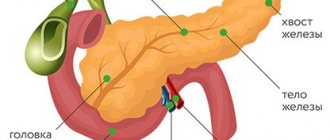
Bacteria Helicobacter pylori, Helicobacter pylori, Helicobacter, HP is a gram-negative microaerophilic bacterium of a curved or spiral shape with many (4 to 6) flagella. Helicobacter pylori (HP) is about 2.9 µm long and 0.8 µm in diameter. Helicobacter pylori , Helicobacter pylori (HP) is found deep in the gastric pits and on the surface of epithelial cells, mainly under the protective layer of mucus lining the gastric mucosa. It can exist in areas of gastric dysplasia with frequent acidification of the duodenal bulb. Directly around the bacterium, the pH is about 7, and the nutrient content is quite sufficient for the life of the microorganism. Helicobacter pylori (HP) is able to exist under conditions of varying concentrations of hydrochloric acid in the gastric cavity. Helicobacter pylori (HP) survives at a pH of 4 to 8. It reproduces successfully at a pH of 6 to 8. The most favorable habitat is the antrum of the stomach. Under unfavorable conditions, Helicobacter pylori (HP) can develop into a coccal form. Helicobacter infection is very dangerous.
Types of analyzes and diagnostics
There are several methods for studying the state of the body. Each of them has its own characteristics.
ELISA for bacteria
An enzyme immunoassay is prescribed to assess the level of concentration of antibodies to Helicobacter pylori in the patient's blood. If there are antibodies to the bacteria in the blood, it means that the immune system developed them at least 1-2 weeks ago. If the body's reaction has not yet been formed, a false negative result is possible.
Reasons for false positive results:
- error by the laboratory technician conducting the study;
- not enough time has passed after eradication, since antibodies are in the blood some time after complete cure.
A positive ELISA result indicates the need for a full examination, studying the results of an extended blood test, which provides more information about the condition of the body.
Immunoglobulins in the blood LgG, LgM, LgA
In the human body, when infections or pathogenic microorganisms penetrate it, immunoglobulins are produced - blood cell proteins that resist foreign viruses. When infected with Heliobacteriosis, the human body produces immunoglobulins LgG, LgM, LgA, which actively resist Helicobacter pylori. If they are detected in a person’s blood, it should be concluded that he is sick with heliobacteriosis.
The peculiarity of immunoglobulins is that in pursuit of pathogenic cells they overtake them anywhere in the human body. These proteins are produced after colonization by bacteria in the gastrointestinal tract.
Analysis transcript:
- A reduced or negative level of LgG immunoglobulin indicates one of the following options: the bacterium is absent in the body and the likelihood of developing a peptic ulcer is low, or only 3-4 weeks have passed since infection. If you have pain in the gastrointestinal tract, you need to repeat the examination in a week.
- An elevated or positive level of immunoglobulin LgG indicates one of two options: the bacterium is present in the body, increasing the risk of developing cancer and stomach ulcers, or the person has recently been cured of heliobacteriosis.
- A reduced level of immunoglobulin LgM, usually detected immediately after infection, or its absence, clearly indicates the absence of bacteria, since this protein is detected in the early period after infection.
- An increase in LgM levels indicates that the gastrointestinal tract is at an early stage of infection, and it is almost undamaged by the infection.
PCR analysis
The results of a polymerase chain reaction study can detect Helicobacter pylori DNA samples in the blood, which puts the PCR test in first place among all diagnostic methods. A positive test result proves the presence of bacteria in the body, a negative test shows its absence. The only thing that is not available when performing a PCR blood test is determining the time of introduction of the bacteria into the body.
If antibacterial therapy or antiseptic treatment was carried out before taking blood for analysis, the results of the study will not be objective. It is better to entrust the interpretation of blood tests to a specialist, although serious laboratories include a table of the norm and deviations from it in the results.
Cytological examination
In this test, swabs are taken from the antrum of the stomach during endoscopy. The area where the smear is taken is the area of edema and hyperemia, but not erosion or ulceration. After taking fingerprints, the laboratory technician dries and stains the smears.
When examined under a microscope, Helicobacter pylori can be found in the form of a spiral or in the form of the wings of a flying seagull.
Cytology reference values:
- (+) — low level of contamination (up to 20 microbial bodies);
- (++) – average level (up to 40 microbial bodies);
- (+++) – high level (over 40 microbial bodies at 360x magnification).
Additionally, in the presence of inflammation, a cellular infiltrate is detected: (eosinophilia, neutrophilia, lymphocytes, plasma cells). Additionally, dysplasia, proliferation, metaplasia, and malignant neoplasms can be detected.
Urease test
The study is performed after taking a biopsy of the mucous membrane during endoscopy. It is placed in a carrier gel containing urea, a pH indicator, and a bacteriostatic agent. Conclusions are made based on the change in color of the biopsy material ranging from yellow to crimson.
Reference values:
- A crimson color appeared in the first hour – severe infection (+++);
- In the first 2 hours – moderate level of infection (++);
- By the end of the day – an insignificant level of infection (+);
- At a later date – the norm or a negative result.
With a low level of infection, a false negative result may be recorded. To increase reliability, it is combined with a histological method.
13C urease test, breath
This testing involves taking background breath samples on an empty stomach. After a light breakfast in the form of juice or milk, the subject drinks an aqueous solution of 13C-labeled urea, the so-called test solution. Then, 4 final samples are taken every 15 minutes.
Analysis of the test based on the presence of a stabilized isotope using a mass spectrometer:
- Mild infection - less than 3.5%;
- Average degree – 3.5-6.4%;
- Severe degree – 6.5-9.4%;
- Very severe degree - over 9.5%.
The norm for Helicobacter pylori in the urease test is 1%.
Hemotest
The analysis is based on determining the titer of antibodies - class G immunoglobulins. Normally, when Helicobacter enters the body, they bind microorganisms, stopping their pathogenic effect. The study is carried out by enzyme immunoassay after taking blood from a vein into a sterile tube. Before the study, you should not eat for 8 hours, drink coffee or alcohol, or smoke. To study the dynamics, the analysis is carried out throughout the entire treatment period.
Helicobacter pylori: analysis, test, methods for detecting Helicobacter pylori
What is the diagnosis of Helicobacter, how to detect the bacterium? Basic methods for detecting Helicobacter pylori (HP) :
1) Analysis for Helicobacter pylori: bacteriological (culture of a biopsy of the mucous membrane on a differential diagnostic medium).
2) Morphological (is the “gold standard” for the diagnosis of Helicobacter pylori; staining of the bacterium in histological preparations of the gastric mucosa according to Giemsa, Ghent, Warthin-Starry, toluidine blue, with the cytological method staining according to Gram, Giemsa).
3) Test for Helicobacter: breath test (determination of 14C or 13C isotopes in exhaled air; they are released as a result of the breakdown of urea in the patient’s stomach under the influence of urease from the bacterium Helicobacter pylori (HP)).
4) Urease test for the bacterium Helicobacter pylori (determination of urease activity in a biopsy of the gastric mucosa by placing it in a liquid or gel-like medium containing a substrate, buffer and indicator).
5) PCR, polymerase chain reaction (extremely sensitive method).
6) Immunological methods (blood test, blood for Helicobacter, antibodies for Helicobacter): enzyme immunoassay and rapid tests based on immunoprecipitation or immunocytochemistry, including determination of IgM, IgA, IgG, their titer (1: 10, 1: 20, 30, 40, 50). ELISA, what is antigen, antibodies, AT?
The Helicobacter result will be ready in a few days (positive - 1, 2, 3, 4 or negative test, positive or negative).
Helicobacter pylori (HP) stimulates the human immune system to produce systemic antibodies, but at the same time helps to suppress local immunity.
Indications for testing
It is difficult to imagine that a person who does not have symptoms of the disease would go for a full-scale examination unnecessarily.
Symptoms predisposing to testing for Helicobacter pylori:
- Gastralgia (pain in the stomach and intestines) of varying intensity. Appear during or after meals, caused by enzyme deficiency and impaired digestion of food and its stagnation in the stomach.
- "Hunger pains." They appear 2-3 hours after eating and disappear during meals. Due to damage to the mucous membrane, its sensitivity worsens, the patient feels how food and water pass through the digestive tract.
- Intense heartburn. It occurs due to the backflow of aggressive gastric juice into the esophagus that is not intended for this, and with frequent repetitions it is a symptom of pathology.
- A feeling of heaviness that occurs even after taking minimal portions of food. The patient has a feeling of a full stomach.
- Frequent nausea not caused by objective reasons (for example, toxicosis of pregnancy).
- A combination of stomach pain with vomiting and inability to eat and drink.
- Slight discomfort in the projection of the stomach, loss of appetite, mild heaviness. Often appear at the initial stage of heliobacteriosis.
- The presence of mucus in the stool.
Any of these symptoms is a reason to immediately consult a doctor for examination and treatment.
How to treat Helicobacter?
Sarklinik knows how to treat and cure Helicobacter , how to get rid of Helicobacter. In the treatment of Helicobacter pylori, effective procedures, antibiotics, means of protecting the gastric mucosa, and medications that normalize acid production in the stomach are used. Unfortunately, homeopathy, folk remedies, medicine, treatment with folk remedies at home, left pills, drugs do not eradicate Helicobacter pylori (bacillus), and anti-Helicobacter therapy will not succeed. On the medical website sarclinic.ru you can read reviews from patients.
gormed.su
Name
Price
Breath urease test
900 rub.
What is Helicobacter pylori?
(cheelicobacterpylori, traditionally spelled helicobacterpylori) is a spiral-shaped bacterium that infects various areas of the stomach and duodenum.
Helicobacter pylori has the ability to form biofilms, which contribute to the bacteria's immunity to antibiotic therapy and protect bacterial cells from the host's immune response. It is believed that this increases its survival in the acidic and aggressive environment of the stomach.
Why is Helicobacter pylori dangerous?
Many cases of gastric and duodenal ulcers, gastritis, duodenitis, and possibly some cases of gastric lymphoma and gastric cancer are etiologically associated with Helicobacter pylori infection. However, according to some authors, from 70 to 90% of people on our planet are infected carriers of Helicobacter pylori, while the classic symptoms of the disease (gastritis, duodenitis, etc.) are not necessarily detected.
Mechanisms of digestion
Fine:
Our stomach is the first “staging point” where the primary mechanical and chemical processing of food takes place and prepares it for digestion in the intestines. Chemical processing is carried out by gastric juice, consisting of enzymes and hydrochloric acid. Hydrochloric acid is necessary to create an optimal environment for the digestion of food, it promotes the breakdown of proteins of incoming food, regulates the motor activity of the stomach, and destroys bacteria that have entered the stomach.
Gastric juice is a rather aggressive environment, so our body has developed protective mechanisms that neutralize the effect of the aggressive environment of gastric juice.
The body's defense mechanisms against high acidity:
- a layer of insoluble mucus that covers the inside of the stomach and prevents the penetration of gastric juice into the stomach cells;
- mucous-alkaline barrier, which is located under a layer of mucus, and the amount of alkali is directly proportional to the amount of hydrochloric acid formed;
- secretion of special protective substances that help reduce the production of hydrochloric acid, stimulate the formation of mucus, help optimize blood flow in the vessels of the stomach and accelerate the renewal of stomach cells;
- rapid renewal of cells of the gastric mucosa - every 3-5 days;
Such a system in a healthy person is in a state of equilibrium. But, if as a result of the disease the balance between the factors of aggression and defense is disturbed, then the gastric mucosa is the first to suffer - inflammation, erosion and ulcers form.
The normal functioning of the stomach depends on many factors.
Risk factors for the development of gastritis, gastroduodenitis, peptic ulcer
- Violation of the regime and nature of nutrition.
- Neuropsychic factor (stress).
- Increased secretion of gastric juice and decreased activity of protective factors of the mucous membrane of the stomach and duodenum (mucoproteins, bicarbonates).
- Smoking
- Alcohol abuse.
- Compounded heredity (presence of gastrointestinal diseases in close relatives).
- Long-term or uncontrolled use of certain drugs, such as non-steroidal anti-inflammatory drugs (NSAIDs).
- Accession of infection
When infected with H. pylori:
Colonization of the gastric mucosa occurs quite quickly due to the presence of special devices in the bacterium. The flagella in her arsenal help her move quickly; specific enzymes - break down the protective layer of mucus and help bacteria colonize the gastric mucosa. Having established itself on the surface of the mucous membrane, the bacterium begins to produce urease (by the way, a high concentration of urease helps to detect bacteria during a breath test), due to which the concentration of ammonia increases in the mucous membrane and the layer of protective mucus near the growing colony and the pH rises. By a negative feedback mechanism, this causes an increase in the secretion of gastrin by the cells of the gastric mucosa and a compensatory increase in the secretion of hydrochloric acid and pepsin, with a simultaneous decrease in the secretion of bicarbonates.
In addition, mucinase, protease and lipase produced by the bacterium cause the protective mucous layer of the stomach to dissolve, allowing hydrochloric acid and pepsin to gain direct access to the exposed gastric mucosa and begin to eat away at it, causing inflammation and ulceration of the mucous membrane.
The role of bacteria in maintaining a constant inflammatory process due to a malfunction in the immune system, as well as the formation of foci of metaplasia in the gastric mucosa has been proven.
How to diagnose Helicobacter pylori?
Usually, the first factor to suspect a Helicobacter infection is the patient’s complaints of pain in the stomach, cramping, stabbing, pressing, etc., which can appear before, during and after meals; at night and/or in the morning; vomiting, nausea, loss of appetite, weakness; bad breath, coated tongue, etc.
The doctor then selects a series of tests that can help confirm or refute the presence of Helicobacter pylori infection. Often, the choice of a doctor depends on the capabilities of the clinic in which he works.
In our clinic, by choosing the method that is most suitable for you, and depending on your preferences, we will be able to select the safest and most effective diagnostic method.
Non-invasive (not requiring endoscopy) tests for Helicobacter pylori infection
- determination of antibody titer in blood to H. Pylori antigens
- determination of the presence of H. pylori antigens in stool
- breath test (urease breath test).
An invasive technique involves a biopsy performed during an endoscopic examination of the stomach and duodenum (gastroscopy, EGDS, FEGDS). Mucosal tissue taken during a biopsy is subjected to rapid testing for the presence of urease and Helicobacter antigens and histological examination.
Despite the fact that this technique is perceived by patients, let’s be honest, “not very well,” endoscopy remains the reference method that allows you to visually assess the degree of damage to the mucous membrane, and often detect a completely different pathology.
In some cases, gastroscopy is impossible due to the presence of physical or psychological barriers or when the picture of the disease is reliably known, when it is necessary to determine the effectiveness of eradicative therapy, in such cases a non-invasive urease breath test is preferred.
What is a HELIC test?
First of all, the HELIC test is a non-invasive respiratory diagnosis of Helicobacter pylori infection. It is used in the practice of gastroenterologists, internists, pediatricians and family doctors. The HELIK breath test can be used for the initial diagnosis of Helicobacter pylori, as well as for monitoring the progress of anti-Helicobacter therapy and checking the effectiveness of therapy already carried out.
Secondly, the HELIC test is safe even for small children and pregnant women, since urea of normal isotope composition is used, unlike other tests that use the C13 isotope or the radioactive C14 isotope;
Thirdly, the HELIC test allows you to get the result immediately after the end of the examination, unlike other breath tests where you have to wait until the sample results come back;
And the last positive fact, but far from the least important, since it often becomes decisive for most patients, is that this technique is absolutely painless. Only exhaled air is tested.
HELIK load test method
The principle of operation of the HELIC test is based on a biochemical method for determining infection with the Helicobacter pylori bacterium by its urease activity, i.e. according to the ability to hydrolyze urea. The patient takes a solution of urea, and the gas formed during hydrolysis enters the air in the oral cavity. The loading method is based on comparing the level of gas content formed during the hydrolysis of urea (load) with the basal, initial level of this gas content. Of all the possible urease producers, only Helicobacter pylori exhibits such high urease activity, as a result of which urea is quickly hydrolyzed. For each patient, his basal level is compared with his load level, due to which the HELIC method provides high accuracy in diagnosing Helicobacter pylori.
Preparing for testing
A breath test for Helicobacter pylori is performed on an empty stomach, with the last meal at least 6 hours before the examination. It is best to carry out the examination in the morning, with the last meal of a light dinner in the evening, no later than 22.00 the day before.
Before the examination it is not recommended:
- Take antisecretory drugs and antibiotics for 2 weeks,
- Analgesics, anti-inflammatory, antacid drugs for 5 days.
- Strong alcoholic drinks – within 3 days
- Eat legumes (beans, peas, soybeans, corn)
3 hours before the examination:
- Stop chewing gum
- No smoking
Before the examination, be sure to brush your teeth and rinse your mouth thoroughly.
Advantages of the HELIC test:
- Completely painless
- Only takes a few minutes
- Diagnostically significant
- Absolutely safe even for children and pregnant women
- Result immediately after the examination
- Possibility to prescribe therapy after testing.
How to fight Helicobacter pylori?
At the first consultation, the doctor will tell you about current problems. What is the importance of nutrition, diet, de-nol, garlic, propolis (propolis), stool analysis (stool). Why are they called pilari - hilary, pillori, pilor, is it necessary to identify it, is it a virus? Where to get tested for Helicobacter in Saratov. What is the most effective eradication regimen? Where is diagnostics carried out, how to donate blood, how to kill bacteria, effective destruction? How to treat gastritis associated with Helicobacter? How to treat without antibiotics? How do people become infected with Helicobacter, where does the bacterium come from, how is it transmitted, how can one become infected, how do they become infected, what are the routes of transmission, methods of infection, is gastritis contagious, what is prevention? Call us, we know how to fight Helicobacter pylori .
Sign up for a consultation. There are contraindications. Specialist consultation is required.
Photo of Helicobacter pylori: (©) Knorre | Dreamstime.com\Dreamstock.ru
Related posts:
Lectins – informative molecular probes
Helicobacter pylori: treatment, symptoms, test, analysis, bacteria, norm
Comments ()
Preparing for analysis
To obtain objective indicators, you need to carefully prepare for testing for Helicobacter pylori.
Necessary preparations:
- Complete smoking cessation the day before the test. Nicotine negatively affects the gastric mucosa, distorting test results.
- Complete abstinence from alcohol. The reason is the same.
- Prohibition on drinking tea and coffee, which negatively affect the gastrointestinal tract.
- Temporary restriction on food intake for 8 hours before analysis.
Since blood for Helicobacter testing is taken from a vein on an empty stomach, you can take some food and water with you to the clinic.
How to prepare for the procedure
The patient is given a special plastic tube, which must be placed in the oral cavity. You just need to breathe for a few minutes - normal breathing, you don’t need to do anything special. Then, at the doctor’s signal, the tube is removed, and you need to drink the so-called urea test solution. Then the tube is inserted into the mouth with the other end and you need to breathe again. The only condition that requires testing for Helicobacter is that saliva does not get inside the tube. The test results are processed on a digital device, which reduces the examination time and guarantees high information content.
Why diagnose and treat helicobacteriosis?
Helicobacter pylori promotes the development of:
- stomach cancer
- mucosal atrophy
- peptic ulcer
- non-ulcer dyspepsia
- gastritis.
* % of cases of development of diseases associated with the bacterium Helicobacter Pylori.
The mechanism of the negative impact of Helicobacter Pylori
Helicobacter Pylori is a gram-negative bacterium that infects the stomach and duodenum. The bacterium secretes toxic enzymes that damage the cells of the stomach and duodenum.
This causes ulceration of the mucous membrane. Helicobacter Pylori delays the healing of ulcers, the unprotected wall of the stomach begins to be exposed to gastric juice, and this contributes to the transition of the disease to a chronic form. The bacterium impedes trophism and microcirculation of tissues, which in turn also interferes with the normal healing of any damage. In this case, therapy that is aimed at eradicating bacteria (their complete destruction) and normalizing regeneration mechanisms can help.