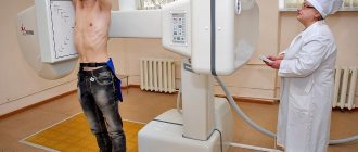
X-ray, due to its accessibility and fairly high information content, has been a leader among various diagnostic techniques for more than 100 years. The main reason for putting X-ray research methods in first place is the use of lung fluorography for preventive purposes, covering the broad masses of the population.
Due to the fact that the radiation dose during fluorography, especially using film technologies, is quite high, the feasibility of mass screening of the population for the purpose of tuberculosis prevention is controversial. However, taking into account the vast territorial extent of Russia, most of which is located in cold regions, as well as the deteriorating epidemiological situation associated with pronounced social stratification of the population, does not allow us to abandon radiation diagnostic methods.
The influence of lifestyle on changes in the lungs
Over time, the lungs reduce their versatility. By the age of 30, the flow rate during inhalation and exhalation decreases, and nutritious foods, vitamins and microelements from the blood begin to flow in minimal quantities. Age-related changes in the lungs are not immediately noticeable during fluorography - closer to 50 years. Sufficient exercise, exercise, and an active lifestyle in most cases help maintain normal respiratory function in old age. However, people who have undergone surgery at retirement age often feel sorry for themselves, observing only bed rest. A long period of recovery of respiratory function after such a regime is a common occurrence, because the lungs work superficially, do not receive a sufficient amount of oxygen, and its consumption and output of carbon dioxide are disrupted.
poor fluorography results
Natalia asks: Hello!!!! In November 2012, she was treated in a private clinic for inflammation of the female part. She was treated with Betadine, Hexicon, Klion. They gave me injections. The doctor said that I had vaginal dysbiosis. After the ultrasound, they said that I had an endometrial polyp, they tried to treat me with duphaston, but it didn’t work, they told me to do a gesteroscopy. After that, I began to worry a lot, plus stress at work. Also poor nutrition. After treatment, I developed thrush. I went to the state clinic with the results of the ultrasound and said that I had a polyp. She said that we would try to treat it this way first. She prescribed me Terzhanin suppositories and methridazole tablets. But the treatment did not help. And she gave me a referral for surgery. But the next day I had bad discharge and I didn’t go for surgery. All this time I was worried about the polyp. I went for an ECG of the heart and the result was coronary heart disease... I got scared and started to worry a lot, my heart was jumping out... The next day I did an ultrasound of the heart ... prolapse... the doctor said there was nothing serious... I calmed down a little. After the corporate party on March 8, the next day my throat was very sore. I lost my appetite. I felt sick. My heart began to pound. I started losing weight every day. I went to the ENT specialist, the ENT specialist said to do an FGDS. He said chronic pharyngitis. FGDS showed fungal exophagitis, reflux gastritis. The hysteria is not tightly closed, the gallbladder is bent. I was treated with fluconazole, I gargled with iodine. But the throat does not go away. My stomach hurts periodically. They told me to do a stool test. Giardia and ascariasis were found. She was treated with nemazole for two days. In addition, I have dysbiosis. and temperature 36.9-37.4. I took a smear test for STIs, everything was normal. Blood tests for chlamydia, ureplasma, mycoplasma, trichomonas, everything was normal. I don’t know what to do, I seem to have an appetite... but I continue to lose weight. lose weight. Tell me what to do... Fungal exophagitis seems to still exist because I periodically see white plaques in my throat. Is it possible to lose weight like this because of parasites. By 4-5 kg. I think about it all the time. I would like to return to my previous weight of 51 kg. Hello. Get a blood test for RV, HIV, hepatitis. Do a fluorography of the chest organs.
The RV analysis was done, everything was normal... I did fluorography 2 times... everything is normal. I tested for HIV in February... I have a regular partner. I did an immunogram. Results:
My Norma
Leukocytes 4.6 4.0-8.0 Lymphocytes 36.3 20-37 abs. h. 1.7 1.2-3.0 t CD3 lymphocytes 68.7 65-76 abs. h. 1.1 0.8-2.3 t helpers CD4 37.9 35-45 t supr/cytotox CD8 28.9 24-34 CD4/CD8 1.3 1.5-2.2 B lymphocytes 16.3 7- 15 abs.h. 0.27 0.1-0.5 NK 14.1 14-23 IgA 2.4 0.9-2.5 IgM 2.8 0.6-2.8 Ig G 7.7 8-18 CEC 10 8 -37 Phagocytosis of bases. 65%-7 40-80% Phagocytosis stim. 60%-8 Complement 62 40-80 WBC 4.6 lymf 1.7 Mid 0.3 gran 2.6 lymf % 36.3 Mid% 7.0 gran% 56.7 RBC 4.49 HGB 139 HCT 38.3 MCV 85.4 MCH 30.9 MCHC 362 RDW-CV 13.1 RDW-SD 40 .3 PLT 273 MPW 8.6 PDW 16.9 PCT 0.234 ESR 5
My temperature dropped. An infectious disease specialist prescribed me Klion against lamblia. I drank it one day, I’m more afraid to drink it because there are more white plaques in my throat... fungi are on the way. Fungal exophagitis and dysbacteriosis started with antibiotics, now they say antibiotics again... I don’t know what to do... Please advise... Thanks in advance...
Problems of age-related changes in the lungs during fluorography
Age-related changes in the lungs during fluorography are typical for the older generation. The ability to fight various pulmonary infections is gradually lost, the cough reflex decreases, less antibodies and immunoglobulin A are produced in the nasopharynx, so pneumonia and all kinds of concomitant diseases often plague even relatively healthy people.
Age-related changes in the lungs during fluorography are manifested by a decrease in O2 levels and the appearance of:
- apnea (sudden stoppage of breathing during sleep);
- pneumonia;
- bronchitis;
- emphysema;
- lung cancer.
The body’s ability to protect itself from infections and external negative factors disappears.
Film fluorography
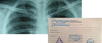
The entire process of fluorographic examination comes down to irradiating the patient’s chest with X-rays, followed by photographing the projection of the resulting image from a fluorescent screen.
Why are x-rays harmful?
As a result, a reduced X-ray image is obtained, the maximum size of which can be 10 cm by 10 cm, and the sides of the minimum acceptable size are 2.5 centimeters.
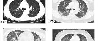
From the point of view of the quality of the resulting image, of course, one cannot talk about its high information content, since the resulting image is not the actual image of the chest, but only a reduced copy of it.
Taking into account that the effective equivalent dose (EDD) of radiation received by the patient as a result of film fluorography is 0.8 m3v on older devices, and 0.4–0.5 m3v on more modern ones, doubts arise about the advisability of this kind of diagnosis in comparison with the risk of developing pulmonary tuberculosis. In order to assess the contribution of fluorography to the total human radiation dose, which is about 2.4 m3v/year, it is necessary to consider medical exposure in the structure of all sources of ionizing radiation.
As can be seen, ionizing exposure carried out for medical purposes is in second place among all background sources accompanying a person throughout life. What is the share of fluorography in the total exposure of a person for medical purposes?
Table: Proportion of radiation exposure for all types of x-ray diagnostics.
| Name of diagnostic method | Share % |
| Radiography | 23,2 |
| X-ray | 31,0 |
| Fluorography (preventive) | 35,6 |
| Fluorography (diagnostic) | 10,2 |
Due to the extremely high radiation exposure on the body, film fluorography is not used in developed countries and is not recommended for diagnosis and prevention in underdeveloped countries. However, despite the fact that the radiation dose for radiography is significantly lower than for fluorography, and is about 0.3 m3v, the widespread use of X-rays for preventive examinations in order to identify the early stages of tuberculosis is not advisable due to the high cost of the procedure.
Considering the size of the country's population and the need for mass annual examinations, the cost of the procedure is a priority for the state. Very often, the quality of images obtained using film fluorography is so low that the doctor has to use a magnifying glass when examining them.
Prevention of age-related changes in the lungs
Age-related changes detected during fluorography can be eliminated with medications. But a more effective way is to prevent such changes. Quitting smoking, physical activity, walking, following doctor's recommendations - all this will help eliminate factors affecting the aging of the respiratory system.
Older people who are actively involved in singing, reading aloud and communicating, intellectual and physical work are less susceptible to various risks. And only the person himself can prevent age-related changes in the lungs, visible during fluorography, by regularly carrying out examinations.
What do the numbers and codes on fluorography mean?
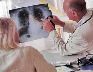
There is no point in self-diagnosis. If a change is detected in the images, the doctor will continue research to identify the cause of the deviations, and then select the appropriate therapy.
For the convenience of specialists, special codes were adopted. How the results are interpreted after fluorography is presented in the table.
| Numeric value | Result |
| 01 | Shadow shaped like a ring |
| 02 | Darkening of the lung tissue |
| 03 | Pockets of Shadows |
| 04 | Expanded shadow in the center, increase in root size |
| 05 | Pleural effusions |
| 06 | Obvious fibrotic changes |
| 07 | Unpronounced fibrotic changes |
| 08 | Lung tissue becomes more transparent |
| 09 | Obvious changes in the pleura |
| 10 | Minor changes in the pleura |
| 11 | Large quantities of large petrificates in lung tissue |
| 12 | Large and numerous petrificates in the roots |
| 13 | Small petrifications in lung tissue in large quantities |
| 14 | Many small petrificates in the roots |
| 15 | One large petrificate in lung tissue |
| 16 | One large petrificate in the roots |
| 17 | One small petrification in lung tissue |
| 18 | One small petrificate in the roots |
| 19 | The diaphragm is changed, but there are no abnormalities in the pleura |
| 20 | Postoperative view |
| 21 | Changed skeleton of the chest area |
| 22 | Foreign body |
| 23 | Pathology of the heart or blood vessels |
| 24 | Other |
| 25 | Norm |
| 26 | Marriage |
The fluorography result looks like this. If the image demonstrates the presence of several pathologies, each of them must be indicated using the appropriate code in a separate column. In the second column, the location and extent are written through a fraction: the numerator is the right lung, the denominator is the left. The third column is a place for the specialist’s personal number.
The most common pathologies
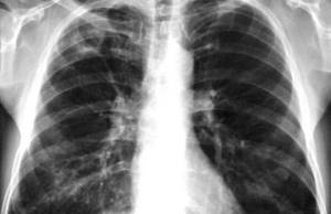
“Bad” fluorography may indicate some serious pathologies.
- The roots are expanded. This result is the most common. It can be seen in photographs of a smoker or asthmatic. In this case, compaction of the roots of the lungs is visualized. Diseases in which a similar defect is observed:
- bronchitis,
- edema,
- pneumonia.
- Heaviness of roots. A pathology characteristic of smokers, patients with bronchitis, or workers in hazardous industries.
- Pronounced vascular pattern. The image clearly shows the vascular pattern of the lungs. This occurs due to the fact that blood flows from the arteries to them more intensely than usual. Signals bronchitis or pneumonia.
- Focal spots. Dimensions, as a rule, are no more than one centimeter. A similar result indicates focal pneumonia. When the contours of the spots become clear, it means that the disease is receding. Shadows in the upper parts of the lungs may be a symptom of tuberculosis.
- Blackouts. They can come in different shapes and sizes. If this result occurs, additional examinations will be required.
- Fibrosis. Occurs after infections, injuries or operations. This kind of tissue takes the place of damaged tissue. Does not pose a threat to health.
- Calcifications. These are cells that have been affected by tuberculosis or pneumonia. When there are a lot of calcifications, the disease has passed.
- Spikes. Lead to isolation of inflammation from healthy areas. No treatment required.
- Scoliosis. The pathology has nothing to do with the lungs. But the picture clearly shows the skeleton, so the doctor can make a diagnosis by measuring the angle of the curvature.
- Pneumosclerosis. Smoking, frequent bronchitis, as well as tuberculosis provoke an increase in connective tissue. The composition of the bronchi changes, and lung tissue decreases. Patients should spend time in the mountains.
- Sealed sinus. Pleural sinus is a cavity formed by pleural folds. In healthy patients these cavities are free. When there are deviations, liquid accumulates in them.
- Changing the aperture. Occurs when:
- disruptions in the gastrointestinal tract,
- liver function disorders,
- pleurisy,
- overweight,
- malignant formations.
Features of interpretation of results
The absorption of X-ray radiation depends on the characteristics of the body. The contrast may be heterogeneous, but in healthy lungs it is always even. In conclusion, the specialist will indicate the presence of the pathologies listed above, which, in turn, indicate diseases.
| Disease | The result in the photo |
| Bronchitis | Compaction in the roots, heaviness, pronounced vascular pattern |
| Tuberculosis | Focal spots, darkening |
| Pneumonia, asthma | Extended roots, intense vascular pattern, darkening |
| Tumor | Pronounced vascular pattern |
Features of decoding fluorography of a smoker
Fluorography with decoding can show whether the patient smokes. But to do this you need to be a smoker with some experience. And although even one smoked cigarette has an effect on the body, the consequences of a one-time episode are not visible in the picture.
Heavy smokers are skeptical about the procedure, although it is mandatory for them, because thanks to the method, serious abnormalities can be diagnosed in a timely manner.
Thickened lung tissue, fluid accumulation, the presence of neoplasms - all this is clearly visible in the image of the lungs of cigarette lovers. If such signs are detected, the doctor will prescribe treatment that will help avoid unnecessary problems. Fluorography is an informative method that allows you to monitor the condition of a person’s lungs. An annual procedure helps to detect pathological processes in a timely manner.