CT is one of the most informative research methods in medical practice. To obtain the result, the device takes layer-by-layer images of the part of the body being examined; small intervals between the layers allow for a three-dimensional reconstruction of the image. A chest CT scan makes it possible to assess the condition of important organs: lungs, heart, esophagus, trachea, as well as the position of nerves and blood vessels. A CT scan of the chest organs displays the location, presence or absence of pathological changes, its size and nature.
Description of the method
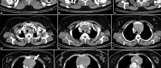
A CT scan of the chest is performed by rotating an X-ray tube. X-ray beams are recorded on a special detector, which displays the image on a monitor. An OGK CT image consists of thousands of layer-by-layer images. The thickness of the sections varies within 10 mm. Tissue density is used to assess the condition of the chest organs on a CT scan. Pathological lesions show a decrease or increase in density in relation to surrounding structures. You can get a chest CT scan at our medical center. At the Yusupov Hospital you can immediately get a doctor’s consultation, an interpretation of the image and treatment.
Indications
The doctor prescribes a chest CT scan as both a primary and an additional examination method. Computed tomography of the chest cavity shows changes in the mediastinum, lungs and heart. A referral for testing is given by the attending physician if the following symptoms are present:
- Suspicions of a tumor of the mediastinum, lungs, pleura;
- Symptoms of tuberculosis;
- Pain in the heart area;
- Abnormal structure of the aorta and other vessels;
- Difficulty in moving the thoracic spine;
- Clarification of the localization of the pathological focus before surgery, radiation therapy;
- Decreased sensitivity, tingling of the upper extremities;
- Difficulty passing food through the esophagus, suspected stenosis;
- Protrusion in the sternum;
- Injuries, fractures of ribs and spine;
- Emergency conditions, including pulmonary embolism;
- Lack of information in other studies;
- Cough, shortness of breath, wheezing in the lungs.
Computed tomography makes it possible to make an accurate diagnosis. Using an image, you can identify neoplasms, their nature, and also differentiate the nature of tumors even of minimal size.
A dynamic chest CT scan shows what has changed in the area being examined over time. This method is not cheap, but the information content justifies its price.
Features of CT scan of the lungs with contrast
The enhancement agent procedure complements the native scan. Always performed after native tomography.
Is CT scanning dangerous?
Human radiation exposure is dangerous when the exposure dose is exceeded. Every year a person receives about 2000 mSv of ionizing radiation from the background of the planet. Gradual action poses no threat. The human body is able to eliminate the negative effects of small doses. The influence of high levels of radiation leads to tissue damage. If acute radiation sickness develops, irreversible organ damage occurs.
CT scanning becomes dangerous when multiple examinations are performed over a short period of time. The approach is used in radiation therapy of tumors. The progression of the tumor is deadly, so it is necessary to stop the disease by any means. Radical treatment of cancer is only possible if it is detected at the beginning of its development. Using computed tomography, it is possible to detect tumors of the lungs and mediastinum measuring several millimeters in size.
Contraindications
Since computed tomography is a highly informative, accessible and fast research method, there are no absolute contraindications to its use. Relative contraindications are as follows:
- Pregnancy. When carrying a child, teaching the mother is dangerous for the life of the fetus, so CT scanning can only be performed if prescribed by a doctor, taking into account all possible consequences;
- Children. A growing body is sensitive to any influences, so this study is not recommended for people under 18 years of age. However, in some cases, doctors decide to choose CT in diagnosing the disease;
- CT with contrast is not performed on people with iodine allergies, kidney failure, or thyroid disease;
- Obesity. Some devices are not designed for body weights over 140 kg.
Contraindications to computed tomography of the lungs
Limitations to the procedure arise due to ionizing radiation of organs. The pulmonary system consists of soft tissues that are prone to mutations. Tomography should not be performed on children under fourteen years of age without strictly analyzing the indications and contraindications. Only when the information content of the study significantly exceeds the harm, is there a need for a computed tomography scan for the child. The condition is met only when pathology cannot be established using alternative non-radiation diagnostic methods.
Contraindications to lung tomography:
- Pregnancy;
- Children under 14 years of age (only for strict indications);
- Kidney failure;
- Patients with chronic diseases of the thyroid gland and kidneys;
- Severe forms of diabetes mellitus;
- Allergic reactions to seafood and iodine;
- Multiple myeloma;
- Patients with twitching of certain muscle groups;
- Mental illnesses;
- Previous x-ray examination with barium injection;
- Limitations of computed tomographs based on human weight (on average no more than 150 kg).
Individual contraindications for the examination are determined by the attending physician. The specialist identifies possible complications and predicts the possibility of performing tomography.
What diseases does a chest CT scan detect?
A CT scan of the chest can reveal lung diseases:
- Lungs' cancer;
- Emphysema, bronchiectasis;
- Interstitial lung diseases;
- Tumors, inflammatory processes of the pleura.
Vascular pathology:
- TELA;
- Aneurysm, aortic dissection;
- Superior vena cava syndrome.
A computed tomogram is performed to identify lymphadenopathy, mediastinal neoplasms, mediastinitis, the presence of foreign bodies in the esophagus, and esophageal neoplasms. Injuries to the thoracic spine and ribs can also be diagnosed using this method. The most important value of CT lies in the possibility of differential diagnosis of inflammation, benign and malignant neoplasms.
What does a chest CT scan show?
It is necessary to separate the different features of tomographs designed for scanning the lungs and mediastinal organs. In the first case, even step-by-step settings can be used. The heart is constantly beating. To obtain high-quality tomograms with classical CT, you should remain still. To visualize the myocardium, the scanning cycle must be synchronized with the heart rate. Multispiral equipment (MSCT) with multiple source-receiver complexes has such capabilities. The picture is formed by software by comparing images at different time intervals.
What does tomography of the mediastinum show:
- Volumetric formations;
- The lymph nodes;
- Large blood vessels;
- Thoracic aorta.
What can be seen on lung images:
- Neoplasms;
- Inflammatory processes;
- Cysts;
- Pleural growths;
- Traumatic injuries;
- Pneumothorax spontaneous;
- Foreign bodies;
- Cancer;
- Hemorrhages;
- Hemothorax.
The list of nosologies can be expanded. Constant improvement of diagnostic technologies leads to an increase in the information content of the method.
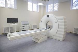
Carrying out
Computed tomography of the OGK is a minimally invasive research method and does not cause harm to health. The patient lies down so that the chest is located under the scanner.
CT chest with contrast
Contrasting is the introduction of a special substance into the human body for clearer detailing of certain structures. The patient assumes a position with his arms raised up. As a contrast, iodine-containing substances are used, which are administered orally or intravenously, depending on the purpose of the study. In case of iodine intolerance and thyroid diseases, the study may be cancelled. To analyze the result, the degree of accumulation of the contrast agent is studied. The study lasts from 10 minutes to an hour. The price of computed tomography of the chest with bolus contrast in the Yusupov Hospital is 13,250 rubles, CT angiography of the pulmonary trunk is 19,150 rubles, MSCT coronary angiography is 23,350 rubles.
Intravenous contrast is used to view blood vessels. MSCT angiography is used to diagnose pulmonary embolism, dissection, coarctation of the aorta, and pathological changes in the coronary vessels.
Intravenous contrast is used to examine the esophagus. The indication is a suspicion of a perforation or tumor of the esophagus.
CT chest without contrast
CT without contrast is prescribed to study the chest and musculoskeletal system. The study usually lasts no more than 20 minutes. In some cases, for differential diagnosis of the disease, the patient is asked to hold his breath.
Progress and duration of the computed tomography procedure
The duration of the CT scan is determined by the area of study. Multispiral CT of the lungs (MSCT) lasts about five minutes. If a procedure with contrast is required, the interval is extended to twenty minutes. The duration is determined by the interval of injection of the drug, observation of the passage of the substance through the vessels.
The need for contrast may arise during the initial examination of the chest cavity if the specialist detects additional foci that require verification. For example, small tumors are not clearly visible on tomograms. After administration of an iodine-containing compound, the nature of the blood supply to the tumor node can be monitored.
To quickly relieve complications of contrast CT studies, radiology rooms are equipped with emergency first aid kits. State medical institutions have intensive care units that provide assistance in life-threatening situations.
If contrast injection is planned, a flexible catheter is first installed in the patient's antecubital vein. When passing the drug, a person feels heat, warmth, and a metallic taste in the mouth. The duration of the sensation is one to two minutes. A few minutes after the procedure, a person may feel the urge to urinate, which is associated with the contrast drug reaching the kidneys. The effect quickly wears off on its own.
When a baby is scanned, parents are given a lead apron for protection. The office is equipped with a communication system with staff, so any complications must be reported to the staff immediately. The clicking and buzzing sounds of the device are natural operation of the equipment.
Side effects of CT scanning
Complications after the procedure are rare. List of CT complications:
- Dizziness;
- Nausea and vomiting;
- Allergic reactions;
- Dizziness;
- Indigestion;
- Increased pressure;
- Dyspnea;
- Metallic taste in the mouth.
Side effects after a CT scan are rare, but you need to be aware of the possibility of them occurring.
Preparation
Preparation for a chest CT scan includes the following:
- During pregnancy, radiation can harm the fetus. Therefore, the patient should inform the doctor even about a small probability of pregnancy;
- Metal objects distort the image of normal body tissues. In this regard, the patient is first asked to remove all metal objects (belts, watches, jewelry, etc.);
- Before starting the study, you must inform your doctor about taking any medications;
- When examining a CT scan with contrast, allergic reactions are possible. To prevent it, the doctor administers special medications;
- If you have chronic diseases of the kidneys, thyroid gland, heart and lungs, you must inform your doctor;
- During the examination, it is necessary to lie motionless in special equipment for a certain time. With claustrophobia, a panic attack may occur. If you are afraid of confined spaces, you must warn your doctor about this.
Carrying out a CT scan
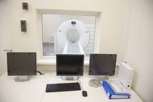
During the tomography, the couch with the patient lying on it slowly enters the tomograph ring. The duration of the CT scan is usually no more than 30 minutes. During the procedure, the patient needs to lie down without moving, this is necessary to obtain the most reliable examination results. The X-ray emitter of the tomograph directs rays at different angles, these rays have different intensities. All signals are sent to a computer and analyzed on it. The diagnostic results are displayed on the screen in the form of two-dimensional sections of the mammary glands. During the procedure, the medical staff is located in the adjacent room.
Decoding the results
The CT image is analyzed by a diagnostician. A comparison of normal and pathological conditions is made based on a comparison of tissue density and their absorption abilities. The image in the image is black and white; bones and calcifications are displayed in white. Fatty tissue, as well as parenchymal organs, are gray in color. The image displays more than a thousand shades of gray, which makes it possible to determine the nature of the pathological focus.
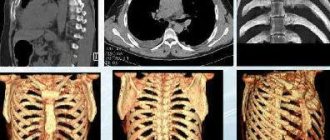
When analyzing the results of the study, the doctor evaluates the contours of the lesion, its density, and size. Foci of inflammation and malignant neoplasms have unclear contours. If the size of the tumor is too small and it is impossible to determine its nature, a repeat CT scan is prescribed. Images can be viewed in several modes: three-dimensional reconstruction to create a three-dimensional object, viewing in “cinema” mode, viewing thick sections.
Advantages of the method
Computed tomography has a number of advantages over other research methods:
- Time. CT is performed in a short time, which is especially important in emergency conditions;
- Clear visualization of bone structures and other tissues of the body, no overlap of objects, the ability to assess the location of formations relative to each other;
- The ability to view the area under study in different planes, 3D modeling allows you to examine “blind spots”;
- Minimally invasive, safe, painless;
- Availability;
- Versatility. Using CT, you can examine the entire body;
- Low radiation exposure. Doctors do not recommend examinations more than once a year, but if indicated, examinations can be carried out up to three times a year;
- High quality images make it possible to make a correct diagnosis.
Breast CT price
Computed tomography for breast cancer with contrast is on average 3 times cheaper than MRI of the mammary glands with contrast. In this case, CT will be able to evaluate not only the mammary glands, but also the lungs and bone tissue in the scanning area for metastases, but MRI will not.
Breast CT is not included in the compulsory medical insurance program, therefore (especially when examined by a competent specialist) be prepared to do it for a fee.
It is better to find out the price of research from the performing doctors:
Specialists at the University Breast Center recommend being examined by trusted specialists.
CT scan of the chest in Moscow
You can get a CT scan of the chest organs at the Yusupov Hospital. Our clinic is located at Moscow, st. Nagornaya, 17, bldg. 6. The medical center has all the conditions for a comfortable stay.
The Yusupov Hospital is equipped with a modern diagnostic center with high-quality equipment. Our doctors are top-level specialists who will establish an accurate diagnosis and prescribe rational therapy. You can get an OGK CT scan in Moscow inexpensively from 7,000 rubles at the Yusupov Clinic.
Preparation for the procedure
Typically, patients do not require any special preparations before undergoing a breast CT scan at a diagnostic center. Often during the examination, 80-100 ml of contrast agent is injected. The patient's last meal should be no later than 5 hours before the start of the CT scan in the clinic. However, in some cases, you may need to fast for about 12 hours before the procedure. The best period for research is from the 5th to the 12th day of the menstrual cycle.