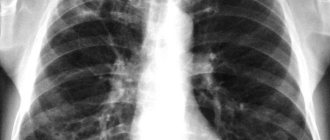
Fluorography is a method of x-ray examination that allows X-rays to penetrate the human body, recording the resulting image on the image screen. The quality and uniformity of a fluorography image is determined by the ability of different body tissues to absorb these rays. In the picture, the heart, bronchioles, ribs and the bronchi themselves appear as light spots. Unevenness of lung tissue and white spots in the wrong place may indicate the presence of various pathologies: fluorography shows pneumonia, emphysema, tuberculosis, obstructive pulmonary disease and other diseases of the pulmonary system.
Doctors at the Yusupov Hospital carefully approach each case, prescribing proven medications and procedures that speed up the treatment process and minimize the risk of complications.

Fluorography for pneumonia
Pneumonia (pneumonia) is an acute lesion of the pulmonary lobes that is infectious and inflammatory in nature. This inflammatory process involves the alveoli, lung tissue, and in case of improper treatment or severe form, other structural elements of the respiratory system. Diagnosis of pneumonia is carried out by auscultation, blood tests, and also using such common methods as x-rays or fluorography. With pneumonia, the clinical picture is characterized by the following symptoms:
- fever;
- cough (dry, wet, with mucus, paroxysmal or continuous);
- shortness of breath or shortness of breath;
- pain in the chest (most often in the right side);
- dizziness and headaches;
- general weakness and weakness;
- increased sweating;
- drowsiness and decreased appetite.
There are a number of factors that predispose to the development of pneumonia:
- old age or under 5 years of age;
- smoking and alcoholism;
- hypothermia;
- insufficient physical activity;
- unbalanced diet;
- diabetes;
- poor immune system function (including HIV);
- chronic heart failure;
- atherosclerosis;
- oncological diseases.
Today, pneumonia is one of the leading causes of death among adults and children. Most deaths occur precisely as a result of untimely treatment. It is very important to start treating pneumonia at the first stages of its development, as this reduces the risk of developing side pathologies several times.
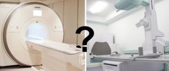
What is an X-ray?
X-ray (radiography) is a method of studying the internal structure of human organs. The main task is to identify whether there are pathologies in them or whether the organs are normal. Also, competently assess the dynamics of treatment. A digital X-ray machine is a device through which such diagnostics are carried out. The idea is that a beam of X-rays first penetrates the internal tissues of the human body, and then is projected onto special paper/film.
What diseases are digital radiography used to diagnose?
- respiratory organs and various chest injuries;
- the patient's cardiovascular system;
- human musculoskeletal system.
Pneumonia: X-ray or fluorography?
It is generally accepted that fluorography of the lungs shows inflammation, and x-rays can only confirm this. In fact, fluorography is more of a preventive method than a method for examining a specific pathology. The answer to the question “Does fluorography show pneumonia and bronchitis?” - controversial, since the resulting picture depends entirely on both the severity of the pathology itself and the thickness of the tissue through which the radioactive rays penetrate. A preventive fluorography image will protect against chronic pneumonia or its atypical form and prevent the development of other pathologies, but it is the x-ray that will help determine the localization of the inflammatory process and its complexity. Moreover, the X-ray method is safer, since its radiation dose is not as high as the radiation dose from fluorography.
Which is better and what are the differences?
Compared to digital x-rays, FLG is an outdated method. Modern, well-established clinics (for example, the Kyiv clinic ACMD-MEDOX) use digital radiography. Difference between digital x-ray and fluorography:
- Accuracy. Digital X-rays are more accurate and give an idea of the condition of the deep tissues of the human body. Consequently, it shows pathologies at an early stage and requires a simplified treatment regimen for the disease in the future. While fluorography is a superficial study, looking at the image of which, you can only identify advanced diseases or pathologies. As for the images obtained with both methods of medical research, then again, x-rays are more accurate and more effective: the specialist (and you as the patient) receives the image on a special film. With FLG, the picture is first displayed on the screen, and only then is it photographed.
- Radiation dose. Performing a FLG study involves a larger dose of radiation to the human body. With the help of digital X-rays, less radiation exposure is given to the body (which is safer for the patient’s health).
- Price. The only advantage of FLG compared to digital radiography is the lower cost of the procedure. But when accuracy, efficiency and your safety are on one side of the scale, and price is on the other, then digital X-ray remains the winner.
You can avoid overpaying for high-quality digital radiography and get study results within the entire 1st working day at the ACMD-MEDOX clinic!
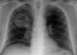
X-ray of the lungs for pneumonia
An X-ray for pneumonia is performed if the doctor has recorded all visible signs of the disease and if laboratory tests (blood and urine tests) have confirmed this. There are no contraindications to x-rays, but it is recommended that women avoid this test during pregnancy. But in cases where the expected benefit to the mother’s health outweighs the harm to the child, x-rays are still used during pregnancy, while doing everything possible to minimize exposure to radioactive rays. There are two classifications of pneumonia, the differences of which are clearly visible on an x-ray: focal pneumonia (clinical manifestations include fever, wheezing during breathing, a slight increase in leukocytes in the blood. In the image, the initial stage of focal pneumonia may not be revealed, since of the obvious signs only the presence of a small infiltrate in the pleural cavity. However, a competent Yusupov hospital is able to determine the inflammatory process in the respiratory system by the following signs: by an increase in the size of the root of the lung due to effusions in the pleural cavity, by the presence of an uneven structure of the lungs, the level of the pleural cavity (pleurisy), dark spots in the lung area , which indicate a decrease in the airiness of the lung tissue) and lobar pneumonia (usually seen in the picture as a large, medium-intensive darkening. Can be localized in one or two pulmonary lobes. The causative agent of lobar pneumonia is most often Friendler's bacillus, which is dangerous to human life and can cause more severe consequences. Signs of lobar pneumonia on fluorography or x-ray include: total or subtotal darkening in the lung area, deformation of the domes of the diaphragm, total or subtotal deformation of the lung structure, pleural effusions, changes in the shape of the roots of the lungs).
Main objectives of the study
Often patients, upon receiving a referral, are indignant about why fluorography is needed or simply do not fully understand the importance of regular examinations. But this diagnosis is one of the simplest, inexpensive and, no less valuable, fastest ways to identify pneumonia, tuberculosis or neoplasms of various types.
We should not forget that pneumonia in almost all cases is accompanied by severe symptoms, such as cough, high temperature, etc. Therefore, this disease is not difficult to determine, and fluorography is necessary only to confirm the alleged diagnosis, which cannot be said about tuberculosis and cancer
Oncological processes and tuberculosis often do not manifest themselves for a long time, that is, they do not give doctors the opportunity to recognize them in the initial stages, when there is a high probability of a favorable prognosis with therapy. The only way out for patients with such diseases is to undergo fluorography, and as early as possible.
Fluorography: pneumonia, photo and diagnosis
A fluorogram is also an effective examination method, especially in the absence of other methods. There are a number of changes that fluorography can show:
- changes in the roots of the lungs and pulmonary contour;
- fibrosis;
- the focus of pneumonia and its localization;
- petrificates and calcifications;
- swollen lymph nodes;
- infiltration and foci behind the heart shadow.
X-ray images are quite informative, but they are not able to fully clarify the diagnosis. For example, it is difficult to see the thin linear shadows associated with pneumonia, as well as some types of pneumonia caused by certain infections.
How often should I be checked?
All adults, with a few exceptions, must undergo fluorography. In addition, there are certain categories of the population who, due to their work activities or life circumstances, need to undergo FLG at least once a year.
These include the following persons:
- Professionals whose activities involve the threat of contracting tuberculosis or infecting others. This group includes medical workers, as well as people working in specialized institutions such as kindergartens, schools, or those employed in trade and the food industry.
- Patients at medical risk. This includes people with serious illnesses that pose a danger to the patients themselves and/or to others. These are people suffering from diabetes mellitus, pulmonary pathologies, immunodeficiency status, including HIV, as well as serious diseases of the digestive system, such as hepatitis and colitis. Due to their weakened general condition, it is very easy for these patients to become infected with tuberculosis, which will rapidly develop in them.
- People who constitute a social risk group. These are persons who abuse alcohol, psychotropic or narcotic substances, lead an antisocial lifestyle, have no fixed place of residence, as well as former convicts and those in prison.
For other citizens, the rules established by the Ministry of Health state that they need to undergo FLG at least once every two years. It also states that if you have been in contact with a patient with tuberculosis for two years, you need to be observed by a TB doctor, undergoing an X-ray examination of the chest organs once every six months.
If a person in contact with a person suffering from tuberculosis has a question about how many times FLG can be done, and whether it will be harmful to the body, then he should explain how difficult and lengthy the treatment of the disease itself is.
Can fluorography show pneumonia?
Pneumonia can be seen both on fluorographic and x-ray examinations. The examination must be carried out by a qualified specialist, because the further treatment of the patient and, as a result, his well-being will depend on how he reads the photo of the lungs during pneumonia.
Experienced doctors at the Yusupov Hospital identify pneumonia and its varieties using both fluorographic and x-ray photographs. Pulmonologists, in turn, will be able to carefully and competently prescribe treatment, including high-quality drugs proven by many years of experience. You can make an appointment by phone or on our website by contacting our 24-hour online service.
Preparation and execution
Diagnostics does not require special or complex tedious preparation. The only thing the doctor will recommend on the eve of the procedure is abstaining from smoking for several hours and a light breakfast if the examination is scheduled for the next morning. The entire procedure, including undressing and dressing, will take no more than 5 minutes.
Step by step it will look something like this:
- the patient is invited to a special room intended for taking the image;
- he undresses to the waist, approaches the apparatus, and rises to a low step;
- the chin is located in a depression approximately corresponding to the average height of a person;
- the subject is warned not to hold his breath for a minute and not move;
- the nurse turns on the device, the picture is taken, the procedure is over.
- In some situations, during fluorography, a protective apron is used, which protects the organs located in the lower part of the abdominal cavity from radiation. The results of the study are usually ready the next day.
Pulmonary tuberculosis
Preparation for fluorography is most often necessary for those whose doctors suspect tuberculosis. However, it is recommended to undergo diagnostics even without the direct instructions of a doctor if you begin to be bothered by shortness of breath, a cough that does not go away for a long time, as well as constant weakness.
Often, for preventive purposes, doctors resort to prescribing fluorography to all family members of a woman who has learned about pregnancy and registers with a public clinic at her place of residence. This is necessary in order to be sure that there is no threat to the life and health of the expectant mother, as well as her child.