An X-ray of the spine can be performed to visualize one or several parts of the spine. X-ray imaging takes advantage of the ability of tissue to transmit x-ray radiation differently depending on the density of the structure. An X-ray of the spine in two projections is prescribed to obtain information about the state of the bone structure of the spine, as well as to determine the degree of deformation or the presence of space-occupying formations. As a rule, frontal and lateral projections are used. If necessary, other projections (oblique, axial) can be used.
Indications
X-rays of the spine (one or more sections) may be ordered to diagnose the origin of back or neck pain, dislocations, fractures, vertebral displacement, vertebral degeneration, tumors, spinal curvatures such as kyphosis or scoliosis, or congenital anomalies.
The doctor may prescribe an x-ray of the spine for other reasons:
- Finding the cause of lower back pain
- Past trauma
- Abnormal curvatures of the spine
- Abnormal wear and tear of the cartilage and bones of the spine, such as bone osteophytes and narrowing of the space between vertebrae
- Cancer (with significant changes in bone structure)
- Fractures
- Signs of thinning bones (osteoporosis)
- Spondylolisthesis
- Preparation and control of surgical treatment
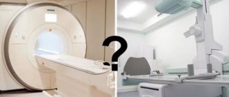
However, X-rays are limited and cannot diagnose conditions such as sciatica, herniated disc or spinal stenosis. In such cases, it is necessary to perform an MRI or CT scan (MSCT).
Who doesn't use X-rays?
- First of all, radiography is contraindicated during pregnancy, since X-rays can cause unwanted mutations in the cells of the embryo. The degree of risk depends on the stage of pregnancy. Sometimes doctors still make an exception and prescribe a test for a pregnant woman.
- X-rays are not performed in patients who are in serious condition, if there is serious bleeding or damage to the chest with depressurization of the pleural cavity.
- The solutions used for X-ray contrast studies contain iodine. It causes allergic reactions in some people. If the patient is allergic to iodine, contrast should not be administered.
- X-ray contrast studies are contraindicated in certain diseases of the thyroid gland, severe pathologies of the kidneys and liver, active tuberculosis, and decompensated diabetes mellitus.
Sign up for an x-ray at the medical office. To make an appointment, call +7 (495) 120-08-07 . If you are scheduled for an X-ray of the lumbosacral region, read about preparing for the study.
Risks
- The patient has the right to find out from the doctor about the amount of radiation that will be received during the study and the possible risks. The patient needs to record data on previous radiological studies, since X-ray radiation has a cumulative effect.
- If the patient is pregnant or suspects that she is pregnant, she should inform her health care provider due to the risk of birth defects in the baby. Therefore, it is better to replace x-rays with other diagnostic methods, such as MRI.
- Depending on the specific disease, there may be other risks.
In what cases is it necessary to take an x-ray? What diseases does it help diagnose?
X-ray examination is one of the most commonly used imaging methods. It is used to diagnose a wide variety of diseases in various fields of medicine.
When a person who has been injured arrives at the emergency room, the first thing the doctor will do is order an x-ray. The study helps to determine whether bones and joints have been damaged and to distinguish fractures and dislocations from less serious injuries. Traumatologists use radiography to check how correctly the bone fragments were reduced (compared), whether they were installed correctly, and whether the pins, screws and other metal structures were displaced.
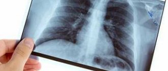
X-rays help dentists assess the condition of the tooth root and the tissues that surround it and the jaw bones. ENT doctors use X-rays to evaluate the condition of the paranasal sinuses. Chest X-ray plays an important role in diagnosing pathologies of the heart and lungs.
X-rays are often used to diagnose spinal diseases. It helps to identify anomalies and injuries (fractures, subluxations) of the vertebrae, assess the condition of posture, intervertebral discs, diagnose scoliosis, osteochondrosis and other diseases. This is an indispensable diagnostic method in the work of neurologists and orthopedists.
Soft tissues are not as visible on x-rays as bones. But they can be “painted” using special radiopaque solutions. X-ray with contrast is used to study blood vessels, organs of the digestive system, bronchi, kidneys and bladder.
Before the procedure
- The attending physician should explain the procedure to the patient and answer any questions they may have about the procedure.
- As a rule, no preliminary preparation is required for an X-ray of the spine.
- It is necessary to inform the radiologist if there is or is suspected pregnancy
- It is important to notify the radiologist of a recent barium study as this may interfere with optimal image quality.
- Depending on your health, your doctor may recommend certain preparations.
Are X-rays dangerous?
X-rays are a type of electromagnetic wave. They are emitted by electrons, which, having greatly accelerated, hit a dense material. X-ray waves are not something created artificially by man, they are natural radiation that reaches the Earth with the rays of the Sun. Every person daily experiences the effects of X-ray and even radioactive radiation in minimal doses.
Yes, scientists classify X-rays as carcinogens, that is, factors that increase the risk of cancer. However, if X-rays are used correctly, the risks are negligible and are not comparable to the benefits that X-rays bring.
It is better to take an x-ray if this is helpful. Making a diagnosis and choosing the best treatment option outweighs the very small risk of having an x-ray.
X-ray is a safe diagnostic method because:
- The level of radiation in X-ray machines is strictly dosed. During the study, the patient receives a safe dose.
- Doctors prescribe a study only if it is necessary, if without it it is impossible to establish a correct diagnosis and prescribe effective treatment.
- Certain periods of time are maintained between x-rays. No doctor will order a test for you every day.
- Modern devices provide a lower level of radiation compared to older models; the body receives a minimal dose.
- If the doctor finds contraindications for you, they will not prescribe an x-ray. Doctors always weigh potential benefits against potential risks.
During the procedure
X-rays can be performed in a clinic or while the patient is in a hospital.
- The patient may be asked to remove clothing and objects on the body that could potentially cause distortion in the images.
- If the patient is asked to remove any clothing, they will be given a gown.
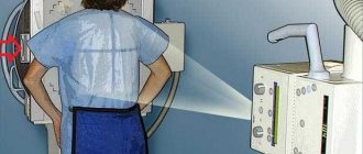
- The patient will be placed on the x-ray table, with the area of interest captured and the x-ray beam passes through the body and hits the film or digital matrix. The attending physician may also order x-rays to be taken with the patient standing.
- Parts of the body that are not scanned may be covered with a protective apron to reduce the harmful effects of X-ray radiation.
- The radiologist will ask the patient to stand or lie still in the desired position for a few seconds while the scan takes place.
- When x-rays are taken after injuries, steps can be taken to prevent additional injury. For example, if a fracture is suspected in the cervical spine, a cervical brace may be used.
- In some cases, an x-ray of the spine can be performed not only in standard positions.
- It is extremely important to remain absolutely still during the scan, as even slight movement can cause the image to become distorted and additional images may be required. The patient may be asked to inhale or exhale during the procedure.
- The X-ray beam will be focused on the area being examined.
- During the scan, the radiological technologist will be in an adjacent room and observe the patient through a protective window.
The X-ray procedure does not result in any pain, but if the patient has recently had an injury or an invasive procedure, discomfort or pain may occur when placing the patient in certain positions for the scan.
Types of X-ray
The first photograph of the hand bones, taken by Wilhelm Roentgen, became a real sensation. For the first time, doctors were able to see what was inside the human body without resorting to an autopsy. Over time, radiography has undergone great changes, its capabilities have grown greatly. Modern clinics use different types of X-ray diagnostics.
During X-ray contrast studies, a solution of a special substance is injected into the organ, which gives a bright shadow in the images and thereby contours its walls. The contrast solution can be drunk, used as an enema, or introduced through special catheters into the bronchi, bladder, ureters and renal pelvis, bile ducts and pancreatic ducts, and other organs. A separate type of X-ray contrast studies is angiography, during which a contrast solution is injected into the vessels.
Radiography can be combined with fluoroscopy, a test during which the doctor observes the operation of an organ on a screen in real time. Certain types of radiography are used for screening - early diagnosis of cancer and other diseases. For example, fluorography helps to detect pathological formations in the chest in time, mammography - in the mammary gland.
If during the examination you move the radiation source and the film in a special way, you can obtain an image with a “slice” of the part of the body being examined at different levels. This type of research is called tomography. A CT scan also uses x-rays.
What is fluorography?
It often happens that a person who needs to examine his lungs thinks about how fluorography differs from an x-ray of the lungs. It is worth knowing that fluorography, just like radiography, is based on obtaining an image based on the passage of ionizing radiation through the human body. However, the radiation dose, the technique, and the information content of these methods vary significantly.
The difference between lung radiography and fluorography is that the latter method is considered much more gentle and safe, since the radiation dose at a time is 0.015 mSv. For problems to arise due to excessive radiation exposure, you need to undergo such diagnostics about a thousand times a year.
The history of fluorographic research
Before you understand what fluorography and X-ray of the lungs are, how the methods differ from each other and which one is better, it is worth familiarizing yourself with the history of the fluorographic diagnostic method. It appeared in 1930 and was actively promoted by the Soviet scientist S. A. Reinberg.
The first fluorography devices still required a fairly high radiation dose, up to 2.5 mSv, and such an examination required significant effort on the part of doctors. If you want to know what the difference is between fluorography and x-rays of the lungs, then keep in mind that modern fluorography machines have become much safer and allow you to obtain very high-quality images.
Types of fluorography
You can understand the difference between X-rays of the lungs and fluorography after familiarizing yourself with the main types of the latter. They are widely used to diagnose inflammation and tuberculosis.
Digital fluorography
Lung X-ray or fluorography: what is the difference and advantages of modern research methods? Digital fluorography is a more advanced method and provides minimal stress on the body. As a result of such a study, an image of the shadow of the organs will appear on the computer equipment monitor, which is created thanks to a special chip located in the receiver. The produced beam will linearly pass through the entire examined area, after which special software will reconstruct the image. When choosing between whether fluorography or x-ray of the lungs is better, we can say that the digital research method is much safer.
Traditional fluorography
It is an outdated technique that differs little from x-rays. The photograph is taken on small film. If you have an x-ray or fluorography of the lungs (which is better in this case, it is unlikely to be determined) of the traditional type, the radiation exposure will be the same.