Fluorography in two projections - what is it?
For preventive purposes, doctors recommend that adults undergo fluorography once every 24 months. Monitoring the condition of the lungs and internal organs allows for timely detection of diseases and treatment of them. Preventive, or classical, radiography of the lungs is also called anteroposterior and is prescribed to assess the general condition of the human body.
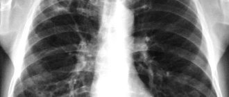
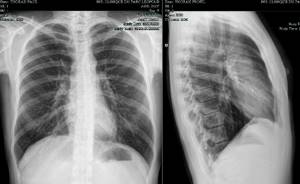
If a doctor or radiologist, having taken a classic photo, sees an area on the screen that requires additional consideration, he takes a picture for the patient in 2 projections. In classical radiography, only the anterior and posterior walls of the lungs are visible, but with the two-projection method, the specialist has the opportunity to examine the organs from the side.
X-ray of the lungs in two projections allows you to examine the organ in detail, enlarging the required section or area. Such examination helps to identify a number of diseases at an early stage.
Basic projections for fluorography
The x-ray is taken in several projections. An anteroposterior image allows you to detect pathologies such as:
- deformation/intensification/thickening of the pulmonary pattern;
- intense darkening due to tuberculosis or pneumonia;
- disruption of the innervation of the diaphragm;
- increased airiness of the lung fields with emphysema;
- collapse of lung tissue;
- pathology of bone and soft tissue;
- expansion of the heart.
If the specialist is not sure that the classic photo showed the full picture, he recommends that the patient also take a side photo. In this case, the subject is turned to the apparatus with the left or right side. It depends on which lung they want to examine in detail.
The lateral view shows the full thickness of the chest and internal organs. The X-ray will determine:
- pneumonia;
- pneumothorax;
- cancer;
- cysts;
- abscesses;
- heart diseases;
- pathological changes in lung tissues and fields;
- inflammatory changes in the bronchi.
A right or left lateral fluorogram is prescribed if a person has a pacemaker or catheter in the heart or pulmonary arteries. The procedure helps control their work.
Indications
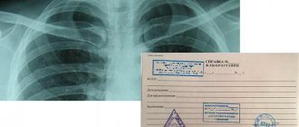
It is recommended that all practically healthy people undergo fluorography as part of an annual medical examination. A conclusion on the results of fluorography is required during an initial visit to a therapist, hospitalization, employment, enrollment in studies, registration of a sanatorium card, enrollment in sports clubs and swimming pools, obtaining a driver’s license, and before planning a pregnancy. If there is a prolonged cough, shortness of breath, weight loss, or weakness, the patient is sent for fluorography immediately.
The purpose of fluorography is a targeted mass examination of the population, identifying latent lung diseases (tuberculosis, pneumoconiosis, nonspecific inflammatory diseases and lung tumors), lesions of the pleura and mediastinum, diaphragm, large vessels, ribs, and heart. If changes are detected, a clarifying chest x-ray is performed.
When is it necessary to make two projections?
A double-sided radiograph is taken when it is necessary to examine the peripheral parts of the lungs. This procedure makes it possible to identify malignant tumors. The specialist also insists on a multilateral image when:
- airiness assessment required;
- there is a suspicion of tuberculosis of the pulmonary fields;
- diagnosed with central/peripheral cancer;
- inflammation of the alveoli is observed;
- you need to determine the size of the heart.
The attending physician prescribes 2 projections in the case when the patient, for no apparent reason, complains of:
- lethargy;
- drowsiness;
- weakness;
- apathy;
- loss of performance;
- shortness of breath;
- lack of appetite;
- persistent fevers;
- a sharp increase in temperature and the inability to bring it down;
- lingering cough with painful spasms;
- blood in sputum;
- chronic cough that cannot be treated.
Fluorography will show the source of the problem and allow you to take the necessary measures for further treatment.
How much does preventive fluorography cost?
Doctors urge the adult population to take responsibility for their health and not ignore preventive medical examinations, which include fluorography.
The cost of preventive x-ray examination depends on the type of equipment used, the pricing policy of the medical institution, as well as the region where the clinic is located. In Moscow, the price for digital fluorography of the lungs starts from 500 rubles. equipped with modern equipment that ensures the effectiveness and safety of the procedure. In addition, we offer our customers the best prices, promotions and discounts. Real professionals work here who diagnose the disease in a timely manner and, if necessary, prescribe the correct treatment. Make an appointment
Author: Serbin Igor Pavlovich
Surgery, neurology, neurosurgery, manual therapy. Doctor of the highest category, colonel of the medical service. Experience 37 years
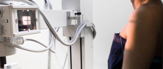
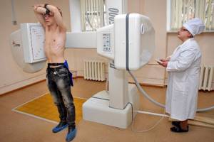
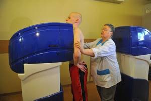
How is the procedure performed?
The examination is carried out under the same conditions as classical fluorography. The patient, entering the office, gives a referral or tells his information to the doctor. After this, he goes into a room where he takes off his clothes from his upper body. In addition, you need to leave jewelry, metal products, belts with buckles, hairpins with iron elements, and watches in the locker room.
The radiologist guides the subject to the machine, setting the desired level of radiation. The patient first stands in front of the device and comes close to it. Then he straightens up, places his chin on the top of the device and spreads his arms to the sides as the doctor shows.
To take a lateral photo, the patient is turned to the device with the desired side. The man raises his hands to his head and presses tightly against the X-ray. In both cases, the subject is asked to take a deep breath and not breathe for several seconds.
Before fluorography, smoking patients are recommended to smoke their last cigarette 3 hours before the examination.
Depending on the equipment on which the image was taken, the doctor announces the results immediately or asks you to wait a few minutes.
The procedure is contraindicated:
- children under 14 years of age;
- pregnant women;
- those suffering from claustrophobia.
On-site medical examination, on-site medical examination in Moscow
Service namePrice
| Medical examinations (preliminary, periodic) without issuing a personal medical record | from 1,000 rub. for 1 person |
| Medical examinations (preliminary, periodic) with registration of a personal medical record | from 2,000 rub. for 1 person |
Where you can do it:
In our other clinics, a medical examination is carried out only to issue medical certificates and medical books.
For the convenience of clients, the MedicalClub network of clinics has entered into partnership agreements with medical centers located in different areas of Moscow and the Moscow region.
This means that medical examination services can be provided at the clinic closest to you at:
Clinic nameAddress
| Admiral Lazarev | Moscow, st. Admiral Lazarev, 72 |
| Balashikha | Moscow region, Balashikha, microdistrict. Yantarny, st. Koltsevaya, 24. room 3 |
| Bratislavskaya | Moscow, st. Bratislavskaya, building 33 |
| Ivanteevskaya | Moscow, st. Ivanteevskaya, 21 |
| Kotelniki | Moscow region, Kotelniki, 2nd Pokrovsky proezd, 4, bldg. 1, room 21-23 |
| Krasnogorsk | Moscow region, Krasnogorsk, st. Shkolnaya, 11, room 2 |
| Semenovskaya | Moscow, st. M. Semenovskaya, 15/17, building 7 |
| Izmailovskaya | Moscow, Izmailovsky Boulevard, 43 |
| Khimki | Moscow region, Khimki, st. Panfilova, 4, room. No. 2 |
Inquiries by phone: 8, 8 800 555-99-64
Mandatory periodic medical examinations of employees by order of the Ministry of Health and Social Development of the Russian Federation No. 302n dated April 12, 2011 are one of the conditions for the normal functioning of a business.
How to conduct a medical examination of employees quickly and efficiently? The most convenient solution is on-site medical examinations, which the MedicalClub network of clinics specializes in. We have more than 9 years of experience in this field.
Our license allows us to conduct medical examinations of employees of any enterprise, including hazardous industries.
In the last year alone, we have conducted more than 30 on-site medical commissions, 2 thousand medical examinations and issued more than 2 thousand medical books and health passports.
In addition to orders from private companies, the clinic has won dozens of tenders for conducting preventive medical examinations, pre-trip and post-trip examinations in state enterprises and institutions.
How to organize an on-site medical examination?
First, you need to contact us by phone at 8 495 660-03-03 (or leave a request on the website) and provide information about the scope of your enterprise, the number of employees, workplace certification (for example, places with hazardous working conditions), etc. .P. Based on the data provided, we determine the required scope of research and the composition of the visiting team.
After this, the date and time of the medical examination are agreed upon and the contract is signed.
Procedure and results of the medical examination
The MedicalClub clinic conducts high-quality medical examinations of employees on the customer’s premises in a short time. For this purpose, a visiting team of doctors is being formed. It includes the following specialists:
- occupational pathologist (our employees have completed advanced training courses in this specialization);
- therapist;
- radiologist;
- dermatologist;
- psychiatrist-narcologist;
- Ultrasound diagnostician;
- dentist;
- otolaryngologist;
- ophthalmologist;
- surgeon;
- gynecologist;
- nurse.
The visiting team is equipped with modern medical equipment:
- mobile fluorograph;
- mobile ultrasound scanner;
- 6-channel electrocardiograph;
- audiometer;
- digital spirograph;
- vibration platform;
- a set of ophthalmological equipment;
- other necessary equipment for conducting examinations
The following examinations and procedures are carried out on-site:
- inspections of enterprise employees by specialized specialists; fluorography;
- Ultrasound examination (ultrasound of the mammary glands is performed only for women over 40 years old);
- collection of blood and smears for laboratory tests (immediately after collection of the biomaterial, the courier delivers it to the laboratory for analysis).
- ECG (electrocardiography);
- spirography (measurement of lung volume);
- audiometry (measurement of hearing acuity);
- dynamometry (measurement of the dynamic characteristics of a person’s step when walking);
- determination of visual acuity, hardware vision examination (examination
- fundus, eye cameras, refractometry, determination of visual fields);
- pallesthesiometry (determination of vibration sensitivity of the hands and feet);
- instrumental studies of the vestibular apparatus.
Based on the results of the medical examination, the customer is given the necessary package of documents: a certificate of medical examination, health passports, and an occupational pathologist’s conclusion on the admission of employees. Also, if necessary, employees are issued or renewed with personal medical records.
At the customer's request, we provide additional services:
- We vaccinate employees (for example, against influenza, typhoid fever, dysentery);
- We carry out additional examinations.
Fluorography in two projections: safety issues
X-ray in several projections is more dangerous than the classical procedure. This is due to the fact that a person receives a double dose of radiation. But doctors note that in this case, the harm to the body is much less than the benefit brought after the examination.
In addition, after a series of studies, experts have proven that during fluorography a person is exposed to a small dose of radiation, which is not harmful to health. In the 21st century Methods have been developed for filming internal organs with a minimum dose of radiation, for example, digital.
To protect the genitals of the subject, a lead apron is put on him, tied at the waist.
Preparation
To do fluorography, no special preparation is needed. Before starting the procedure, the patient must undress to the waist, remove glasses, jewelry, and leave metal objects outside the room that may interfere with X-rays. The procedure lasts several minutes. It is painless, discomfort can only be associated with touching the chest to a cold metal plate.
If you are looking for where to get fluorography done inexpensively in Moscow, a medical one is waiting for you. Our center is equipped with modern diagnostic equipment, allowing us to perform diagnostic procedures quickly, safely and obtain accurate results.
You can make an appointment for fluorography in Moscow by phone or fill out a special form on the website.
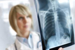
What does the examination show?
On an image of the lungs, a specialist may find:
- fluid in the pleural cavity;
- diaphragm defects;
- cavity in the lung;
- malignant oncological formation;
- focal eclipses;
- large round shadow.
A cavity in the lung, for example, is necrosis of lung tissue. Small focal eclipses indicate pneumonia or tuberculosis. Major eclipses – cancer. A large round shadow on the lung may be progressive tuberculosis or a tumor.
In addition, by analyzing the images, the radiologist can exclude suspicion of pleurisy or malignancy.
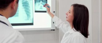
Decoding the results
Studying the images can lead a specialist to different conclusions. Eclipses or white spots may indicate tuberculosis, pleurisy and occupational diseases. Black spots on the lungs indicate chronic inflammation. In addition, such a nuance may indicate malignant tumors in the examined organ.
With pneumonia, an x-ray shows global spotty formations over the entire surface. The more distinct the spots, the more advanced the process.
The doctor diagnoses bronchitis if the photo shows root deformation, changes in the pattern and lamellar lesions. In addition, after irradiation, areas of fluid accumulation are read on the monitor.
If the trachea is pulled forward or displaced, and a thin darkened strip is visible on the picture above the edge of the lower costal arch, then this is evidence of exudative pleurisy. Pulmonary edema will be indicated by unevenly distributed shadows, similar to flakes.
Having seen the “butterfly wings” formed by the expansion of the roots in the image, the specialist diagnoses venous congestion of the pulmonary circle. If there are no light areas under the dome of the lungs, and an accumulation of gases is visible in the abdominal cavity, then this indicates peritonitis.
When a darkening of the posterior mediastinum is visible on a lateral X-ray, this indicates a collapse of the lung lobe or atelectasis. Heart disease can be recognized by the enlargement of the rounded border of the heart shadow. Shadowing on the right side means problems with the left ventricle and vice versa.
Having seen on the monitor in frontal and lateral projections at the apex of the right lung a mid-focal infiltrative shadow up to 0.6 cm in size and a path extending from it to the right root, the doctor concludes that this is pneumonia.
In addition, the right lateral projection image shows small, focal shadows in S1 and S2. What convinces the specialist about the presence of infiltrative tuberculosis in the patient’s upper lobe of the right lung. At the same time, no changes in the contours of the diaphragm and sinuses were detected in the subject. The cardiac shadow does not depart from the norm.
A note in the patient’s referral that he complains of a persistent cough and sputum with blood, and wheezing in the right lung identified by the therapist confirms the radiologist’s diagnosis.
Interpretation of results
When assessing the result of fluorography, the radiologist evaluates the condition of the pulmonary pattern, roots of the lung, pleural sinuses and mediastinal shadow. An enhanced pulmonary pattern, determined by fluorography, occurs with acute inflammation in the lung of any origin. The presence of fibrous tissue in the lung indicates a penetrating injury, surgery, or acute infectious process resulting in fibrosis (tuberculosis, pneumonia). Focal shadows in the middle and lower lobes often indicate focal pneumonia, in the upper parts - tuberculosis. Calcifications detected on fluorography serve as evidence of an isolated, undeveloped focus of infection (tuberculosis, helminthic, bacterial) and do not pose a danger.
The compaction, expansion and heaviness of the roots of the lung suggests pneumonia or bronchitis, bronchiectasis. Adhesions and pleural layers indicate a previous inflammatory process. The condition of the sinuses during fluorography is important for determining the presence of pleural effusion in pleurisy. The expansion of the mediastinal shadow primarily indicates changes in the heart, but does not have serious diagnostic significance. If the mediastinum is displaced according to fluorography, the accumulation of effusion or air in the pleura, large lung tumors should be suspected, and the patient should be immediately referred to a thoracic surgeon.
Where to get diagnosed and how much does it cost?
A fluorogram can be done either in a public hospital or clinic, or in a private medical institution. Patients who cannot walk to the X-ray room on their own can undergo the procedure at home. Portable portable devices are provided for this purpose.
The cost of fluorography depends on the region and location. The price indicator varies from 110 to 1300 rubles.
Timely examination helps to identify diseases in the early stages and treat them. Doctors recommend taking a photo of the lungs every 1.5-2 years.
Possibilities of the procedure at home
When a radiologist is called to your home, the examination will be carried out using a portable device. Such equipment, despite its size, provides all types of radiography, with the exception of those that require contrast display of the result.
Our center carries out almost any x-ray examination, both stationary and mobile. This service option will be convenient for people
those with limited mobility, as well as those who do not want to waste time getting tickets and queuing. In addition, we can provide the following services:
- ECG at home;
- Ultrasound of the abdominal cavity at home;
- Ultrasound of the kidneys at home.
We carry out a full-fledged ultrasound examination of the pelvic organs and respiratory system using the same portable equipment as radiography. Trust our specialists, saving time and nerves!
Medical equipment server MedCom
The main differences between film and digital X-ray machines. What to choose? Answers to frequently asked questions.
Which fluorograph should I choose for the clinic?
Digital, film? Is it possible to upgrade film to digital? Scanning or charge-coupled? Stationary, mobile or portable? Experts answer questions about choosing fluorographs. The film fluorograph is well known to many generations of radiologists.
To work on it, no additional knowledge of a personal computer or special programs is required. The downside is the presence of a laboratory for developing the film and the delay in obtaining shooting results, but given the significantly lower initial cost of the equipment, this may not be significant. Unlike the previous model of the 12F7 fluorograph, the new 12F9 fluorograph is equipped with a mid-frequency power supply unit URP-SCH with a power of 30 kW. This made it possible to reduce voltage pulsations on the X-ray tube, thereby increasing its service life and improving the accuracy of maintaining the anode voltage to 2% under all destabilizing factors. In addition, the exposure and, accordingly, the radiation dose received by the patient during the image were reduced by approximately 20%. We have developed a new X-ray protective cabin, which is made from modern materials. It significantly reduces the radiation load on personnel, so the device can be placed next to doctors even in such limited volumes as the van of a mobile fluorographic complex. Some parts of the device were fundamentally changed taking into account the wishes of radiologists, and additional functions appeared, such as, for example, a remote control with which you can control the patient lift, diaphragm curtains and take an image. Therefore, we are confident that while experts are arguing about the future of film fluorography, if you choose our 12F9 film fluorograph, you will not have the feeling that you are working on equipment of the last century.
Digital fluorographs allow you to see the completed image almost instantly; it is possible to process it with special filters, if necessary. Patient data and fluorograms are easily archived. They are always available for comparison and analysis. They can be sent via electronic communication lines for consultations. It is efficient, convenient and modern. But for this you need to pay approximately twice as much as for a film fluorograph. There is an opinion that despite the fact that a digital fluorograph is more expensive than a film one, it will quickly pay for itself due to the lack of consumables. This is not entirely true and depends on three parameters. Firstly, digital fluorographs have their own consumables: disks for archiving, cartridges and thermal paper for printers. This means that to the amount initially spent on the device, you will regularly add an amount for consumables, just like with a film device. Secondly, on the price of a digital fluorograph. Obviously, the more expensive it is, the greater the likelihood that by the end of its operation it will never become cheaper than film. Thirdly, it all depends on the number of studies per year. If you plan to take no more than 15,000 photographs per year, then, in terms of 10 years of operation, a digital fluorograph (for example, at a price of 65,000 EUROS) will cost you more than any film one, even taking into account consumables and costs for a darkroom. If it is more, then you will either be equal in price, or digital will actually be cheaper, for example, film with a 100 mm camera. But in any case, the benefits of working with a digital fluorograph are probably worth the cost.
The most widespread in our country are scanning fluorographs and fluorographs with CCD (Charge-Coupled Device). We produce both.
Let's consider the process of obtaining an image in our CCD fluorograph (APTSF-01). X-ray radiation passes through the patient's chest and hits a re-emitting screen (400 by 400 mm2). It converts X-ray radiation into a visible image. Using a system of lenses (lens), the image of the lungs obtained on the re-emitting screen is reduced to the size of the detecting area of the CCD (about 25 by 25 mm2). From the CCD, the signals are read by electronics and transmitted to the computer. After appropriate processing, an X-ray image appears on the monitor screen. The main advantage of a digital CCD fluorograph is that for a doctor, taking an image with this device is no different from the procedure with a film fluorograph. Only obtaining an image is greatly simplified, since it almost instantly appears on the monitor screen.
The disadvantages are the following:
- Due to light losses on the re-emitting screen and in the lens, it is not possible to significantly reduce the patient’s radiation dose (relative to the 12F9 film fluorograph);
- X-ray radiation scattered in the patient's body forms false images on the re-emitting screen. To eliminate them, a raster is used, which suppresses this phenomenon, but does not completely remove it. In this case, the use of a raster forces an even greater increase in the radiation dose. The negative result of the scattering effect is an additional increase in dose and deterioration of contrast sensitivity;
- Spatial resolution depends on the quality of the re-emitting screen and lens and, naturally, deteriorates as light passes through these media.
Our devices are equipped with digital CCD cameras providing spatial resolution of 1.6 or 2.8 line pairs per mm.
At the workplace of the radiologist and laboratory technician, the latest generation computers and professional graphic monitors measuring 21 inches are used. The medical graphics printer provides 256 shades of gray and high spatial resolution. At the same time, the image is equally informative both on the monitor and on a hard copy. To print periodic reports, the device is equipped with a laser office printer.
The ProScan program for fluorograph control and image processing complies with the international DICOM-3.0 protocol, which includes the latest changes to the 2003 standard (with JPEG2000 image compression), which allows, if necessary, to integrate it into any modern medical information system. The program is created in close collaboration with radiologists, therefore it contains a unique development that makes it easier for the doctor to write a report using a formalized protocol, as well as a huge number of periodic reports. The program contains almost unlimited possibilities for processing the resulting image with special filters. Archiving of images is carried out on a DVD disk with a capacity of 4,700 MB (about 4,000 images). Today this is one of the most reliable ways to store such information.
In a scanning fluorograph, an image is performed as follows: a linear detector about 400 mm in size moves simultaneously with a fan-shaped beam of X-ray radiation along the patient’s chest. The detector contains several thousand independent elements. During the movement of the detector, the electronics reads signals from all elements several thousand times, transmitting the data to the computer. At the end of the movement, the data is processed and the image appears on the monitor screen. The device has the same set of radiologist and laboratory assistant workstations and software as a fluorograph with a CCD matrix.
The disadvantages of the scanning system include the exposure duration of several seconds (up to 5).
The advantages of scanning fluorographs are as follows: · Dose is tens of times lower than in a film machine or a digital one with a CCD. This is achieved due to the absence of a raster, re-emitting screen and optical system (lens). A direct image is obtained at only 150 µR in the patient plane. · Radiation scattered in the patient’s body cannot reach the detector and create a false image , therefore a raster is not needed and contrast sensitivity is determined only by the characteristics of the detector; · Spatial resolution is not degraded by additional means and is determined only by the parameters of the detector itself. For example, in the Proscan-7000® device it reaches 3.2 pairs of lines per mm. · The linear silicon detector does not require periodic maintenance . · The mid-frequency power supply device URP-30-SCh-"AMIKO" in the ProScan-2000® and ProScan-7000® versions is connected to a single-phase electrical network of 220 V ± 10% and a resistance of up to 1 Ohm. (For normal operation, most fluorographic devices require a three-phase network of 380 V ±10% with a resistance of no more than 0.3 Ohm. Including APTSF-01 with CCD)
This year we are supplying ProScan-2000® fluorographs with a new detector that provides an image format of 1800 by 1800 elements, with a spatial resolution of 2.2 pairs of lines per mm.
Our new ProScan-7000® fluorograph, which has unique characteristics, is currently undergoing certification: a spatial resolution of 3.2 line pairs per mm is achieved at only 400 μR in the plane of the receiver. The device is equipped with a professional liquid crystal medical monitor EIZO (Japan). There are two operating modes of the fluorograph: preventive and diagnostic. The standard preventive mode with a spatial resolution of 2.2 pairs of lines per mm allows 95% of examinations to be performed with the lowest possible radiation dose to patients (about 200 μR). If it is necessary to perform a diagnostic examination with the highest possible spatial resolution, the device is switched to diagnostic mode. Mobile fluorography rooms. More than 150 mobile fluorographic cabinets produced at our enterprise operate in our country, Ukraine, Kazakhstan and Bulgaria. We were the first organization to develop and certify film fluorographic cabinets based on the ZIL-5301EO (“Bychka”) and digital fluorographic cabinets based on the “Bychka” and the KAMAZ-43114 all-terrain vehicle.
All mobile fluorographs have a treatment room, separate from the office with medical staff. To increase capacity, up to 3 patients can be changed in it at the same time. Possible contact between patients and medical staff is eliminated thanks to independent entrance and exit from the treatment room, a public address system and video surveillance between the medical staff’s office and the treatment room. An antibacterial lamp is constantly running to disinfect the room. High ceilings, modern furniture and finishing materials, air conditioning and additional heating devices create comfortable working conditions. Fluorographs in box stacking. For hard-to-reach areas, we produce film and digital fluorographs in box packaging. The fluorograph is transported disassembled in cases that serve as structural elements of the device. The weight of one piece is no more than 80 kg. The number of seats for the basic configuration is 7. The minimum opening width for loading is 580 mm. The device can be transported by any type of transport, including a helicopter and horse-drawn vehicles. The fluorograph can be installed by two people within one hour. Modernization of film fluorographs to digital ones. In addition to supplying new equipment, our organization is upgrading film fluorographs to digital ones.
In most cases, it is enough to replace the fluorographic camera with our digital one and install an automated radiologist workstation, consisting of a computer and software. The cost of modernization is half that of a new digital fluorograph. Useful article?
Share with your friends from social networks! Return to list