When should a chest x-ray be taken?
Plain radiography of the chest organs is carried out:
- During an annual preventive health examination to identify risk factors and life-threatening diseases such as cancer or tuberculosis.
- For pain in the chest and heart area.
- With painful or bubbling breathing, sensations of lack of air.
- With loss of appetite and a sharp decrease in body weight.
- For swelling and high blood pressure.
- For injuries, to identify foreign objects in the esophagus, lungs, and respiratory tract.
- If you suspect lung diseases: pneumonia, abscess, pleurisy, silicosis, etc.
Contraindications for the study
If you familiarize yourself with how fluorography occurs, what this type of research is and what it is based on, it will become clear that this procedure is not so harmless. Therefore, there are a number of restrictions on its passage.
Age up to 15 years
It will not be possible to find out what fluorography is or how such a procedure is done if the patient is under 15 years of age. This is due to the fact that the effect of X-ray radiation on a child’s body is much stronger than on an adult. In this regard, a doctor can prescribe fluorography only as a last resort, when there is a threat to life.
Severe respiratory failure
Respiratory failure is a serious contraindication to fluorography. The thing is that radiation can have a negative effect on the human body and worsen an already bad condition. It is best to opt for a more gentle diagnostic method, for example MRI. Fluorography is safer than x-rays, but it is still extremely undesirable in such a situation.
Pregnancy
If you are thinking about how to undergo a fluorography procedure if you are pregnant, there is only one answer - nothing. Pregnancy is an absolute contraindication, since X-rays can affect the body of both the expectant mother and the baby. The child is at much greater risk; the harm can be so significant that it can lead to the loss of the fetus.
We can talk about how to prepare for a fluorography procedure during pregnancy only when there is a threat to life. In this case, the doctor can determine whether there are other research methods, and, if there is no other way out, try to reschedule fluorography to the third trimester. In later stages, x-ray examination is less dangerous, since all the vital systems of the child have already been formed.
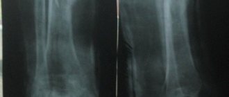
When is it necessary to do a plain radiography of the spine?
X-ray of the spine: coccyx, cervical, thoracic, lumbosacral regions is used:
- For pain, impaired mobility, and a feeling of stiffness in the back.
- For inflammatory diseases such as arthritis or arthrosis.
- For curvature of the spine: scoliosis, kyphosis, lordosis.
- For back injuries, vertebral displacements.
- For osteochondrosis and osteoporosis.
- Suspected tuberculosis, metastatic lesions and spinal tumors.
X-ray and general information about it
Many health problems cannot be identified without an accurate diagnosis or x-ray. For example, when we suspect pneumonia, we often hear from the doctor: “we need to do a fluorography.” What is an X-ray examination? And why do doctors often recommend it?
Fluorography is the most popular type of x-ray.
What is an x-ray? In professional medical terms, an x-ray is a detailed study of the internal structure of the body by shining through it with x-rays and recording the image on a special film or digital detector, i.e. x-rays penetrate through the tissues of the body without damaging them and form a picture of the condition of human organs.
What does an x-ray show? In the pictures you can see (depending on the purpose of the device) various pathologies: inflammation, fractures, neoplasms (tumors), degenerative-dystrophic changes, destructive changes, developmental anomalies, etc. X-ray methods are used to examine the lungs, bones, soft tissues, and internal organs (stomach, kidneys, etc.).
After an X-ray examination, the doctor can make an accurate diagnosis in a number of complex diseases.
The color of the image of organs depends on their density. Different tissue captures X-rays differently. Bones, muscles, lungs - will appear differently; the denser the tissue, the lighter it will be on the x-ray image.
How often can an x-ray be taken?
X-rays can be preventive or diagnostic. For preventive purposes, fluorography or radiography of the chest organs is done (at least once a year), mammography (at least once every two years). Diagnostic x-rays (including fluorography) are done if the presence of any diseases is suspected; it is prescribed by the attending physician. Radiation dose limits for patients (and, accordingly, the number of x-ray procedures) for diagnostic purposes are not established (SanPiN 2.6.1.1192-03).
What norm is acceptable?
The standard for preventive radiation during preventive medical X-ray examinations is 1 mSv per year. Radiation dose limits for patients for diagnostic purposes are not established (if the doctor considers it necessary, then it is necessary). When the accumulated dose of medical diagnostic radiation to a patient reaches 500 mSv (the average dose for one x-ray examination is approximately 0.001-0.5 mSv), measures should be taken to further limit his radiation exposure if radiation procedures are not dictated by vital indications.
Is it necessary to remove radiation from the body after an X-ray examination?
After X-ray examinations, there is no need to remove radiation, since the radiation dose is negligible. Even after scintigraphy, in which a radioactive drug is injected into a vein, it is only recommended to drink more fluids.
An important role is played by high-quality modern equipment and competent work with a specialist’s apparatus.
At Sanas MC, X-rays are taken using the best new generation Japanese equipment, Shimadzu SONIALVISION G4. This is the best in its class and the only multi-complex in the Far East, which, in addition to standard radiographic functions, has unique functions - tomosynthesis (layer-by-layer examination) and SLOT radiography (panoramic image of the spine or lower extremities). Provides the highest quality images and detailed transmission of information with a minimum radiation dose.
7 indisputable advantages of Shimadzu SONIALVISION G4 over other devices:
- SONIALVISION G4 is a premium universal remote-controlled X-ray diagnostic complex . The all-in-one multi-purpose system sets new standards for flexible imaging systems, increasing radiology room productivity compared to conventional systems.
- SONIALVISION G4 recognized as the best in its class universal X-ray unit. Independent analysis company KLAS awarded Shimaszu Medical Systems the “2015 Best in KLAS award” in the X-ray equipment segment.
- The world's first remote-controlled device with the function tomosynthesis is a radiographic research method that produces a layer-by-layer image of the area under study with a slice thickness of 0.5 mm, which allows you to see the smallest pathological changes up to 1 mm. The diagnostic capabilities of this method are much wider than with conventional digital radiography.
Tomosynthesis significantly expands the detection limits of smaller pathological changes than traditional radiography. 74% of focal-like shadows (focal-like shadows can occur with tumors, metastases, tuberculosis and other pathological processes) are detected with tomosynthesis compared to 25% with standard radiography, which indicates a threefold increase in detection sensitivity with tomosynthesis. Digital radiography failed to detect metastatic changes in the lungs in 21.3%, which were determined by tomosynthesis. The information value of tomosynthesis in detecting peripheral lung cancer has been proven by scientists at the Research Center for Cancer Prevention and Screening (Tokyo, Chiba).
- The low radiation dose allows tomosynthesis to be used as a screening method, in contrast to computed tomography. In the low-dose mode (20 sections), the dose does not exceed 0.001 mSv, which corresponds to radiation safety standards.
- Another advantage of tomosynthesis over computed tomography is the ability to examine patients with metal implants without artifacts.
- SLOT radiography – (also known as panoramic radiography, slit radiography, axial radiography, teleradiography). This method allows you to produce a panoramic image of the entire spine including the pelvis or lower extremities including the pelvis in one image with one pass of the X-ray tube. The image is obtained with true anatomical dimensions, in contrast to the image stitching method. Slot radiography is effectively used to diagnose: scoliosis, shortening and deformation of the lower extremities, distortion and rotation of the pelvic bones. This method is necessary for the work of orthopedic doctors and chiropractors.
- The well-thought-out design of the device ensures that all studies are carried out without moving the patient; the head-to-foot coverage is 202 cm.
The Sanas MC employs experienced doctors - radiologists and x-ray technicians who will take and describe high-quality X-ray images of the desired organ and give truthful information about the state of your health.
When can an x-ray be prescribed?
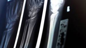
A traumatologist or surgeon may prescribe digital radiography of bones and joints (skull, bones of the shoulder, forearm, foot, hand, femur, hip bone, etc.; temporomandibular, elbow, knee, wrist, hip joints, etc. ):
- If the patient experiences pain, crunching, movement blocking, or joint instability.
- In case of injury or suspected injury: dislocation, fracture, sprain.
- In order to make or exclude a diagnosis: longitudinal flatfoot.
- For the diagnosis of growths and calcified areas, metastatic lesions and tumors.
Is it possible to do fluorography in a T-shirt?
This question is perhaps one of the three most frequently asked questions regarding fluorographic examination, so we answer:
– Yes, you can undergo fluorography in a T-shirt.
Naturally, the T-shirt should be the simplest and most ordinary, without pockets, rhinestones and buttons, then this will in no way affect the quality of the pictures and their information content.
Many girls are also interested in the question: are breasts visible on fluorography? This question can also be considered a frequently asked question.
– No, the breasts are not visible on the fluorographic image.
Fluorography is done to detect tuberculosis and displays changes in the lungs, and the breast, however, as well as lumps in the breast, can only be seen by doing an ultrasound of the breast or on a mammogram.
How to prepare for an x-ray examination?
Before examining the lumbosacral spine, pelvic bones and hip joint, a cleansing enema is performed in the evening before the procedure. A light breakfast is allowed in the morning.
Before examining the abdominal organs, preparation may be required (do not take any food or medications 6-8 hours before the procedure, do not drink or smoke, etc.). People suffering from increased gas formation are recommended to exclude brown bread, baked goods, dairy products, cabbage, apples, carrots, etc. from their diet 2-3 days before the examination.
People suffering from increased gas formation are recommended to exclude brown bread, baked goods, dairy products, cabbage, apples, carrots, etc. from their diet 2-3 days before the examination.
Before examining the lumbosacral spine, pelvic bones and hip joint, a cleansing enema is performed in the evening before the procedure. A light breakfast is allowed in the morning.
In all other cases, no preparation is required.
Preparation and performance of fluorography
If you have not encountered fluorography of the lungs before, or do not know how often such a procedure can be done, it is worth knowing that it does not require preparation. All you need to do is make an appointment and visit the doctor at the appointed time. The patient enters the office, undresses to the waist and removes all metal accessories and jewelry that may affect the quality of the image. Doctors often ask the patient to hold the chain in his teeth so as not to unfasten or remove it. After this, you need to go into the machine’s booth and follow the doctor’s instructions. You will need to take a special position and hold your breath for a few seconds at the doctor’s signal. If everything is in order and no pathologies are found, an appropriate certificate will be issued. This is often enough to get a job or obtain a driver's license.
What device do you use for radiographic examination?
The Clinic on Komarova uses the latest generation universal X-ray complex - AGFA DX-D 300 (2016), the only one in the Far East. Modern digital technologies make it possible to obtain images of the highest quality with minimal radiation exposure, which have undeniable diagnostic effectiveness.
For comparison, when performing digital chest radiography AGFA DX-D 300, the radiation exposure is only 0.03 mSv (millisievert) per procedure, which is 17 times lower than with conventional fluorography (0.5 mSv), 10 times lower than with conventional radiography (0.3 mSv) and 2 times lower than with digital fluorography of the chest organs (0.05 mSv).
One of the unique features of the AGFA DX-D 300 is an individual approach to research. The German company has thought through everything to improve the level of patient comfort. The maximum flexibility and motorization of the AGFA DX-D 300 allow the equipment to independently take a position in space relative to the patient during the examination. This unique feature makes it easy to carry out a wide variety of examinations on any population, be it children, the elderly or patients with limited mobility.
Is it possible to do x-rays for children?
According to research by the World Health Organization, radiation from x-rays is quite dangerous for the health of minors. But at the same time, this fact does not mean that such medical research is not carried out on children who have not yet reached 16 years of age.
Similar procedures are carried out, but in this case, medical specialists must use radiation protection equipment. In addition, the dose of treatment that a child will be exposed to is calculated based on his health, age and other characteristics.
Possible consequences of an X-ray when diagnosing a child usually include burns and even the possibility of developing leukemia due to changes in blood composition. But it must be taken into account that negative consequences for the child’s health occur with prolonged exposure and incorrectly selected dosage. And modern equipment makes it possible to minimize such risks, so there is no need to be afraid of an X-ray examination.

What to shoot with fluorography?
I think that with regard to whether it is possible to do fluorography in a T-shirt and why you need to take off your bra, everything has become clear. But what about jewelry, earrings, chains, etc., which of these need to be removed for fluorography? And what to do with long hair, won’t it interfere with a high-quality photo?
All jewelry that gets into the chest area, and these are beads, chains, pendants, crosses, must be removed. There is no need to remove the earrings as they will not be included in the photo area.
As for your hair, it is better to put it in a bun and pin it at the top of your head.