Prices

Fetometry
Ultrasonic method for determining fetal size
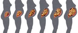
Fetometry is intrauterine measurements of the fetus using an ultrasound machine. The purpose of this study is to determine how the body develops and to detect visible disorders.
Ultrasound is a safe method and is periodically performed on every pregnant woman. Thanks to the information content, the doctor can determine the gestational age as accurately as possible, find out whether the baby’s size corresponds to this period, and identify existing pathologies.
When conducting a study, the doctor is guided by special tables indicating the parameters to be measured. The numbers in mm obtained during the measurements are compared with the norm for each week in order to accurately assess the correspondence of the baby’s development to the gestational age.
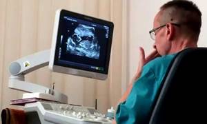
The research is carried out in two ways:
- Transvaginal (in the early stages or for the purpose of obtaining additional information). A condom is placed on the ultrasound machine's sensor, after which it is inserted into the vagina.
- Transabdominal. The sensor is placed on the woman's abdomen, lubricated with gel.
| Duration of the procedure | 60-70 minutes |
| Recommended course | 1 session |
| Periodicity | — |
| Recovery period | 2-3 days |
| The effect is noticeable after | — |
| Equipment | Cocoon (Spain) |
What is the time frame for the research?
According to the plan, fetometry is prescribed to pregnant women three times in each trimester (12th, 22nd, 32nd weeks).
Unscheduled examinations are prescribed by the doctor if indicated. The first fetometry checks the expected due date, the presence of chromosomal abnormalities, and developmental defects.
Second, possible intrauterine development disorders are identified. If the position of the fetus is successful, the doctor can determine the sex of the unborn child.
Third, the baby’s health is checked, height and body weight are determined. These indicators are necessary to choose the method of birth (natural or cesarean section).
What factors determine the weight of the unborn child?
- Floor. According to statistics, boys are born larger.
- Nutrition for the expectant mother. If she consumes a large amount of carbohydrates, fats, and flour products, then the child may be born large.
- Heredity.
- Multiple pregnancy.
- Bad habits in a pregnant woman.
- Chronic diseases of a pregnant woman (diabetes, etc.).
- The presence of fetal malformations and placental insufficiency.
- Presence of hemolytic disease in a child.
- The presence of severe toxicosis, stress during pregnancy.
- Number of pregnancies and age of the expectant mother.
Measured parameters, standard values and deviations
The physiological characteristics of the fetus are individual and depend on the height and physique of the parents. This factor must be taken into account when analyzing fetometry results.
There are the following forms of developmental delay:
- Symmetrical. All indicators are below normal for this stage of pregnancy.
- Asymmetrical. Some parameters are normal, while others are below standard.
The lag of parameters is also divided into 3 degrees:
- I - difference of two weeks;
- II - at 3-4 weeks;
- III - more than 4 weeks.
What does it mean if fetometry repeatedly shows deviations from standard indicators? If there is a large difference, additional diagnostics using other methods are prescribed. For example, a rapidly growing size of the head circumference raises suspicions about the presence of hydrocephalus, additional data is required (Doppler results, CT), and monitoring is carried out over time. In any case, the verdict about developmental delay and malnutrition must be confirmed by two specialists: a gynecologist and a geneticist. The gynecologist should decipher the indicators of fetometric studies.
Developmental delays are usually caused by lack of oxygen and nutritional deficiencies. Possible reasons:
- chronic diseases of the mother;
- genetic defects of the fetus itself;
- lesions of the placenta;
- unbalanced diet of a pregnant woman;
- immune incompatibility of the fetus and mother.
Parameters are monitored gradually, depending on the duration of pregnancy:
- Body weight by week. One of the most important parameters is monitored throughout pregnancy. The growth dynamics should be positive.
- KTR (coccygeal-parietal size). Based on this indicator, the gestational age is determined in the first trimester. The distance in mm from the crown to the coccyx is measured.
- BDP (biparietal head size), measured in the second trimester. Shows brain development.
- Thigh length. Allows you to identify the risk of skeletal dysplasia.
- Abdominal circumference. The doctor studies the correct development of internal organs, the umbilical vein and the ductus venosus.
- Chest volume.
Photometry standards by week
| Obstetric week of pregnancy | Fruit weight, g | KTE, cm | EG (DHA), mm | DB, mm | BPR, mm |
| 11 | 11 | 6,8 | 20 | 7 | 18 |
| 12 | 19 | 8,2 | 24 | 9 | 21 |
| 13 | 31 | 10,0 | 24 | 12 | 24 |
| 14 | 52 | 12,3 | 26 | 16 | 28 |
| 15 | 77 | 14,2 | 28 | 19 | 32 |
| 16 | 118 | 16,4 | 34 | 22 | 35 |
| 17 | 160 | 18,0 | 38 | 24 | 39 |
| 18 | 217 | 20,3 | 41 | 28 | 42 |
| 19 | 270 | 22,1 | 44 | 31 | 44 |
| 20 | 345 | 24,1 | 48 | 34 | 47 |
| 21 | 416 | 25,9 | 50 | 37 | 50 |
| 22 | 506 | 27,8 | 53 | 40 | 53 |
| 23 | 607 | 29,7 | 56 | 43 | 56 |
| 24 | 733 | 31,2 | 59 | 46 | 60 |
| 25 | 844 | 32,4 | 62 | 48 | 63 |
| 26 | 969 | 33,9 | 64 | 51 | 66 |
| 27 | 1135 | 35,5 | 69 | 53 | 69 |
| 28 | 1319 | 37,2 | 73 | 55 | 73 |
| 29 | 1482 | 38,6 | 76 | 57 | 76 |
| 30 | 1636 | 39,9 | 79 | 59 | 78 |
| 31 | 1779 | 41,1 | 81 | 61 | 80 |
| 32 | 1930 | 42,3 | 83 | 63 | 82 |
| 33 | 2088 | 43,6 | 85 | 65 | 84 |
| 34 | 2248 | 44,5 | 88 | 66 | 86 |
| 35 | 2414 | 45,4 | 91 | 67 | 88 |
| 36 | 2612 | 46,6 | 94 | 69 | 89,5 |
| 37 | 2820 | 47,9 | 97 | 71 | 91 |
| 38 | 2992 | 49,0 | 99 | 73 | 92 |
| 39 | 3170 | 50,2 | 101 | 75 | 93 |
| 40 | 3373 | 51,3 | 103 | 77 | 94,5 |
Based on the results of the second fetometry, the doctor can give a referral to a geneticist. If correction is not possible with therapeutic methods, artificial birth is induced. If the pathology is reversible, a course of treatment is immediately prescribed to save the child.
In the third trimester, fetometry makes it possible to identify pathologies such as cleft lip or cleft palate. If abnormalities are detected, the pregnant woman is hospitalized. If the dynamics are negative, the obstetrician-gynecologist can stimulate premature birth. At 38 weeks, additional testing may be ordered to decide whether a cesarean section is necessary.
Fetometry indicators are especially important for women with a narrow pelvis, during the first birth, in the presence of genetic abnormalities, and chronic diseases. For example, if the weight does not increase, a diagnosis of “frozen pregnancy” is possible.
Newborn weight. Criteria and factors influencing it
March 26, 2018
Normal birth weight for babies born between 37 and 42 weeks of pregnancy is between 2500 and 4000 grams.

With such a wide range of "normal weights", it is difficult to predict how much a baby will weigh at birth. However, there are factors that can help understand the baby's future weight:
1) Heredity / genetics
If the child’s parents were born large, then the child himself is more likely to be born large, but if the parents weighed little at birth, then there is a high probability that the baby will weigh little. If one parent was large and the other was not, a more important indicator is the mother’s build.
2) Pre-pregnancy weight
Women with a low pre-pregnancy weight (body mass index less than 19) are more likely to have a low birth weight baby. In contrast, overweight women (body mass index greater than 24) have an increased risk of having overweight babies.
3) Maternal age
For reasons that are likely related to a confluence of medical problems, especially gestational diabetes, women with advanced maternal age (age 35 years or older at the time of delivery) tend to give birth to larger babies compared to younger women. Mothers under 20 years of age are more likely to have a low birth weight (less than 2500 grams) and very low weight (less than 1500 grams) baby.
4) Mother's behavior during pregnancy
The weight of a newborn also depends on the amount of nutrients he receives while in the womb. Insufficient amounts of nutrients and essential vitamins have a strong influence on the formation of a child’s weight in the direction of decrease. Also, low and very low weight babies are born to mothers who abuse smoking and alcohol before and during pregnancy.
5) Pre-existing medical problems
Chronic diseases (anemia, kidney disease, pulmonary disease, diabetes mellitus, systematic connective tissue disease, heart and vascular disease) usually lead to low birth weight infants, which is most likely due to a decrease in the delivery of nutrients to the fetus.
6) Gestational diabetes mellitus
The development of glucose cravings during pregnancy, also known as gestational diabetes, allows for increased glucose availability to the fetus. Increasing the amount of "fuel" can lead to more weight for the baby.
7) Gestational age at birth
Most weight gain occurs during the third trimester, especially during the last four weeks before giving birth. Over the past month, weight gain of as much as 200 grams per week is possible. Therefore, timing of delivery definitely affects birth weight.
 Multiple pregnancy
In multiple pregnancies (monozygotic and dizygotic twins), the average weight of each child at birth is less than with the birth of one baby.
9) Location of placenta/uterine fibroids
Abnormal placentation, especially placenta previa, can lead to decreased perfusion (blood supply) to the fetus. The presence of uterine fibroids may reduce perfusion by “stealing” blood from the baby. Decreased perfusion results in decreased access to nutrients, leading to decreased birth weight.
In any case, the main thing is that the baby is healthy, so lead a healthy lifestyle during pregnancy.
Technique for calculating weight using ultrasound diagnostic data
Weight calculations are carried out by medical professionals. With the ultrasound data, the patient should visit her gynecologist. It is impossible to determine body weight in the first trimester. At this time, all the doctor can determine is:
- diameter of the fertilized egg;
- coccyx-parietal distance;
- Head circumference.
The listed data is not enough to determine weight. That is why weight is not established in the first trimester.
The doctor can determine the weight of the fetus using a number of indicators
It is possible to determine a child’s weight using ultrasound data only if the following fetometric indicators are available:
- embryonic gestation period of a baby in weeks;
- fronto-occipital size;
- Head circumference;
- BPR;
- abdominal circumference;
- length of the femur.
Additionally, information about the size of the shin bone, humerus and forearm may be required. Such indicators are measured after the 19th week. Ultrasound diagnostics allows you to most accurately determine the baby’s weight. Small errors are possible if the baby is not presented correctly.
It is worth understanding that the baby’s weight according to ultrasound and at birth may differ. No calculator or formula can give 100% correct results. However, in most cases the error does not exceed 100–200 g.
Knowing the fetometry data of the child, you can calculate the weight yourself. To do this, you will need to use specialized calculators into which all available data is entered. However, in this case one cannot be sure of the correctness of the result obtained.

It is important for the obstetrician to know the weight and size of the fetus in order to plan the birth
Self-calculation of weight is needed only for self-development. Without ultrasound diagnostics, determining body weight is impossible.
Need to set weight
The main purpose of establishing the weight of the fetus before delivery is aimed at understanding whether the baby meets accepted age standards. Weight is not calculated until the 8th week. This is due to the fact that a child is comparable to a large orange seed.
Until the 8th week, the baby weighs 1 g. There is no need to calculate weight. This indicator is entered into the fetometry data a little later, when internal organs and systems begin to form.
By calculating the weight, the doctor is convinced that the child does not have any intrauterine disorders or defects.
Any deviations can also indicate pathologies in the expectant mother. The main goals of determining the baby’s body weight are listed in the table.
| Gestation period | Based on the weight of the fetus, it is possible to determine the expected gestational age. This is especially important when there is no data on the first day of the last menstruation. |
| Baby development | By weight, the doctor determines how much the baby meets the required standards. To do this, you also need to know your gestational age. |
| Diabetes | Increased weight in the baby in the womb may indicate diabetes in the mother. In this case, the child will be recommended additional diagnostic measures. |
| Progress of delivery | It will be necessary to establish the exact weight of the child if the mother has a narrow pelvis. This will help you choose the most appropriate tactics for managing pregnancy and childbirth. |
| Risk of developing disorders | You need to know the weight if you plan to have a caesarean section. The higher the weight and gestational age, the greater the risks of developing various disorders, such as respiratory distress syndrome. |
Weight allows timely diagnosis of a large pregnancy. This will help you successfully carry your baby.

Deviations in fetal weight often indicate pathology