- Home /
- Branches /
- MRI /
- MRI of the pelvic organs
One of the most informative methods today for diagnosing the causes of diseases of the reproductive system, bladder, rectum and sigmoid colon is magnetic resonance imaging.
MRI allows you to detect developmental anomalies, pathological processes and neoplasms at an early stage, which helps to make the correct diagnosis and prescribe timely treatment. In women, MRI of the pelvic organs successfully replaces more dangerous invasive research methods, such as hysteroscopy, laparoscopy, etc. This technique is used for a thorough assessment of the uterus with appendages, vagina, bladder and rectum with surrounding soft tissues, vessels and lymph nodes .
In men, MRI of the pelvic organs allows one to evaluate the structure of the prostate gland, as well as identify pathological processes occurring in it. For example, for diseases such as prostatitis, prostate adenoma (benign hyperplasia) and prostate cancer, early diagnosis is very important, which is what MRI allows. For examining the prostate, the MRI method is unique in its information content. In addition to the prostate gland, the examination visualizes the seminal vesicles and vas deferens.
A huge advantage of the technique is its safety: reproductive organs are not exposed to any harmful effects during MRI.
To study bone structures, it is recommended to conduct a computed tomography scan of the pelvic bones.
MRI of the pelvis requires special preparation; please read the preparation information before conducting the study.
Indications for pelvic MRI in women
- Developmental anomalies of the pelvic organs;
- injuries to the pelvic area;
- inflammatory diseases of the pelvic organs;
- chronic pelvic pain;
- menstrual irregularities;
- uterine bleeding of unknown etiology;
- infertility;
- suspicion of the presence of neoplasms;
- assessment of the effectiveness of the treatment, including postoperative observations;
- suspicions of diseases of the rectum and bladder.
MRI OMT with contrast: indications and contraindications

There are many indications for MRI; this type of diagnosis has improved the detection of pathology of the pelvic organs at an early stage
Indications for pelvic MRI with contrast include the following:
- the need to clarify the diagnosis in case of ambiguous data from previous diagnostic methods;
- tracking changes in dynamics;
- preoperative planning;
- suspicion of a pathological process in the pelvic organs.
Complaints that are considered as indications for MRI of the pelvic organs with contrast in a woman, taking into account the general clinical situation:
- absence of a desired pregnancy within 12 months, subject to regular sexual contact without contraception;
- dyspareunia (pain during intimacy);
- repeated self-termination of pregnancy with normal tests;
- the appearance of bloody discharge in the interval between regular menstruation, a change in its character: more abundant, scanty, with large blood clots, the appearance of an unpleasant odor;
- pain in the lower abdomen of unknown etiology;
- dysfunction of urination: the need to strain, a feeling of incomplete emptying, urine mixed with blood, frequent urges, etc.;
- irregular bowel movements: diarrhea, constipation, bloating, bloody stool.
Common symptoms that are suspicious of a neoplastic process in the pelvic organs: weakness, fatigue, unexplained fever, sweating, weight loss, anemia. MRI of the pelvic organs with contrast is performed as part of the general diagnosis if changes in the hormonal background are detected: increase/decrease in FSH, LH, prolactin. According to indications, the search can be expanded: the brain (pituitary gland), which is responsible for the production of biologically active substances that regulate the functioning of the female reproductive system, is additionally examined.
Contraindications for MRI

The risk of MRI in some cases outweighs the benefits; before going to the clinic, read the contraindications
There are a number of contraindications to MRI of the pelvic organs with contrast:
- Metal objects in the body. Any implants with electrical, magnetic or mechanical activation (cardiac and neurostimulators, insulin pumps), cochlear devices, ferromagnetic clamps, brackets, orthopedic structures will cause artifacts to appear on the images during the procedure. There is a risk of failure of equipment providing vital functions. Metal shrapnel, shavings or a bullet under the influence of a magnetic field can begin to move, as a result of which neighboring tissues are injured. The intrauterine device, as a means of contraception, can also lead to image defects.
- Inability to remain still during pelvic MRI with contrast. Some neurological pathology (Parkinson's disease, epilepsy, etc.), psychiatric illnesses in the acute stage, severe curvature of the spine, severe pain are an obstacle to magnetic scanning. For tremors and obsessive movements, the examination can be performed after the administration of sedatives, which is also important for patients suffering from claustrophobia. Immersion in medicinal sleep is one of the ways out in this situation, but the issue is always resolved on an individual basis.
- Obesity. If you have a high body mass index, it will not be possible to do an MRI of the pelvic organs for technical reasons: too much weight (120 kg or more) will not allow the patient to move inside the device. For this category of people, scanning is performed on open-type tomographs, which significantly reduces the quality of diagnosis due to low power, or they resort to other testing methods. Sometimes it is recommended to follow a diet for 1.5-2 months in order to lose weight and undergo a full examination.
An MRI of the pelvic organs with contrast in a woman has an additional obstacle to performing: pregnancy in the first trimester. In world practice, there is no data on the teratogenic effect of a magnetic field on a developing embryo, but it is more rational to conduct research after the 12th week of gestation, when the main formation of organs and systems of the fetus is completed. According to vital indications, magnetic resonance scanning can be performed at any time. MRI of the pelvic organs helps in diagnosing intrauterine malformations of the fetus, but there must be compelling reasons for carrying out the procedure, for example, if ultrasound detects abnormalities that are suspicious of a serious pathology in both the mother and the unborn child. Compared to other research methods - cordocentesis, amniocentesis - magnetic resonance scanning has no side effects.
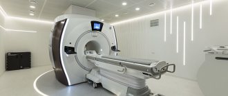
Expert equipment
The Clinical Hospital on Yauza uses the latest generation 1.5 T Philips Ingenia fully digital magnetic resonance imaging scanner, which provides:
- Reduced scanning time (less time to lie in the magnet)
- The maximum possible diameter of the tunnel (73 cm) - the patient’s waist circumference (circumference) is up to 225 cm
- Highest quality images
- Full range of studies due to maximum equipment
- Additional information through advanced software
- Environmental control system (music from the patient’s playlist, choice of lighting, personal climate control)
- Automated quality control system for research and descriptions
- IT platform with triple control of research results - with the support of professors, leading specialists from Russia, Europe and Israel (“second opinion”)
Risks
MRI does not use radiation. To date, no side effects have been found from magnetic fields and radio waves.
The most common contrast agent is gadolinium based. It's quite safe. An allergic response to the substance is rare. But gadolinium can be dangerous for patients with kidney pathologies.
The strong magnetic fields created by an MRI scanner can cause pacemakers and other devices to stop working, and if there is a piece of metal in the body, it can become dislodged during the scan.
Alternative research methods
- Ultrasound of the pelvic organs is a more accessible and cheaper method than MRI, but it does not convey a complete picture of the anatomical features. It has its functional limitations.
- CT scan of the pelvic organs is an indispensable method for assessing the pelvic bones; soft tissue structures and organs of the reproductive and excretory system can only be assessed with the use of a contrast agent.
Contraindications Preparation
How to scan
The patient lies on his back on a narrow table. The table slides into the middle of the MRI machine. Coils are typically placed around the hips. If detailed visualization of the prostate and rectum is desired, a small coil may be inserted into the rectum.
When performing a pelvic MRI with contrast, a contrast agent is injected into a vein.
During an MRI, the radiologist will monitor the patient from another room. The scan usually takes 30 to 60 minutes, but may take longer if contrast is used.
Price
You can see prices for services
Preparation for MRI of the pelvis in women for gynecological diseases
| Cycle day | 7-24 days of the cycle (the first day is the day of the beginning of the last menstruation) | |
| Diet for 2 days | Avoid foods that increase gas formation in the intestines:
| Use foods that reduce gas formation:
|
| Diet on the day of the study |
| |
| Drugs |
| |
| Bladder |
| |
| Appearance at the MC |
| |
| Take with you |
| |
Preparation for MRI of the pelvis in men regarding the prostate gland
| Analyzes | PSA (PSA) | |
| Abstinence | 3 days before the study, abstain from sexual intercourse and ejaculation | |
| Diet for 2 days | Avoid foods that increase gas formation in the intestines:
| Use foods that reduce gas formation:
|
| Bowel preparation |
| |
| Drugs |
| |
| Bladder |
| |
| Take with you |
| |
Advantages
MRI has deservedly found widespread use in diagnostics. Computed tomography, hysteroscopy, and laparoscopy are inferior to it - they are more traumatic, painful, and cause radiation exposure. MRI has no restrictions on age or number of times it is performed. The procedure itself is completely painless and harmless to the patient. Thin sections with different projections give a clear picture; it is easy to track the dynamics of changes and the effectiveness of treatment. The price of MRI of the pelvis and abdominal cavity is affordable, so doctors recommend undergoing tomography during preventive health checks.
What are the indications?
A referral for an MRI is given by the attending physician, this can be a urologist, gynecologist, surgeon, oncologist or proctologist. There is no age limit for the study.
The general list of indications is as follows:
- Detection of tumors and metastatic growths;
- Anomalies and structural pathologists;
- Pain in the pelvic area;
- Cyst perforation;
- Acute pathologies of surgery;
- Diseases associated with the genitourinary system;
- Diagnosis of stones and sandy sediment in the excretory system;
- Diagnosis of infertility;
- Congestion and intestinal pathologies and so on.
In addition to the general ones, there are indications that differ by gender. Women undergo an MRI with the following symptoms:
- Suspicion of endometriosis;
- Internal bleeding;
- Inflammatory diseases;
Men are referred for an MRI in the following cases:
- Inflammatory processes of the genitourinary system - vesiculitis, prostatitis, etc.;
- Diagnosis of neoplasms in the seminal sac and so on.
In addition, magnetic resonance examination is a necessary step in monitoring treatment, clarifying studies already carried out, and is also carried out with the aim of choosing the right treatment tactics or surgical intervention.