That is why gynecologists are more attentive to the condition of the expectant mother: she will have to visit a specialist every 10-14 days. In addition, the woman needs to undergo several additional tests.
One of these tests, which is prescribed to the expectant mother in the later stages, is third trimester screening. Anna Marchenko, chief physician of the Doctor ANNA clinic, explained what research indicators are considered normal, what a pregnant woman should pay attention to and how to properly prepare for the procedure.
Third ultrasound during pregnancy
The purpose of ultrasound in the third trimester of pregnancy
Third ultrasound during pregnancy
(the period for which it is carried out will be discussed later) pursues a number of goals:
- detection of fetal malformations;
- determination of fetal weight;
- determining the amount of amniotic fluid;
- examination of the condition of the placenta;
- blood flow study;
- detection of fetal entanglement with the umbilical cord.
When are 3 ultrasounds performed during pregnancy?
The third screening ultrasound examination is recommended to be carried out at 30-34 weeks of pregnancy. These deadlines are enshrined in the resolution of the Ministry of Health of the Russian Federation. In cases where the data from the second ultrasound did not provide an accurate picture of the development of the fetus or the pregnant woman complains about her condition and does not feel active movements of the fetus, the third ultrasound can be performed earlier.
What does a screening test consist of?
In the third trimester, screening is scheduled for weeks 31-34. The main goals of its implementation include:
- assessment of fetal developmental status;
- identification of developmental defects that were not previously noticeable or had formed due to this; assessment of the work of the placenta, the organ responsible for feeding the fetus;
- preparation for childbirth and determining its method, taking into account the size of the baby and the birth canal of the pregnant woman.
The main components of the third screening are a biochemical blood test, cardiotocography, ultrasound and Doppler of the fetal vessels.
Biochemical screening
This blood test can detect chromosomal abnormalities in the fetus. It is prescribed only if it was not carried out at earlier stages or if previous studies revealed any abnormalities requiring monitoring.
Biochemical screening allows you to assess the level of placental lactogen (somatomammotropin), hCG, AFP and free estriol.
Placental lactogen level
This glycoprotein, synthesized by the placenta, is the main indicator of its normal functioning. Its concentration in the blood increases in proportion to the growth of the placenta and fetus and reaches a maximum at 36-37 weeks of gestation.
With placental insufficiency, malnutrition and hypoxia of the fetus, the level of somatomammotropin decreases sharply. Its increase should also be treated with special attention: an upward change in the indicator often occurs with kidney disease, Rh conflict, multiple pregnancies, as well as an enlarged placenta in diabetes mellitus.
Human chorionic gonadotropin (hCG)
Responsible for the development of the child and its preservation in the womb. The normal hCG level is 1880-59000 mIU/ml. A decrease in its level in the blood indicates a malfunction of the placenta and can cause premature birth.
Alpha fetoprotein (AFP)
This is a glycoprotein, the concentration of which indicates whether the embryo receives enough nutrients. Normally, the AFP level should be at least 141-423 IU/ml. An increase in this parameter indicates the risk of developing a defect of the neural tube or the anterior abdominal wall of the fetus, kidney pathologies, intestinal and esophageal atresia.
Free estriol
A parameter indicating the presence of chromosomal abnormalities. The norm for free estriol is considered to be 19.5-81 nmol/l.
A decrease in hormone concentration is one of the signs that allows early diagnosis of fetal development disorders. To confirm such a diagnosis, the results of other studies are required: both laboratory and instrumental.
The level of free estriol may decrease with fetal growth retardation, adrenal hypoplasia of the child, preeclampsia, anemia, postterm pregnancy and intrauterine infection of the fetus.
Third screening ultrasound
An ultrasound examination allows you to evaluate not only the size and structure of the baby’s internal organs, but also the condition of the amniotic fluid and placenta. At this time, the examination makes it possible to identify developmental anomalies that require urgent help immediately after the birth of the child (for example, surgical intervention for heart defects), to exclude entanglement of the fetus with the umbilical cord and the threat of premature birth.
Ultrasound allows you to evaluate the following indicators:
- fetal head with determination of biparietal size;
- chest (average diameter) with assessment of the structure of the heart and lungs;
- spine in two planes (transverse and longitudinal);
- internal organs of the abdominal cavity and retroperitoneal space of the fetus (kidneys, liver, spleen, etc.);
- abdomen (its average diameter and circumference) and anterior abdominal wall;
- child’s limbs (hip, shoulder length).
The doctor assesses the condition, quantity and transparency of amniotic fluid, the structure and thickness of the placenta. In the third trimester, the amniotic fluid index is normally 144-219 mm, the degree of placental maturity is II.
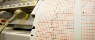
Cardiotocography
This study allows you to evaluate the cardiac activity of the fetus. At this stage, the baby’s heart rate is normally 110-150 beats per minute.
Dopplerography
This study allows us to assess the nature and speed of blood flow in the vessels of the fetus, placenta and uterus. Doppler ultrasound shows the blood flow to the baby, supplying it with oxygen and nutrients.
What does the third ultrasound show during pregnancy?
Third planned ultrasound
– a study during pregnancy that allows you to identify fetal malformations that were not apparent on previous ultrasounds. The doctor also needs to assess the general condition of the child, his vital activity, the level of blood flow in the uteroplacental and fetoplacental complexes.
The third ultrasound will show the exact presentation of the fetus. Using ultrasound, you can confirm the presence/absence of gross pathologies, genetic or chromosomal abnormalities in the fetus. Also, screening at 30-34 weeks of pregnancy will allow you to predict the upcoming birth and understand whether there is a risk of bleeding or not. Perhaps the third ultrasound will show that natural delivery is impossible (for example, oligohydramnios has been diagnosed, which may result in weak labor). In this case, a decision is made to perform a cesarean section.
The gender of the child can be determined much more accurately on the third ultrasound than on the second. Although this does not in any way affect the upcoming birth, most expectant parents want to know the gender in advance.
Third ultrasound during pregnancy: what do they look for?
When 3 ultrasounds are performed during pregnancy, they look at the anatomy of the fetus and examine its almost fully formed internal organs. The size of the fetus is assessed, its weight is measured, and its sex is determined. Just as with the second ultrasound, the fetus is examined for the presence of various pathologies, chromosomal or genetic disorders.
An important point for the third ultrasound is Doppler sonography. This study allows you to evaluate the blood supply to the umbilical cord, the filling of its veins and arteries with blood. This assessment makes it possible to determine in advance the development of fetal hypoxia and take the necessary measures.
Doppler ultrasound examines the blood flow between the uterus and placenta. Blood flow in the child’s cardiovascular system is also assessed.
Preparation.
Screening does not require any special preparation. But you must take into account that with CTG, the baby’s motor ability is assessed from 30 minutes to 2 hours, depending on his activity. Therefore, to prevent your baby from falling asleep at the wrong moment, you can eat something sweet. It will give the amniotic fluid a pleasant taste, and the baby is unlikely to want to sleep.
It is also recommended to take a walk or take deep breaths. This way the baby will be saturated with oxygen and will be more active.
Back to list of articles
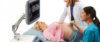
How is a third trimester ultrasound performed?
Ultrasound in the third trimester
performed transabdominally. The woman lies on her back on the couch and slightly bends her knees. The doctor applies a special gel to her stomach.
A typical 2D ultrasound in the third trimester lasts approximately 20-25 minutes. The duration of the procedure depends on the qualifications of the doctor, the power of the ultrasound machine, and the position of the baby in the womb.
If the third ultrasound is performed in 3D or 4D mode, the expectant mother needs to be patient, as such procedures last about an hour.
Neither the mother nor the child experience any unpleasant or painful sensations during the examination.
Preparing for screening
Each individual research procedure requires individual preparation. The exceptions are ultrasound and Doppler ultrasound.
Blood is taken for analysis in the morning on an empty stomach. A couple of days before collecting biomaterial, you should stop eating fatty, spicy, fried foods, marinades and fast food. Possible allergens should also be excluded from the diet.
Cardiotocography is carried out 1.5-2 hours after eating during the period of maximum activity of the baby. According to statistics, this time is from 9.00 to 14.00 and from 19.00 to 24.00. Before the study, you should not smoke or subject your body to serious physical activity.
Only a doctor can interpret the results of the third screening. Women are strongly advised not to attempt this on their own. Identifying any deviations from the norm is not a diagnosis. The gynecologist discusses all cases with the expectant mother and, if necessary, prescribes additional studies and consultations with specialized specialists.
Third ultrasound during pregnancy: interpretation
Only a qualified specialist can decipher 3 ultrasounds during pregnancy, no matter what stage it was performed. Interpret ultrasound data based on established standards - average indicators. Deviations from the average may indicate a lack of fetal weight, intrauterine growth retardation (of varying degrees of severity), or individual characteristics of its structure. All ultrasound data are entered into a protocol, which, after the study, is transferred to the doctor observing the pregnancy.
Ultrasound standards for the third trimester
A number of fetometric measurements can indicate the level of fetal development and its compliance with the established gestational age.
- BPP – bipaterial head size. In the third trimester at 32-34 weeks, this figure can reach 78-82 mm.
- Head size from forehead to nape. The standard for the same periods is 104-115 mm.
- Fetal head circumference. The norm at 32-34 weeks is 310-322 mm.
- The abdominal circumference at the same period can be 285-316 mm.
- The length of the thigh bone is 61-66 mm.
- The length of the tibia bone is 55-62 mm.
- The length of the forearm bone is 50-53 mm.
Small errors are allowed in measurements.
Uncomfortable positioning of the fetus may make measurements difficult. If some parameters of the fetus during a two-dimensional ultrasound examination raise doubts among the doctor, a 3D or 4D ultrasound may be prescribed to clarify the picture and identify possible pathologies. An ultrasound scan in the last trimester examines in detail not only the fetus, but also the placenta. Its location is clarified, its thickness is measured, its structure is studied and its maturity is assessed.
Research:
Absolutely all pregnant women are prescribed ultrasound and CTG at the 3rd screening. The doctor may also prescribe additional tests. But what exactly will be included in the 3rd screening is decided by the treating gynecologist separately in each specific case. The decision is based on the data from the first two screenings, the woman’s well-being, her medical history, and the characteristics of pregnancy.
At screening 3, unlike previous screenings, they also look at the location of the baby in the uterus. If it is in the wrong position, the likelihood of a caesarean section increases. Although there is no need to worry too much - the baby still has a few weeks to turn over. The ideal position is head down, back of the head to the stomach.
Also, don't worry if there are slight deviations from the standards. After all, every child develops differently and has its own characteristics of appearance. Therefore, some people may have larger body parts, and some may have smaller ones. It is dangerous if the baby is behind or ahead of development standards by more than 14 days.
Cardiotocography (CTG) . Here, with the help of sensors, the baby’s heart function, the frequency of contractions of the uterine walls, fetal movements and their intensity are examined. After CTG, you can say with a high degree of probability how the baby feels.
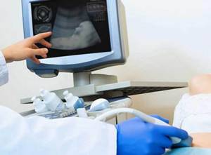
Ultrasound diagnostics. Ultrasound is usually performed abdominally (that is, through the abdominal wall).
Evaluated:
- bone length,
- internal organs of the child,
- dimensions of the head and chest,
- position of the baby in the uterus,
- degree of maturity, thickness and location of the placenta,
- umbilical cord condition,
- cervical condition,
- volume of amniotic fluid,
- abdominal circumference.
Dopplerography. This type of diagnosis is not mandatory, but is recommended for all pregnant women. It can be performed during an ultrasound examination, and the principles are the same.
The only difference is that the monitor does not display the fetus, but multi-colored lines indicating vessels with different blood flow rates. Thanks to this, it is possible to understand whether the child receives enough nutrients and oxygen, and whether metabolic residues are eliminated well. Doppler examines the umbilical cord, placenta and umbilical cord vessels (normally there should be 3 vessels).
Blood analysis. Typically, a blood test at the 3rd screening is prescribed only to those women who had a high risk of genetic pathologies or fetal development abnormalities based on the results of the first two screenings.
Is it necessary to do a third planned ultrasound?
The third ultrasound is included in the list of mandatory diagnostic tests during pregnancy for all women (at what time it is best to do it, the doctor will determine). It is very important to undergo it, as it helps to assess the condition of the fetus as accurately as possible and predict the course of the upcoming birth.
There are often cases when the first two ultrasounds showed that the condition of the fetus is satisfactory and it is developing according to the norm. The third ultrasound could show negative dynamics, to the point that a decision is made about delivery/caesarean section at an earlier date. Of course, in this case the child will be born premature, and he will need to be provided with special conditions for nursing. Nevertheless, this option is better than if it simply stopped developing in the womb and died due to lack of nutrition or other pathologies.
What is examined at the final screening?
This procedure is designed to assess the condition of not only the fetus, but also the mother’s organs, as well as the placenta - the entire system is involved in the birth process, and the baby’s health is influenced by its environment. During the third ultrasound, the following is examined:
- fetal fetometry;
- anatomical structure and development of internal organs;
- motor activity of the baby;
- fetal presentation;
- gender of the child;
- volume of amniotic fluid;
- baby’s breathing – it is extremely important to identify signs of hypoxia and eliminate them in a timely manner;
- blood flow and condition of the umbilical cord;
- structure of the placenta;
- the final date of birth is determined.
During screening in the third trimester, special attention is paid to examining the placenta - the baby’s health depends on this organ.
What pathologies are detected?
Ultrasound screening in the 3rd trimester can identify heart defects, which can be successfully operated on immediately after childbirth (the main thing is that the surgical team is ready to provide emergency care). In addition, the procedure determines the structure of the lungs, liver and spleen.
Using an ultrasound in the 3rd trimester of pregnancy, intrauterine growth retardation can be detected - the condition requires immediate medical intervention. Prenatal diagnosis of pathologies of the kidneys and ureters - hydronephrosis and megaurethra - allows for their effective treatment in the uterine cavity.