How to get to us?
Address: St. Petersburg, Kalininsky district, st. Rustaveli, 66 lit. G (metro station Grazhdansky Prospekt)
Phone, 8-967-342-13-11
From metro station Grazhdansky Prospekt
Free parking, plenty of parking spaces.
Art. m. Grazhdansky Ave. 7 minutes walk
Getting to us: Minibuses: 92, 121, 188, 199, 205, 208, 214, 284, 288,
Trams: 51, 100. Buses: 93, 121, 139, 177
Stop - Prosveshcheniya Ave. 106\ st. Rustaveli or Murino railway station
Metro Devyatkino
The MRI center and RIORIT clinic are a 30-minute walk away
or 10 minutes drive from Devyatkino.
By train it’s just one stop at the railway station. Murino.
Minibuses No. 205, 205a., 619 also go to us from Devyatkino
How the research works
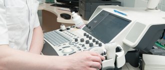
Ultrasound examination is carried out using a special device that records the echo reflected from the organs and sends data to the transducer. Information is displayed on the monitor screen in the form of a two- or three-dimensional image in real time.
An ultrasound takes no more than 15–20 minutes. The patient is in a supine position. The specialist moves a special sensor connected to a monitor over his skin. After the procedure is completed, the patient can continue to lead his normal lifestyle.
What is included in the cost of an ultrasound?
The cost of your research already includes:
- scanning on SAMSUNG SONOACE R7 release 2021;
- written opinion of a sonologist;
- consultation with a diagnostician based on the results of the study.
| Service | Price according to Price | Promotion Price |
| Ultrasound of the abdominal organs | 1500 rub. | |
| Ultrasound of the abdomen and kidneys | 1700 rub. | |
| Ultrasound of the abdomen, kidneys and bladder | 2000 rub. | |
| Ultrasound of the adrenal glands | 800 rub. | |
| Kidney ultrasound | 800 rub. | |
| Comprehensive ultrasound (ultrasound of the abdominal organs + ultrasound of the kidney + ultrasound of the thyroid gland) | 2400 rub. | 1999 rub. |
| Comprehensive ultrasound (ultrasound of the abdominal organs + ultrasound of the kidneys + ultrasound of the thyroid gland + ultrasound of the pelvis with an abdominal probe + ultrasound of the mammary glands) | 4200 rub. | 2999 rub. |
| Comprehensive ultrasound (ultrasound of the abdominal organs + ultrasound of the kidneys + ultrasound of the thyroid gland + ultrasound of the prostate gland with an abdominal probe) | 3300 rub. | 2499 rub. |
| Comprehensive body diagnostics (MRI of the thoracic spine, MRI of the lumbar spine, ultrasound of the abdominal organs, ultrasound of the kidneys, ultrasound of the bladder, consultation with a neurologist, consultation with a therapist) | 11700 rub. | 7000 rub. |
| Service | Price according to Price |
| Ultrasound of the bladder with determination of residual urine | 800 rub. |
| Ultrasound of the pelvis in women and men with an abdominal sensor | 1200 rub. |
| Ultrasound of the pelvis in women with a vaginal probe | 1300 rub. |
| Comprehensive pelvic ultrasound with abdominal and vaginal probe | 1400 rub. |
| Ultrasound of folliculogenesis | 750 rub. |
| Ultrasound cervicometry | 1000 rub. |
| Ultrasound of the prostate gland with a rectal probe | 1500 rub. |
| Ultrasound of the scrotum (testicles, appendages) | 1000 rub. |
| Ultrasound of the prostate and bladder with an abdominal probe | 1900 rub. |
| Service | Price according to Price |
| Ultrasound of the mammary glands | 1400 rub. |
| Ultrasound of the thyroid gland | 1500 rub. |
| Ultrasound of lymph nodes (one zone) | 1000 rub. |
| Ultrasound of soft tissues of one area (face, neck, arms, legs, abdomen, groin) | 800 rub. |
| Service | Price according to Price |
| Ultrasound of one small joint | 1000 rub. |
| Ultrasound of one large joint | 1500 rub. |
| Ultrasound of two small joints | 2000 rub. |
| Ultrasound of two large joints | 2500 rub. |
| Service | Price according to Price | Promotion Price |
| “Healthy Legs” program (duplex scanning of the veins of the lower extremities with assessment of valves and perforating veins and duplex scanning of the arteries of the lower extremities) | 3200 rub. | 3000 rub. |
| Triplex scanning of N/C veins with assessment of valves and perforating veins | 2500 rub. | |
| Triplex scanning of the veins of the upper extremities | 2000 rub. | |
| Triplex scanning of the arteries of the lower extremities with measurement of the brachial-ankle index (BMI) | 2000 rub. | |
| Triplex scanning of the arteries of the upper extremities | 2000 rub. | |
| Duplex scanning of the arteries of the aorto-iliac segment | 1900 rub. | |
| Duplex scanning of the abdominal aorta and visceral arteries | 1900 rub. | |
| Duplex scanning of the lower half of the vein | 1700 rub. | |
| Duplex scanning of the iliac vein | 1700 rub. | |
| Duplex scanning of the renal arteries | 1700 rub. | |
| Intracranial duplex of cerebral vessels | 2500 rub. | |
| Ultrasound of neck veins | 2500 rub. | |
| Ultrasound of the brachiocerebral arteries (BCA) | 2500 rub. | |
| Heart ultrasound or echocardiography for adults | 2400 rub. | |
| Comprehensive head diagnostics (MRI of the brain, MRI of cerebral vessels, ultrasound of neck vessels, consultation with a neurologist) | 10900 rub. | 7500 rub. |
| Comprehensive neck diagnostics (MRI of the cervical spine, MRI of neck vessels, ultrasound of neck vessels, ultrasound of the thyroid gland and soft tissues of the neck, consultation with a neurologist) | 13200 rub. | 9500 rub. |
Forms of payment
Payment is made after the study. Payment in cash and by card is possible.
Ultrasound during pregnancy
Ultrasound examination during pregnancy is a modern and safe way to determine the condition of the fetus and its proper development.
Why do an ultrasound during pregnancy?
Ultrasound examinations are necessary during pregnancy: with their help, the intrauterine development of the child is assessed and the health of the expectant mother is monitored. Ultrasound scanning can be carried out even at the earliest stages.
The value of ultrasound technology lies in the fact that this method guarantees the most complete, comprehensive examination of the fetus being carried by a woman. The accuracy of all data obtained using ultrasound is incredibly high. Thanks to ultrasound, you can accurately determine the gestational age, the number of embryos, the presentation of the fetus, and find out the sex of the unborn child. An ultrasound examination determines the amount of amniotic fluid and the condition of the pregnant woman’s cervix. Ultrasound is indispensable in identifying the features of the development and structure of the placenta and umbilical cord, determining the intensity of blood flow in the placenta and umbilical cord.
Pregnancy management using ultrasound is especially important in multiple pregnancies. Regular ultrasound examination allows you to monitor any unfavorable changes in the condition of the mother and fetus and immediately take appropriate medical measures.
Data obtained using ultrasound waves enable the doctor to effectively identify any problems during pregnancy and conduct highly accurate diagnostics of a wide range of fetal malformations and other pathological conditions.
Ultrasound screening
Ultrasound is a highly informative and completely safe diagnostic method, as a result of which it is used for prenatal screening. Every woman expecting a child must undergo three routine screenings during pregnancy. Its accuracy using ultrasound is very high.
The first screening ultrasound at 11-13 weeks is of particular importance for further pregnancy management. It is during this period that markers of the most common congenital defects and anomalies can be identified in the fetus. A qualitative study allows you to evaluate 20 different indicators, including specific markers of chromosomal abnormalities.
The second (at a period of 20-24 weeks) and the third screening ultrasound 9 at a period of 30-34 weeks) are carried out in combination with the Doppler procedure. Doppler is a very important study that is designed to assess blood flow in the umbilical and uterine arteries, middle cerebral artery and fetal aorta. Dopplerometry has a high diagnostic value, as it can detect any circulatory disorders. Carrying out Doppler measurements makes it possible to detect placental insufficiency, intrauterine hypoxia and take all necessary measures to eliminate these phenomena.
How many times can you have an ultrasound during pregnancy?
During the entire pregnancy, planned screening tests are carried out three times, but most often there are more of them. Additional ultrasound examinations are ordered for medical reasons, as prescribed by the supervising physician. Ultrasound scanning allows you to notice even the slightest unfavorable signs in the condition of the expectant mother and baby.
Is it harmful to have an ultrasound during pregnancy?
Ultrasound is absolutely safe for both the woman and the fetus. The method itself is based on high-frequency ultrasonic vibrations, which do not provide any safety.
Are there any contraindications to ultrasound during pregnancy?
Contraindications to ultrasound during pregnancy are only very severe conditions of the woman, in which emergency delivery is necessary. If the patient has any signs of such a condition, she will learn about it from the doctor managing the pregnancy.
When can an ultrasound determine the sex of a child?
Despite the fact that the genital organs are finally formed in the fetus by the 15th week of pregnancy, the most favorable time for determining the sex of the child is 20-24 weeks of pregnancy.
Preparation before ultrasound?
To undergo an ultrasound, you do not need any preparation. The study takes place in a comfortable environment for the expectant mother without any pain or discomfort.
Dude A.V. Ultrasound diagnostics department doctor
How to prepare for an ultrasound?
Most ultrasounds require no preparation. The exceptions are ultrasound of the abdominal organs and ultrasound of the pelvic organs. Preparation for abdominal ultrasound requires examination on an empty stomach. The minimum break in eating should be 4-6 hours before the examination. The day before the examination, exclude gas-forming foods - legumes, cabbage, black bread, carbonated drinks. You can take Espumisan the night before and on the day of the test.
A pelvic ultrasound will require bladder preparation. To do this, we recommend going to the toilet an hour and a half before the examination, then drinking 3 glasses of water and not urinating again until the diagnosis.
It is better for women to undergo an ultrasound examination of the pelvis on days 5-7 and 20-24 of the menstrual cycle, if women are still having a cycle. If the doctor has not given other recommendations on the day of the cycle. The study is not carried out during menstruation.
Ultrasound of the mammary glands is best performed on days 4-14 of the cycle.
Ultrasound of the pelvic organs
- Ultrasound of the pelvic organs in women
- Ultrasound of the pelvic organs in men
- Ultrasound of the uterus
- Ultrasound of the prostate
- Ultrasound of the scrotum and penis
- Ultrasound of the bladder
Abdominal ultrasound
- Ultrasound of the liver
- Ultrasound of the gallbladder
- Ultrasound of the spleen
- Ultrasound of the pancreas
- Ultrasound of the retroperitoneum
- Ultrasound of the kidneys and adrenal glands
Vascular ultrasound
- Ultrasound of the vessels of the neck and head
- Ultrasound of vessels of the extremities
- Ultrasound of the abdominal aorta
- Ultrasound of renal vessels
Ultrasound of joints
- Ultrasound of the ankle and foot
- Ultrasound of the knee joint
- Ultrasound of the elbow joint
- Ultrasound of the shoulder joint
- Ultrasound of the hip joints
- Ultrasound of the hand and wrist
Ultrasound of soft tissues
- Ultrasound of soft tissues of the neck
- Ultrasound of the thyroid gland
- Ultrasound of the mammary glands
- Ultrasound of soft tissues of the face
- Ultrasound of the soft tissues of the thigh
Ultrasound of the spine
- Ultrasound of the lumbosacral region
- Ultrasound of the cervical spine
- Ultrasound of cervical vessels
Diet before ultrasound
Authorized products:
- lean meat (beef, fish, chicken);
- no more than 1 egg per day;
- hard low-fat cheese;
- porridge;
- baked apples.
All of the listed products are allowed to be consumed boiled; frying them is not recommended.
Prohibited products:
- legumes;
- bread and flour;
- dairy and fermented milk products;
- sweet;
- raw vegetables and fruits;
- alcohol;
- soda;
- coffee and coffee-containing drinks;
- chewing gum.
Indications and contraindications for ultrasound examinations
Ultrasound is one of the most common methods for diagnosing gynecological pathologies, diseases of the gastrointestinal tract, endocrine, cardiovascular, lymphatic, and genitourinary systems. In addition, the technology is actively used when scanning the condition of joints, to monitor the course of pregnancy, and if the development of tumors in the mammary glands is suspected.
The following symptoms may indicate the need for an ultrasound scan:
- The appearance of pain;
- The presence of an inflammatory process in the body;
- Pathological neoplasms;
- Injuries;
- Genetic pathologies;
- Significant deviations from the norm in urine and blood tests.
In addition, there are medical indications that are a direct basis for prescribing an ultrasound examination. These include:
- For early diagnosis of pathologies, preventive screening of the structure and functioning of various organs is used.
- In cases where it is necessary to confirm or refute a preliminary diagnosis, patients are also recommended to undergo an ultrasound examination.
- In order to monitor the effectiveness of the treatment and rehabilitation of the patient.
- Internal surgical procedures (for example, puncture biopsy) are performed under the control of ultrasound diagnostic equipment.
- At various stages of pregnancy, ultrasound allows you to monitor the growth and development of the fetus.
An important advantage of ultrasound examination is the absence of contraindications. The method is absolutely safe for patients of any age; it is prescribed to pregnant women and even newborn babies.
However, there are certain limitations that are not related to the risk to the patient, but to the accuracy and information content of the diagnosis:
- It is not recommended to apply the device sensor to inflamed, scarred or otherwise damaged skin. Such defects can distort the results of the study.
- Transrectal and transvaginal ultrasound is not performed after some gynecological operations, as well as after surgical treatment of pathologies of the last section of the intestine.
- An ultrasound scan of the abdominal organs should not be performed if the patient suffers from flatulence or less than 6 hours have passed since the last meal.
- For women, diagnosis of hormone-dependent organs is carried out on certain days of the cycle (selected by the doctor depending on the indications).
How is the examination carried out?
At the appointed time, the patient comes to our clinic in Kaliningrad. Depending on the type of test, the doctor will ask the visitor to remove any clothing that may interfere with the procedure. After this, the patient lies down on a couch covered with a disposable sterile diaper. In this case, the diagnostician may ask you to take the position that will provide optimal access to the organ being examined.
The average duration of the procedure is 20-30 minutes. It is absolutely painless. It is carried out using a special sensor, tightly, but without excessive force, pressed against the skin.
For some types of gynecological diagnostics, a transvaginal sensor, which is a small tube, can be used. Before use, the device is placed in a special disposable latex case, ensuring maximum sterility and safety. A similar device is provided for diagnostics performed through the rectum.
The skin at the site of contact with the sensor is lubricated with a hypoallergenic gel, which facilitates the sliding of the device and enhances the transmission of sound waves.
