
When monitoring pregnant women, great importance is attached to ultrasound examination. During an examination of the internal organs, various anomalies are revealed that interfere with the normal development of the fetus. If you do not perform an ultrasound during this period, there is a risk of not noticing the pathology and not taking action in time.
Since ultrasound examination is harmless, doctors advise undergoing it as a preventive measure. Thanks to this, it is possible to detect many diseases in the early stages and prevent them from becoming acute.
Indications for ultrasound of the kidneys in pregnant women
Carrying out this procedure during pregnancy must be strictly justified. The woman is explained all the nuances of the study and the possible consequences if she refuses the procedure. The main indications for kidney ultrasound are:
- chronic diseases of the urinary organs;
- the patient complains of pain in the lumbar region;
- abnormalities in the general urine test;
- suspicions of the presence of an inflammatory process in the genitourinary system;
- lower back and back injuries;
- the presence of prolonged low-grade fever;
- swelling;
- increased blood pressure.
It is very important to listen to your body during pregnancy and if there is the slightest deviation in its functioning, notify your doctor. Painful urination with a burning sensation, discharge from the genitals, pain in the lower abdomen are also reasons for prescribing an ultrasound of the kidneys.
You can find out more about the service and its cost here >>
What may be a reason to refer a pregnant woman for an ultrasound scan
A pregnant woman can be referred for an ultrasound of the abdominal organs by a general practitioner or gynecologist who is managing the pregnancy. The reason for this direction could be:
- the patient has a history of chronic diseases of the liver and other gastrointestinal tract organs;
- patient complaints;
- test results.
In particular, the doctor will insist on undergoing an ultrasound procedure in the following cases:
- complaints of abdominal pain of any localization;
- the appearance of signs of intoxication: weakness, dizziness, nausea;
- decreased hemoglobin;
- increased content of leukocytes in the blood;
- elevated blood sugar levels;
- increased body temperature in the absence of signs of acute respiratory infections;
- signs of toxicosis in the second half of pregnancy;
- bowel dysfunction;
- sudden weight loss;
- abdominal trauma.
Such signs may indicate the development of diseases such as: pancreatitis, cholecystitis, hepatitis, cirrhosis, cholelithiasis, tuberculosis, pyelonephritis, appendicitis. In the latter case, even if the patient is expecting a child, emergency surgery may be required.
Thus, ultrasound of the liver and other abdominal organs during pregnancy is necessary in order to:
- promptly diagnose problems the solution of which requires immediate action, for example, an inflamed appendix, blockage of the duct with a stone, accumulation of fluid in the pockets of the abdominal cavity;
- understand the cause-and-effect relationship of “bad” tests. For example, low hemoglobin can be a consequence of both pyelonephritis or hepatitis, and a consequence of hormonal changes. And if in the second case it will be enough to prescribe an iron-containing drug, then if we are talking about problems with the kidneys or liver, a course of such a drug will not be effective - an integrated approach will be required;
- monitor the condition of the expectant mother in order to take the necessary actions and assist in childbirth.
Importance of this examination
Many pregnant women believe that a general urine test is enough to detect kidney pathology. However, this assumption is incorrect and can lead to many unpleasant consequences.
Ultrasound scanning of the kidneys makes it possible to visually determine the condition of the organ, assess its structure, position, and size. Additional Doppler examination of the renal vessels and arteries will tell about the presence of developmental anomalies or the movement of blood through them. Even an ordinary inflammatory process that occurs in a latent form will be visible on the monitor screen of an ultrasound machine.
Unfortunately, it is impossible to study these parameters and make an accurate diagnosis using clinical tests. Therefore, due to the high information content of the method, doctors prescribe it at any stage of pregnancy in order to prevent possible complications.
How to prepare for the procedure?
Ultrasound can be performed routinely or urgently. You should know in advance how to prepare for going to the doctor. If the examination of an organ is carried out as planned, you must adhere to certain rules. Doctors advise diagnosing the pancreas in the morning. Since it is in the morning hours, if all recommendations are followed, that it is possible to achieve the most accurate results. How to prepare and what to do before an ultrasound. The answers to these questions will be given by a specialist who will diagnose the organ.
What can you eat before an ultrasound scan of the pancreas? 3 days before the scheduled diagnosis you should follow a diet. It is not advisable to consume foods that cause bloating or those rich in fiber. Your doctor will provide you with a list of foods you should not eat.
What is needed for an ultrasound of the pancreas. One day before the diagnosis, you should drink a laxative to cleanse the gastrointestinal tract. You must abstain from liquids and food for 12 hours before the procedure. You should also not take medications or smoke. What not to eat before the event. Consuming carbonated drinks can significantly worsen the visibility of results, as they contribute to increased gas formation. Therefore, it is necessary to strictly adhere to all doctor’s recommendations. This is the only way to obtain the most reliable research results.
If the patient has a sudden attack or a sharp deterioration in health, unscheduled ultrasound is used. In case of an emergency procedure, the patient does not need preparation. However, the reliability of the study results is reduced by approximately forty percent. The specialist who will conduct it will tell you how to properly prepare for an ultrasound of the pancreas.
Is it safe to have a kidney ultrasound during pregnancy?
When prescribing any examinations during pregnancy, a woman immediately perceives them with hostility. This reaction is partly correct and is associated with the presence of a maternal instinct, designed to protect even an unborn baby. However, it is important not to forget the point that a doctor during pregnancy never prescribes anything for nothing.
The fear of once again “irradiating” the child inside herself makes the expectant mother refuse an ultrasound. And this happens due to ignorance of the fact that this research method is absolutely safe for both the woman and the fetus, which is under the reliable protection of the placenta. Ultrasound waves are directed by the doctor strictly to the organ being examined and do not systemically affect the entire body.
In certain situations, a pregnant woman faces a huge choice, but how minuscule the doubt should be if we are talking about the normal development of the fetus in the womb. An incorrect diagnosis based on a urinalysis can be very costly. Be prudent and responsible for your health!
Find out the cost
Preparation for ultrasound during pregnancy (first trimester)
POSITION
about the processing of personal data
This Regulation defines the policy, procedure and conditions of the Operator regarding the processing of personal data, establishes procedures aimed at preventing and identifying violations of the legislation of the Russian Federation, eliminating the consequences of such violations related to the processing of personal data. All issues related to the processing of personal data that are not regulated by these Regulations are resolved in accordance with current legislation
Russian Federation in the field of personal data.
1) directly personal data.
regarding the processing of personal data, with this Regulation and amendments to it. Training of these employees is organized by the structural unit for advanced training in accordance with schedules approved by the Operator.
subject to the prior consent of the subject of personal data. Consent may be oral or written.
The specified processing of personal data is considered to be carried out without the prior consent of the subject of personal data, unless the Operator proves that such consent has been obtained.
ensuring the protection of personal data processed by the Operator from unauthorized or accidental access to it, destruction, modification, blocking, copying, provision, distribution of personal data, as well as from other unlawful actions in relation to personal data;
exercise internal control over compliance by his subordinates with the requirements of the legislation of the Russian Federation in the field of personal data, including requirements for the protection of personal data;
bring to the attention of the Operator’s employees the provisions of the legislation of the Russian Federation in the field of personal data, local acts on the processing of personal data, requirements for the protection of personal data;
organize the reception and processing of requests and requests from personal data subjects or their representatives, as well as monitor the receipt and processing of such requests and requests;
in case of violation of the requirements for the protection of personal data, take the necessary measures to restore the violated rights of personal data subjects.
have access to information regarding the processing of personal data entrusted to him and including:
purposes of processing personal data;
categories of personal data processed;
categories of subjects whose personal data is processed; legal grounds for processing personal data;
a list of actions with personal data, a general description of the methods used by the Operator for processing personal data;
description of the measures provided for in Art. Art. 18.1 and 19 of the Federal Law of July 27, 2006 N 152-FZ “Personal Data”, including information on the availability of encryption (cryptographic) means and the names of these means;
date of commencement of processing of personal data;
term or conditions for termination of processing of personal data;
information about the presence or absence of cross-border transfer of personal data during their processing;
information on ensuring the security of personal data in accordance with the requirements for the protection of personal data established by the Government of the Russian Federation;
involve other employees of the Operator in the implementation of measures aimed at ensuring the security of personal data, assigning them the corresponding duties and assigning responsibility.
executive power, internal documents of the Operator, as well as the reasons and conditions that contributed to the commission of the violation.
What are the ultrasound signs of an incipient miscarriage?
- An ultrasound sign showing thickening of one of the walls of the uterus can be deceptive, since there is a physiological asymmetry of the walls of the uterus, which is detected even with ultrasound of the LRS. Short-term contractions of the uterus may occur due to the pressure of the vaginal sensor on the area of the isthmus of the uterus. Such a short-term contraction can be taken as a sign of an impending miscarriage. Long-term hypertonicity is distinguished from short-term hypertonicity using a transabdominal sensor with an empty bladder. Congestive hypertonicity, indicating a threat of miscarriage, exists for a long time, and short-term hypertension soon disappears.
- A change in the configuration of the fetal egg, turning its shape into a scaphoid or drop-shaped, a change in the outer contour of the uterus (a tubercle is raised above the even contour of the uterus above the contracted area of the myometrium).
- The most ominous sign of a threatening and incipient miscarriage is bloody discharge formed due to the fact that a certain amount of blood is poured into the uterine cavity next to the fertilized egg - subchorionic hematoma (gravid hematometra). When the fertilized egg invades the uterine wall, it destroys small vessels, while the increasing hematoma puts pressure on the fertilized egg, as a result of which the connection between it and the uterine wall is lost. Ultrasound determines the volume and localization of gravid hematometra, the time of its formation and the tendency to progression. Thus, the cause of pain and bleeding during a threatened miscarriage can be determined by ultrasound, which will help plan a treatment strategy (for hypertonicity of the uterus with and without a hematoma, it will be different and even mutually exclusive). But in the absence of pain in the lower abdomen, bleeding and other signs of a threatening miscarriage, ultrasound data indicating a threat must be interpreted as a purely hardware phenomenon. An analogue of the expression “threat according to ultrasound” can be the expression “headache according to urine analysis”.
However, spontaneous miscarriage occurs without pain and hypertension.
This case is called a failed miscarriage (“anembryony”, “non-developing” or “frozen pregnancy”). During a frozen pregnancy, the vital activity of the embryo stops, and the contractile activity of the uterus, aimed at expelling the non-viable fertilized egg from its cavity, is absent. The embryo and all elements of the embryonic complex are not visualized by ultrasound at the 5th week of pregnancy in the case of anembryony. This indicates that the development of the embryo stopped before the embryo reached a size of 1-2 mm. Repeated examination at weekly intervals if anembryonia is suspected will help clarify the diagnosis. When the embryo is clearly visible (for example, with a short amniotic leg and a parietal position of the embryo), it becomes possible to exclude aneibryony, in which the fertilized egg grows due to the fluid accumulating in it, but images of the embryo still cannot be obtained.
In the case of anembryony of one of the fertilized eggs during twins, the so-called biamniotic monoembryonic pregnancy, one of the embryos does not develop (failed twins). An “empty” amniotic cavity is found next to the normal one, then, as the fetal egg grows, it crescent-shapedly bends around the image of the normal amniotic cavity and then merges with it completely. An ultrasound phenomenon described as a “double contour of the ovum” or “amniotic thread in the uterine cavity” is a sign of failed twins. This sign does not disrupt the course of a singleton pregnancy.
A miscarriage that occurs after 5 weeks or more is called a frozen pregnancy. In a frozen pregnancy, the embryonic complex is visible (unlike anembryony). However, the embryonic complex consists of poorly differentiated linear objects, in which there are no signs of vital activity - heartbeat and motor activity, characteristic of a normal embryo during a progressive pregnancy.
Normally, with a 5-week pregnancy, the embryo reaches a size of 7-8 mm, with a 6-week pregnancy - 12-13 mm and 18-19 mm - with a 7-week pregnancy. The “growth” of the embryo is called the coccygeal-parietal size (CTD). The “waist circumference” of the embryo also increases - from 2-3 mm to 6-8 in two weeks. “Embryonic pulsation” - heartbeats are detected starting from the 5th week, but the heart on the screen cannot yet be distinguished. At 5-6 weeks of pregnancy, the frequency of contractions is 120-130 beats/min, by 7-8 weeks it reaches up to 200 beats/min. At this time, ultrasound examination already shows the extension movements of the embryo.
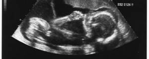
The head end can be distinguished from the pelvic end by the 5th week after conception, and by the 6th week tubercles appear in the place of the future limbs. After the 8th week of pregnancy, the internal organs of the fetus are visible, the spine and skull bones are visible by the end of the 7th week. A living, healthy and mobile embryo will meet with the expectant mother and doctor in the ultrasound diagnostic room at 10-14 obstetric weeks (that is, 8-12 weeks from conception). The story about this meeting is yet to come. During pregnancy, the embryo faces many events and dangers, which it can successfully avoid with the help of the mother, the doctor and, of course, medical ultrasound. Soon he will become not an embryo, but a fetus, and a little later - a newborn!
How to examine the pancreas, except for ultrasound
There are many ways to study the pancreas: ultrasound, MRI, CT and others. For example, endoscopy allows you to see organ tumors that are not particularly large in size, which are not visible to conventional ultrasound or MRI. A puncture is performed to sample tumor tissue for further microscopic examination. In this case, a benign or malignant tumor and its other characteristics are determined. Also, thanks to the results of the study, the attending physician draws up a plan for further treatment of the disease, based on the diagnosis obtained in this way.
Price
| Code | Name | Price |
| 10.03 | Ultrasound of the pancreas | 900 rub. |
Question: Which is better: ultrasound or MRI?
Answer: It should be noted that each of these methods of studying the pancreas is quite common and informative. However, in the presence of chronic pancreatitis, MRI is a more effective method for examining the organ. Since with ultrasound of an organ, the results mainly depend on the qualifications of the doctor. And MRI analyzes are processed by a computer, and errors are almost impossible.
How much does the first ultrasound and subsequent screenings of pregnant women cost at a medical clinic?
Finding out about pregnancy using an ultrasound will cost from 800 rubles. The price will vary depending on the method of the procedure (transabdominal or transvaginal) and the month of pregnancy.
| Services | Price in rubles |
| Ultrasound of the uterus, appendages (examination with transvaginal and abdominal sensors) | 1 200 |
| Ultrasound of the uterus, appendages (examination with a transvaginal sensor) | 1 200 |
| Ultrasound of the uterus, appendages (examination with an abdominal sensor) | 800 |
| Control of the uterine cavity after m/a | 500 |
Decoding the results
The results of the examination are delivered 10-15 minutes after its completion. If any changes are detected, the diagnostician will give detailed comments in an accessible form and recommend further observation by a specialized specialist. The conclusion reflects the following parameters:
- Size, shape, structure of the abdominal organs (liver, spleen, gall bladder, pancreas). Ultrasound also involves assessing the condition of the kidneys.
- Proliferation of organ tissue.
- The presence or absence of fluid in the abdominal cavity.
- The presence or absence of gallstones.
- Aortic diameter.
At the multidisciplinary clinic “Zdorovye”, ultrasound of the abdominal organs is performed using modern equipment. Expert class equipment has high resolution. This allows doctors to accurately determine the location of pathological changes in the area being examined.
Our team consists of the best professionals in the city. Doctors have many years of experience and regularly improve their skills in specialized courses. Diagnosticians detect diseases with high accuracy even in the early stages of development.
Where can an ultrasound of the pancreas be done in Moscow?
Ultrasound examination is the most common type of diagnosis of the pancreas and its work in general. You can always get an ultrasound of the pancreas at the Men's and Women's Health Clinic. Pancreatic ultrasound is performed for patients at an affordable price. The cost is quite varied. In different centers, prices vary from 750 rubles to 3,600 rubles. To find out how much a pancreatic ultrasound costs, it is better to contact a clinic. The website has a detailed price list for all medical services and procedures provided. You can also contact a consultant by calling the clinic’s contact number. In Moscow, the procedure can be performed in almost any medical center or clinic. Today, this procedure is very popular and common for diagnosing all internal organs. In addition, it is available to almost all patients, since it is not particularly expensive.
Determining the sex of a child by ultrasound
Theoretically, a doctor can tell the gender by ultrasound starting from the 15th week of pregnancy, however, in practice, patients will have to wait until 22-23 weeks. In the early stages, identification is difficult for objective reasons. It often happens that the child does not turn his “face” to the scanner, so his gender remains a mystery until the very birth.

If we take into account the timing from which the gender is visible on an ultrasound, then most often parents find out it during the second screening, which is very convenient, since it does not require an additional visit to the doctor.