Electrocardiography (ECG) is one of the simplest, fastest and safest diagnostic procedures used to assess the condition of a baby’s heart, be it a newborn or an older child. An ECG measures and records changes in the electrical activity of the heart that characterize its performance. ECG data shows doctors the speed and regularity of the heart's contractions, the size and position of the heart's chambers, the amount of blood flowing into the heart muscle, and whether there are any abnormalities in its normal functioning. With the help of electrocardiography, it is possible not only to identify acute or chronic diseases that have already developed, but also to predict their occurrence, which means to begin treatment on time and prevent them.
Physical foundations of electrocardiographic research.
An electrocardiograph is a galvanometer device for recording the difference in electrical potentials between two areas of the heart’s projection onto the surface of the child’s body. During operation, the electrical activity of the heart constantly and rhythmically changes. For each stage of contraction or relaxation of the heart muscle of each section, there is its own unique “electrical” pattern, which is recorded by an electrocardiograph and issued in the form of a characteristic graph - a cardiogram on a strip of paper or a computer screen. The shape and parameters of the characteristic elements of the cardiogram (main intervals, amplitudes, shapes and sizes of teeth) carry information for the doctor about the normal or pathological physiology of the heart. The number of waves per minute on the graph shows the heart rate, the distances between identical elements of the cardiogram - heart rate. ECG waveforms show how the heart's electrical impulses are formed and how well the individual parts of the heart work together. Characteristic changes in the shape of the size and position of the waves on the cardiogram carry information about any damage to the heart.
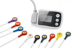
Electrical activity from the surface of the body is recorded through small metal plates (electrodes) that are placed in specific places on the child's chest, arms and legs. The electrodes are connected to the cardiograph by wires through which information is recorded. The electrocardiograph records the electrical activity of the heart, all parameters that are measured, analyzed and printed for the doctor.

What does an electrocardiogram show?
The result of electrocardiography is a graph drawn on thermal paper, which is called an electrocardiogram and consists of teeth, segments and intervals, based on the height and location of which the cardiologist receives the following data about the state of the child’s cardiac activity: sinus rhythm of the heart, heart rate, impulse conductivity and others characteristics.
Evaluating these data, the pediatric cardiologist identifies:
- abnormal heart rate (the norm for children under 3 years old is up to 110 beats per minute, up to 5 years old - 100, up to 8 years old - 90-100, up to 12 years old - 70-85);
- heart rhythm disturbances - extrasystole;
- violation of sinus rhythm - arrhythmia;
- decreased normal heart rate - bradycardia;
- excess heart rate - tachycardia;
- impaired conduction of the heart muscle - depression of cardiac function, heart block;
- increase in myocardial mass - hypertrophy of the heart;
- inflammation of the heart muscle - myocarditis;
- acute disruption of the blood supply to the heart muscle - myocardial infarction;
- violation of the anatomy of parts of the heart muscle - congenital or acquired heart disease, etc.
What diseases can be detected using electrocardiography in children?
Screening (preventive) electrocardiography of the heart, carried out in infants aged 4-5 days and 3-4 weeks of life, allows timely identification of electrocardiographic signs (arrhythmias, prolongation of the ventricular contraction phase), indicating the possible development of sudden death syndrome (one of the leading causes of infant mortality in the first year of life), and start preventive treatment on time.
- An ECG can detect hypertrophy of the baby's heart, which can be caused by congenital heart defects, abnormal heart valves, high blood pressure, insufficient cardiac contractility, or conduction problems.
- Electrocardiography can detect abnormalities in the electrical conductivity of the heart tissue, which can cause a child's heart to beat too fast, too slow, or at an uneven speed (arrhythmias), causing discrepancies in the rhythm of the atria and ventricles.
- An electrocardiogram will show how well the heart muscle is supplied with blood, and whether there are any disturbances in the blood supply to individual areas, both at rest and during physical activity.
- An ECG can be used to judge the functioning of the heart valves and disturbances in blood flow due to their dysfunction.
- Based on the ECG results, we can conclude that there is an imbalance in the level of electrolyte substances in the child’s blood that are necessary for the proper functioning of the heart muscle, such as potassium, magnesium and calcium.
- Electrocardiography can reveal signs of inflammatory heart diseases (endocarditis, myocarditis, pericarditis).
- Based on the results of the ECG, one can judge the presence of cardiomyopathy - an abnormality in the heart muscle itself.
Clinical examination: ECG - is it necessary and how often should an electrocardiogram be taken for a child?
Electrocardiography is a method of studying the heart that does not lose its value over the years.
During the procedure, the cardiograph records the electrical potentials that appear during the work of the heart muscle. All information is sent to the doctor in the form of a graph on a paper tape. It is not at all necessary to be ill with something in order to do an ECG of the heart; on the contrary, it will be considered as disease prevention. But it’s no secret that preventing a disease is much easier than treating it.
An ECG is prescribed to children to study the condition and functioning of the heart. Cardiac diagnosis using ECG in children remains the most popular and widespread technique. What explains this?
Firstly, this technique is informative, safe and completely painless, so an ECG can be prescribed even to a newborn child.
Secondly, an ECG of the heart for children does not require any special preparation. The method is surprisingly easy to use and does not force the doctor, the child, or the parents to strain. The very procedure of an ECG performed on an infant does not differ from the ECG of a one-year-old child, or from the ECG of a teenager.
Thirdly, due to the simplicity of the technique, even in a private hospital it will not cost a lot of money - so it is quite accessible to everyone. Everyone can afford to have their child have an ECG.
Fourthly, the speed of the procedure. During the examination, the small patient lies on the couch, electrodes are attached to his chest and limbs, with the help of which an electrocardiogram is recorded. An ECG for children usually lasts no more than 5 - 10 minutes . The procedure usually takes longer only if we are talking about a stress examination or daily monitoring.
Fifthly, the technique monitors the results over time during repeated studies, after which a clinical picture of the diseases, if any, is drawn up. If you have any doubts, you can repeat the ECG after some time to fully clarify the circumstances.
ECG in children has its own characteristics that differ from adult indicators. What explains this?
Each age period is characterized by its own characteristics that must be taken into account during the ECG. Therefore, it is very important that the child’s ECG result is deciphered and explained by an experienced doctor who specializes in functional diagnostics specifically in young patients. ECG indicators in children at newborn age, at one year old, at five years old and in adolescence, for example, may differ ; there is no single norm in this case. The features of a children's ECG are influenced by a number of parameters, including the anatomical position of the heart in the chest, heart rate, autonomic-endocrine factors, changes in the rate of excitation in the myocardium, changes in the ratio of muscle masses of the left and right ventricles, and so on. Thus, throughout the entire process of growth and development of the child, the morphological structure of both blood vessels and the heart is constantly improving, which is reflected in the ECG results. All this is taken into account by the doctor who conducts the ECG, and the interpretation of the results is based on the parameters that should be observed in children of a given age, while the norm can vary and sometimes quite noticeably.
Cardiac automation is well deciphered using an ECG; waves associated with contraction of the atria and ventricles are visible on it; the distance between them makes it possible to estimate the delay time of an impulse moving “from node to node,” and the magnitude of the wave indicates the state of the heart muscle.
The main method for identifying and assessing arrhythmias is ECG . But right away, in order to dispel unnecessary doubts about the ECG of your children, I will note that often when performing an ECG in children, changes such as sinus arrhythmia and isolated incomplete block of the right bundle branch are detected, which are a variant of the age norm. In these cases there is no cause for concern.
Rhythm disturbances in children are often asymptomatic, which makes it difficult to accurately determine the time of their onset. In approximately 40% of cases, arrhythmias are detected by chance (on an ECG) or during examination in connection with a history of acute respiratory viral infection. Children are much less likely than adults to complain of palpitations, a feeling of interruptions in the activity of the heart, and its freezing, even with severe forms of arrhythmia. Along with this, in prepubertal and pubertal age, rhythm disturbances can have a strong emotional coloring, caused by psycho-vegetative disorders, and be accompanied by other cardiac and extracardiac complaints: pain in the heart, increased excitability, sleep disturbances, and meteosensitivity. With arrhythmias, weakness, dizziness and fainting are possible. In newborns and young children, rhythm disturbances can be asymptomatic or severe, with complications. In older children, the prognosis for arrhythmias is usually favorable, but persistent arrhythmias, especially severe forms, can also lead to an unfavorable outcome.
To identify rhythm disorders, parents only need to count the child’s pulse. In a newborn it is 120-140 per minute, at 1 year - 110-120, at 5 years - 100, at 10 years - 80-85. It is also easy to detect pulse irregularity. If there are significant deviations, an ECG should be done.
In order to timely detect arrhythmias, it is advisable to conduct regular ECG monitoring (ideally once a year), and especially during periods of greatest risk of their development (in newborns, at 4–5, 7–8 and 12–13 years).
The heart rhythm can change under the influence of changes occurring directly in the heart, as well as under the influence of extracardiac (extracardiac) causes (affecting the heart muscle from the outside).
“Cardiac” causes of arrhythmia include congenital and acquired heart defects, rheumatic carditis and non-rheumatic carditis, infective endocarditis, cardiomyopathies and other heart diseases.
In childhood, Ari origin. In this case, perinatal pathology plays an important role (unfavorable course of pregnancy and childbirth, prematurity, intrauterine malnutrition, infection, etc.), leading to disruption of morphogenesis and functional immaturity of the cardiac conduction system. The formation of rhythm disturbances is influenced by emotional and physical overload, as well as vegetative dystonia syndrome and psychogenic disorders. The heart rate accelerates with excitement, fear, physical activity, increased temperature, and with the release of adrenaline into the blood during stress. Extrasystoles of functional origin are most often detected in the pre- and pubertal periods; they are not constant, usually disappear or are significantly reduced with changes in body position and physical activity. Arrhythmias can develop during acute and chronic infectious pathology and intoxication. With any infection, an acceleration of the pulse is a common occurrence; the frequency of contractions increases by ten per minute with an increase in body temperature by 1 °C. But with severe infections, toxic damage to the heart muscle can develop. Very often, somatic conditions play a role in the occurrence of rhythm and conduction disturbances, in particular, diseases of the gastrointestinal tract, organic and functional diseases of the spinal column, endocrine diseases, diseases of the nervous system, overdose or inadequate reaction to medications, deficiency of certain microelements (magnesium, selenium ).
In this context, screening diagnostics and in-depth diagnostics, including instrumental research - Holter ECG monitoring, Holter blood pressure + ECG monitoring, which make it possible to discover inaccessible rhythm and conduction disturbances (hidden, transient - transient (depending on the time of day, physical condition) become relevant etc.)) if it is impossible to register them on a regular ECG. 24-hour monitoring or Holter monitoring of an ECG for a child (Holter) helps to assess the activity of the heart during the day during the normal activity of the child, including during emotional or physical activity. Holter complements the usual ECG performed for children and is often used to identify the causes of fainting and increased fatigue in the child. For the same purpose, an ECG is performed with a load, during which the child will need to perform some physical actions in order to be able to assess the change in the work of the heart in a calm state and in an active one.
Each parent wants their children to be healthy, and therefore you should not postpone checking your child’s heart until the moment when it is necessary. When carrying out a medical examination, it is necessary for the child to have an ECG, even if there seem to be no concerns about health at the moment. Sometimes the rapid growth of the body, a previous history of a severe infectious disease as a child, etc., causes various abnormalities in the development of the heart muscle, which can only be seen with the help of an ECG. A timely ECG of a child’s heart can prevent dangerous diseases.
- Author - Polukhina Anastasia Aleksandrovna
- 01.02.2015
- All consultations by the author
Preparing a child for an ECG
Preparing your child for an electrocardiogram does not require restrictions on food and drink: your child can eat and drink as usual. It is recommended to feed the baby a few minutes before the test to calm him down. For an older child, you can take his favorite toy with you for research.
Your baby's skin should be clean: You should not apply any cream, lotion, powder or baby oil to your baby's skin on the day of the test.
The child's clothing should allow electrodes to be attached to the child's chest, wrists and ankles. Infants will need to be swaddled as directed by the nurse to ensure they remain still during the examination.
How is an ECG performed?
The total time of the electrocardiographic examination takes from 5 to 10 minutes, including attaching and detaching electrodes. During the examination, your child will lie on a special changing table or couch. Several electrodes (small plastic patches with metal strips) will be attached to your baby's chest, and one electrode will be attached to each arm and leg. The electrodes will be connected to the electrocardiograph by wires. During the ECG recording, the child will not feel anything unpleasant. Electrodes are just highly sensitive sensors that respond to changes in the electrical impulses of the heart. Under no circumstances will any current pass or pass through the child’s body.
It is important that your child lies still during the examination and does not speak during the procedure; for infants, it is important that the baby is calm and does not cry. You, as a parent, can be present to support and reassure your child during the procedure.
Once the recording of the study from the sensors is complete, the doctor or nurse will disconnect the wires and remove the electrodes from the skin. After the procedure, the child can lead his normal lifestyle without any restrictions. The research results will be analyzed by the electrocardiograph computer, and then by the doctor who performed the diagnosis, your pediatrician or pediatric cardiologist.
Due to its characteristics (in the 1st year of a child’s life, there are 12 different periods of age norms for ECG), the electrocardiogram of infants must be analyzed by the doctor who conducted the study.
When is an ECG of a child’s heart necessary?
According to statistics, today every second resident of large cities consults a cardiologist. Cardiovascular disease remains a dismal leader in the list of the most common health problems. Therefore, in civilized countries, the practice of mandatory electrocardiographic examination of a baby in the first year of life has been introduced. In addition, an ECG is always performed for the child:
- before entering preschool and school educational institutions;
- during medical examination;
- before visiting the sports section;
- before planned operations;
- regularly with hereditary predisposition and chronic ENT diseases (for example, tonsillitis);
- after acute purulent tonsillitis, bronchitis, pneumonia;
- when detecting a heart murmur.
| This is important to know ! If your baby gets tired quickly when sucking or his lips and skin around his mouth turn blue, call a pediatric cardiologist. If a child loses consciousness, swells, gets tired quickly, complains of dizziness, pain in the heart and joints, call a pediatric cardiologist. If your baby is bothered by frequent sore throats, call a pediatric cardiologist. |
Special types of electrocardiography
In some cases, as prescribed by a pediatrician or pediatric cardiologist, special electrocardiographic studies may be prescribed for older children.
Bicycle erogmetry (stress test): when a cardiogram is taken during dosed physical activity (rotating the pedals of an exercise bike). This study allows you to see how a child’s heart reacts to physical activity.
Holter monitoring : For an in-depth study of arrhythmias, a long-term ECG recording of 15-20 minutes may be required. To identify hidden disturbances in the blood supply to the heart and the rhythm of its work over a long period of time, ECG Holter monitoring is used. In this case, electrodes are attached to the child's skin, and a small computer-cardiograph is hung on the child's neck or belt, which constantly records the ECG.
Decoding the results
ECG results are available almost immediately. When performing electrocardiography on a traditional device with recording the results on paper or when analyzing the ECG of a baby under the age of 1 year, the doctor will need some time to decipher the results. When using a computer electrocardiograph, a conclusion with calculations of all the main parameters of the cardiogram is issued almost instantly. ECG recordings in this case are stored in the form of computer files that can be viewed and printed. In the case of analyzing the cardiogram of children under 1 year of age, if deviations from the norm are found in the cardiogram, the help of a cardiologist may be required to decipher and interpret the cardiogram.
In some cases, a repeat electrocardiographic study or a special type of ECG may be required, accompanied by other types of studies (ultrasound of the heart, Doppler sonography of blood vessels).

Electrocardiographic monitoring of the heart condition in a child
As your baby develops from fetus to newborn, infant to child, teenager to adult, the body's growth and development causes major permanent changes in the size and position of your baby's heart. The most dramatic of these changes occur at birth and during the first year of life. Unfortunately, not all children undergo such significant changes in the body without complications. Therefore, doctors recommend regular preventive electrocardiographic monitoring of your child’s heart function. Early detection of pathology or a predisposition to it will allow you to choose and carry out the required treatment with maximum effectiveness, or make sure that your child is healthy and his body is developing without deviations from the norm.
Remember that if any abnormalities are detected on the ECG, further examination of the child’s cardiovascular system is recommended. Also, all children with detected changes on the ECG are subject to dynamic monitoring by a pediatric cardiologist.
Electrocardiography is an informative, fast and safe method of studying the condition of a child’s heart, which allows you to accurately establish a diagnosis and carry out the required treatment with maximum efficiency, or get rid of worries and doubts about the state of your baby’s health.
To obtain additional information about electrocardiography for a child, you can sign up for diagnostics at a medical center by calling +7 (812) 331-24-22.
Essence and advantages of the method
The method of obtaining information for drawing an electrocardiogram graph is based on recording the electrical potentials that arise during the work of the heart muscle. Registration is carried out using sensors that are attached to certain points on the child’s body. Despite the emergence of new methods for studying the heart, ECG is used for preventive and diagnostic purposes at the first stage of examining the cardiovascular system of children, since it has the following advantages:
- efficiency in obtaining survey results;
- high information content;
- high accuracy of results;
- absolute safety for a child of any age - from several months to 18 years;
- the ability to take readings of heart function over time;
- the ability to diagnose a range of cardiac pathologies in one procedure;
- the ability to conduct diagnostics in a clinic or at home for a young patient;
- affordability