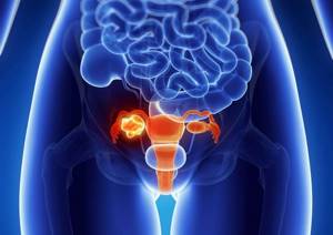
Kidneys are one of the most important organs in the human body. Their function and importance are difficult to overestimate. The main tasks of the kidneys are: filtration (filtration of blood), excretory (removing metabolic products and toxic substances from the body), maintaining the basic acid balance. They also take part in maintaining blood pressure. These are not all the functions of these organs. However, many people develop various kidney diseases during their lifetime. Sometimes they have a characteristic, pronounced clinical picture, and sometimes they can be asymptomatic.
What is research

An examination using radiation exposure to the human body helps to obtain an image of an organ, evaluate its size, location, structure, and see pathological formations, for example, tumors.
If a contrast agent is used during an x-ray examination, the patency of the natural pathways to and from the organ can be assessed. The results obtained help to draw a conclusion about the functionality of the organ and violations of its activity.
The examination is carried out in specialized diagnostic centers or hospitals. The result of the study is given to the patient or forwarded to the attending physician if it was done in the same institution where the patient is observed. The results come the next day.
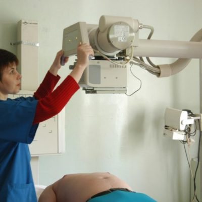
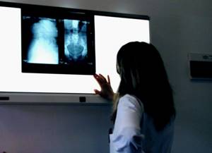
Carrying out X-ray diagnostics
Best materials of the month
- Coronaviruses: SARS-CoV-2 (COVID-19)
- Antibiotics for the prevention and treatment of COVID-19: how effective are they?
- The most common "office" diseases
- Does vodka kill coronavirus?
- How to stay alive on our roads?
During pyelography, a person sits on a couch with his knees bent. The patient's position is fixed with stirrups, after which anesthesia is administered. A catheter is inserted into the renal pelvis through the bladder. Next, the organ is filled with contrast agent to the appropriate level. Using an X-ray machine, radiographs are taken in posterior, anterior, semi- and lateral projections.
For antegrade pyelography, the patient lies on the table with his stomach down. A needle with a tube of seven or eight centimeters is inserted into the area of the twelfth rib. A contrast agent is injected through them, then the calyx and pelvis are photographed.
After the appropriate manipulations, the radiologist gives the resulting images, along with a detailed transcript and description, to the attending physician who referred for diagnosis. This procedure can last from one to one and a half hours.
Types of X-ray examination
Medical practice uses several types of x-rays that are used to diagnose kidneys. The choice of technique remains with the doctor, who knows what results this or that procedure will show. This is taken into account when it is advisable to confirm a particular diagnosis.
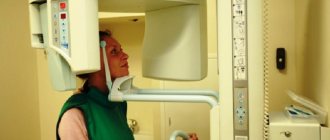
Survey radiography
Among the simple types of examination is digital x-ray. This is a common type of procedure, it is performed quickly and is available in almost every medical institution.
The irradiation area is placed in front of an X-ray screen, and the rest of the body is protected by a lead apron. The procedure is painless.
A survey X-ray examination makes it possible to see the organ as a whole and determine deviations. The disadvantage of such an image is that not all pathologies can be examined and identified.
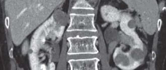
CT scan
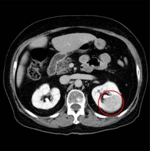
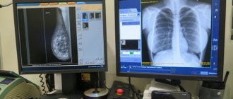
A modern examination for kidney diagnostics is computed tomography. The research is carried out using special high-precision equipment. With a CT scan of the kidneys, the image is displayed on a computer monitor. Using a special program, they work with images that are of high quality.
Therefore, when an X-ray image is enlarged, the doctor sees those pathologies that are hidden from the view of a specialist with a regular X-ray. Layer-by-layer imaging is extremely effective for identifying pathologies - tumors, cysts, stones.
The doctor receives not only the image itself with the localization of the pathology, but also data on the size of the tumors, their structure and other characteristics.
X-ray with contrast
Contrast x-ray of the kidneys
Another type of examination is an x-ray of the kidneys with a contrast agent. The patient is specially injected with a substance that transmits rays through itself differently than tissues and organs.
Once in the cavities and vessels, the contrast agent is distributed into them and displayed on an x-ray. This helps the doctor to see pathologically enlarged cavities, places of protrusion of vascular walls, thrombosed vessels and other results.
For each type of examination, with the exception of computed tomography, preparation is made for radiography of the kidneys. The doctor tells the patient about this when he gives a referral for diagnostics.
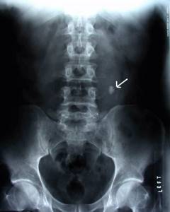
Kidney image without contrast: information content and significance of the study
An X-ray of the kidneys without contrast is usually prescribed at the initial stage of the examination. This diagnostic option is widely used by specialists to determine such basic data as the location of the kidneys, their size, volume, contours, density and other characteristics. This allows you to present a general picture of the condition of the kidneys and suspect pathological processes. An X-ray of the kidneys without contrast, although not highly informative, can provide the attending physician with general data. The advantages of this diagnostic method are:
- fast execution;
- no discomfort;
- cheapness;
- general availability;
- does not require the introduction of special drugs (contrast).
If the clinical picture is unclear or blurred, the patient has nonspecific complaints, and the attending physician has minimal suspicions, it is not advisable to immediately resort to contrast radiography. To begin with, it is better to examine the kidneys without contrast, and if suspicions are confirmed, we can talk about ordering other examinations.
Indications for x-ray examination of the kidneys
X-rays are performed for clear indications when the patient has symptoms of pathology of the urinary system. Such pathologies arise both during the process of intrauterine development and are considered congenital, but they can also be acquired.
Therefore, according to indications, x-ray examinations are carried out for both adults and children, and for young children - in cases of extreme necessity, if other methods fail to establish a diagnosis.
If a person’s kidney function is impaired, serious consequences that can be fatal develop, so you should never refuse the procedure if the doctor insists on performing it. The referral is issued by a general practitioner, nephrologist, surgeon, or oncologist. Data on the condition of the kidneys is valuable for cardiologists and gynecologists, so they can also refer the patient for an X-ray of the kidneys.
Indications for this procedure are:
- signs of renal pathology;
- abnormalities in urine test results indicating improper kidney function;
- the appearance of blood in the urine;
- pathological changes in the urinary organs detected using other studies, for example, ultrasound;
- kidney injuries;
- urolithiasis disease;
- visualization using ultrasound of tumors, cysts;
- renal colic;
- frequent exacerbations of infectious pathologies of the urinary tract;
- control after surgery.
Pathologies can occur without pronounced symptoms. If there are no signs of pain, patients rarely go to doctors. If the patient’s well-being worsens, a urine test is prescribed.
The following symptoms may alert a doctor when visiting a patient:
- patient complaints of pain in the lower back, cutting or aching pain;
- the patient experiences swelling in the morning - on the face, in the lower extremities;
- change in the amount of urine excreted per day;
- the acquisition of uncharacteristic signs by urine - a brown or dirty yellow color, the appearance of sediment or flakes in the urine, an unpleasant sweetish odor or, conversely, a pungent odor;
- patient complaints of pain when urinating;
- general weakness - increased fatigue, headaches, insomnia, increased body temperature.
With all these signs, careful diagnosis is required, since kidney pathologies affect the condition of the body. If the pathology is not detected in a timely manner, you can miss the acute period, during which it is best to treat the disease, but it is difficult to deal with chronic pathologies.
When is the study scheduled?
X-rays of the kidneys are prescribed according to special indications from the attending physician. It is he who makes the decision to prescribe one or another diagnostic method and evaluates and analyzes the data obtained.
Let's consider the most common situations in which an X-ray examination of the kidneys is performed.
Blood in the urine (gross hematuria)
Macrohematuria is a visual determination of blood in the urine without additional studies. Most often observed with kidney disease or passage of stones
along the urinary tract.
Changes in urine analysis that persist for more than 2 months
These changes may indicate chronic or prolonged kidney pathology.
Suspicion of renal hypertension
A steady increase in blood pressure may raise suspicions among your doctor. As part of a comprehensive diagnosis, fluoroscopy of the kidneys may be performed to identify the cause of hypertension.
Trauma to the abdomen and lumbar region
For penetrating and non-penetrating injuries to the kidneys, an examination is necessary to assess the damage caused.
Pain in the lower back and abdomen
Pain in the lower back and lower abdomen (projection areas of the kidneys) may be a sign of the development of the disease. If symptoms are erased, radiography of this area may be necessary.
What will a kidney x-ray show?
X-ray examination of the kidneys helps to identify the following pathologies:
- stones and sand;
- compression of the urinary tract by stones or tumors;
- incorrect position of the organ (more often – prolapse);
- cystic formations;
- benign or malignant tumors;
- abnormalities in the structure of organs (for example, the presence of only one kidney);
- polycystic disease;
- hydronephrosis;
- pyelitis;
- pyeelectasia;
- glomerulonephritis;
- kidney injuries (ruptures);
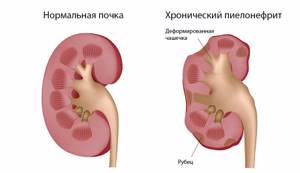
- pyelonephritis;
- tuberculous kidney damage;
- ureteral rupture.

Based on the data obtained, doctors make a diagnosis and begin treatment of the patient. This is extremely important, because when kidneys fail, patients experience severe intoxication - poisoning with products of their own production, which will inevitably lead to death without qualified medical assistance.
X-ray of the kidneys and urinary tract in Moscow
We invite you to urography and other examinations at our clinic. Our priorities in work:
- Caring for the patient and his health.
- The quality of services provided.
- High diagnostic accuracy thanks to advanced equipment.
We are confident in the professionalism of our doctors - each of them has extensive work experience, regular advanced training in courses and trainings, and specialized training in the use of the clinic’s diagnostic equipment.
All this allows you to quickly and accurately make a diagnosis and determine the optimal treatment method for a wide variety of pathologies.
Want to learn more about kidney x-rays? We are ready to answer your questions and make an appointment at a convenient time by phone: +7(495) 478-10-03.
Contraindications for the study
If it is necessary to conduct a study, doctors may refuse the patient and offer alternative diagnostic methods if there are contraindications to the procedure.
Such contraindications may be:
- pregnancy;
- patient's age;
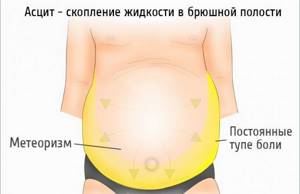
- presence of obesity;
- ascites;
- acute infectious diseases occurring in the body;
- exceeding the radiation dose recommended for the year.
If the patient does not currently have the opportunity to resort to alternative diagnostics, and the condition is critical and requires immediate decision-making, then doctors, even in the presence of the above contraindications, will perform fluoroscopy of the kidneys.
If it is necessary to take an x-ray with a contrast agent, the situation is different. Contraindications to contrast X-ray are an allergic reaction to the injected substance, renal failure, tendency to bleeding, tumor formations of the adrenal glands, acute period of glomerulonephritis, decompensated cardiac pathologies.
In this case, kidney urography is undesirable, but if there is an urgent need, doctors take responsibility for conducting the study and do everything necessary to minimize the risks of complications. In this case, data on the patient’s condition is recorded extremely carefully in the accompanying medical documentation, and the decision on diagnosis can be made collectively.
Conducting a CT scan of the kidneys with contrast
A kidney CT scan with contrast is performed in a special room, into which the patient enters after careful preparation. The patient removes jewelry and other metal objects and changes into the provided robe. After this, the patient fills out a consent form for a CT scan of the kidneys and learns about the possible risks that he may encounter during the study.
After signing the documents, the procedure begins with the following sequence:
- the patient is placed on a couch, which is moved into the tomograph;
- During computed tomography, personnel are in the next room and take control images;
- 30 minutes after the examination, the couch is pulled out and the tomograph is turned off;
- the specialist clarifies the presence of possible allergic reactions to iodine and contrast agent;
- a contrast agent is injected into the femoral vein, after which a specialist monitors the patient’s condition for several minutes and places him in a tomograph;
- after turning on the device, the ring begins to rotate along the axis and a three-dimensional image is sent to the computer;
- The contrast scan can take up to 30 minutes, after which the patient is removed from the scanner.
After the examination, the doctor evaluates the condition of the kidneys and their structures. The results of computed tomography are stored electronically and are displayed as a series of images for inclusion in the medical history. A kidney CT with contrast is a painless procedure, but the patient needs the psychological attitude to spend time in a confined space, while not moving and breathing calmly.
The tomographs used at the Yusupov Hospital are equipped with an emergency button, which the patient can use if their health worsens. After pressing it, the study stops, so specialists warn patients about this before the study.

Preparation rules
The doctor will instruct the patient in advance about preparing for a kidney x-ray.
To obtain clear images, it is necessary to ensure that the kidneys are not covered by the shadow of a crowded intestine. Therefore, it is important for doctors that when performing an x-ray, the intestines are freed from digested food and gases.
It is recommended to carry out preparation two to three days before the x-ray. The patient needs to reduce the amount of heavy foods that cause gas formation and take a long time to digest in the intestines. These are beans, cabbage, baked goods, cucumbers, and dairy products.
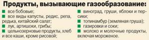
To reduce bloating, it is recommended to take sorbents - “Simethicone”, “Enterosgel”, “Polysorb”. A day before the x-ray, it is necessary to cleanse the intestines with Fortrans, and before the examination, either have a light dinner or not eat at all.
In the morning before the procedure, you can drink some water and take a walk in the fresh air. For patients with constipation or those who ate before the x-ray as necessary, a cleansing enema is performed.
Types of pyelography
Kidney disease requires an individual approach. This applies not only to treatment, but also to examination of patients, which is confirmed by the recommendations of many doctors. X-ray diagnostic methods are divided into several types:
- pneumopyelography using carbon dioxide and oxygen (to detect renal tuberculosis, frontal bleeding, etc.);
- double contrasting with the combined use of a contrast component and gas;
- ascending or retrograde method using a catheter cystoscope and a dye;
- excretory urography with the introduction of a dye through a needle intravenously. It helps determine the structure and structure of the ureters, urethra, kidneys;
- antegrade pyeloureterography with percutaneous puncture, nephropyelostomy or pyelocaliceal system.
Sometimes x-rays of organs can be performed in parallel with intraoperative intervention. In severe cases, when the functionality of the urinary system is impaired and it is impossible to obtain an overall picture for diagnosis, alternative methods are resorted to. For example, if the excretory capacity of the kidneys is reduced, and the contrast agent does not enter the cups due to the lack of dynamics of the process, the optimal solution would be to install a cystoscope (retrograde study).
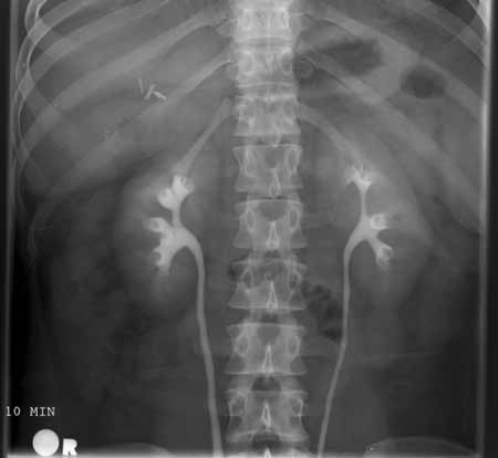
Carrying out urography
Urography is performed in a horizontal position - the patient lies either on his back or on his stomach. To determine the mobility of the organ, the patient will need to stand up. If there is a need to localize a tumor, stones or cyst, urography is performed in the patient’s lateral position.
The X-ray emitter is installed approximately one meter from the area being examined. The procedure of a survey x-ray along with an excretory study is informative.
At the first stage, the patient takes a regular X-ray, and at the second stage, a radiopaque substance is injected into the blood. A series of pictures of the kidneys are taken at certain intervals so that the contrast has time to fill the cavities.
How the human urinary system works and works
The main functions performed by the urinary organs are maintaining water-salt balance and removing metabolic products.
Content:
- How the human urinary system works and works
- What diseases can affect the urinary system and why excretory urography is prescribed
- What is an X-ray of the urinary organs?
- Indications and contraindications for the procedure
- Requirements for preparation for the procedure of excretory urography
- How is an X-ray of the urinary system performed?
- Possible complications and risks of the examination
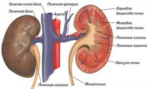
The kidneys are paired organs shaped like beans. They are located in the retroperitoneum, in the lumbar region. Through the renal gate, which is located on the inside of the organ, the inferior vena cava and the aorta enter the kidney. The ureters emerge from them in the same place. The membrane of the kidneys consists of adipose and connective tissue, and the structural units of the organs are nephrons - a collection of glomeruli and excretory tubules. Of the entire urinary system, only the kidneys are involved in the detoxification processes of the body, the rest of its organs are responsible only for the accumulation and excretion of urine.
The ureter looks like a hollow tube with a lumen thickness of about 12 millimeters. Its length is 30-32 centimeters, although it can vary depending on the human body.
The walls of the ureter consist of three layers - internal mucous, middle muscular and external connective tissue.
The bladder is a hollow, sac-like organ that stores urine until it is excreted from the body. The capacity of the bubble is about 300-400 milliliters. When the volume of liquid accumulates from 200 milliliters, a person feels the urge to urinate.
Anatomically, the organ is divided into apex, neck, body and bottom. Its walls have a three-layer structure, consisting of:
- serous membrane, which is located outside;
- middle layer consisting of muscles;
- inner layer: the bladder is lined from the inside with a mucous membrane of transitional epithelium.
In addition, the bladder contains glandular epithelium and lymphatic follicles. At the bottom of the bladder there is a narrowing - the sphincter.
From the bladder comes the urethra - the urethra, which has the shape of a tube through which urine is removed from the body.
Urography in children
Urography is performed for vital indications, so even in childhood, the study can be performed at any time, regardless of the patient’s age. For young children there are some special features when performing x-rays:
- Before the study, a gas tube is placed; enemas are recommended for older children;
- X-rays are performed using digital machines to minimize radiation exposure;
- during carrying out unnecessary areas are covered with protective aprons;
- Before an x-ray of the kidneys with contrast is taken, small children are given anesthesia, the administration of drugs is calculated according to the weight and age of the baby;
- Older children can only be given sedatives and, if necessary, antiallergic drugs.
How is the research conducted?
X-rays of the kidneys are performed in a specially equipped room. After the preparatory measures, the patient needs to take the correct position, and further actions directly depend on the x-ray method. Before contrast urography of the kidneys, a special substance is injected intravenously.
After this, a series of x-rays are taken.
Alternative Research
Diagnosis is carried out according to strict indications, since radiation exposure is unsafe for the patient. During irradiation, the patient does not receive a large dose of radiation, but for safety reasons, they prefer to reduce the number of x-ray procedures to a minimum.
If it is necessary to detail the data, doctors resort to other methods, for example, ultrasound diagnostics. In some cases, the results of these two studies complement each other and help clarify the diagnosis.
In other cases, magnetic resonance imaging of the kidneys will be an informative examination. Diagnostics has different principles for obtaining results, but the data is no less reliable, so it can also be used as an alternative to x-rays.
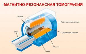
MRI
Kidney CT scan without contrast: price
CT scanning of the kidneys without contrast agent is less expensive because it is faster and does not require the use of expensive contrast agents. If the patient is indicated for a kidney CT scan with contrast, the price in Moscow may be higher. On average, the cost of a diagnostic procedure without contrast is 3,000 rubles.
Patients of the Yusupov Hospital can familiarize themselves with the current price list and find out the cost of highly accurate and safe diagnostics. Diagnosis and treatment of patients with kidney diseases is carried out after a detailed treatment plan and cost of procedures have been presented.
Decoding the research results
A radiologist interprets the results. The conclusion will indicate the size of the kidneys, structural features, and the presence or absence of pathological formations. If there are stones, cysts or tumors, the doctor will definitely indicate their location and size in the test results.
After the conclusion is formed, it is transmitted to the attending physician. Here, a nephrologist or surgeon compares the results of the decoding with other patient tests - blood tests, urine tests, and ultrasound data.
Taking into account all the indicators, the doctor makes a diagnosis and outlines a further treatment plan, taking into account the indications for surgical intervention - planned or urgent.
What can be seen in the photo?
Below we will describe what can be determined from the image obtained as a result of the examination.
Developmental anomalies
Various anomalies of kidney development (changes in their configuration, structure, anatomy, etc.).
Nephroptosis
Nephroptosis is a pathological condition characterized by prolapse of one or both kidneys due to injury or pathological processes.
Kidney cyst
Characterized by the development of cavity formation in this organ.
Tumor
It is possible to detect tumors and metastases of various origins and localizations. If there is a suspicion of the presence of tumors, as a rule, additional laboratory and instrumental examinations are prescribed.
Possible complications of the procedure
Complications after the kidney x-ray procedure are minimal. Most complications are associated with intolerance to the contrast agent. Before conducting the study, doctors test for allergies to contrast, and most patients do not experience problems with routine x-rays.
Minor deterioration in health, weakness, nausea, allergic reactions may occur in patients who were diagnosed without preparation - when it was necessary to quickly obtain data on the condition of the kidneys. In this case, doctors try to minimize complications and alleviate the patient’s condition.
X-ray of the kidneys is an extremely important test for health. It is prescribed for diagnosing diseases of the excretory system, which is responsible for the evacuation of waste products from the body, maintaining metabolism, and water-salt metabolism.
These processes ensure the normal functioning of the body. Therefore, when ordering an x-ray, you should not delay the procedure - it helps to make a diagnosis and save the patient’s life.
general characteristics
If there are suspicions, as well as specific and nonspecific signs of the development of kidney pathology, specialists refer patients to a series of clinical and instrumental examinations that can more clearly show the picture of pathological processes. The data obtained can be decisive in making the correct diagnosis. The methods of treatment and their effectiveness directly depend on this.
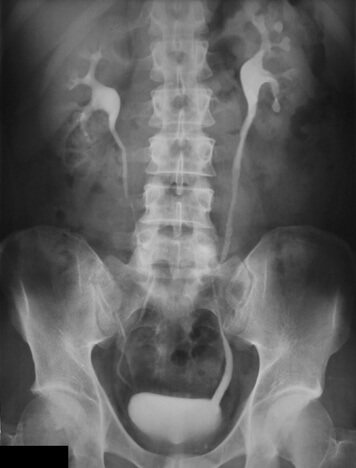
One of the most common diagnostic examination methods is radiography of the kidneys. Its essence lies in obtaining the necessary data on the condition of the kidneys and urinary tract through the action of X-ray radiation, which tends to linger in denser tissues and formations. Visually, this appears on x-ray film in the form of an image and makes it possible to characterize and analyze the condition of the examined area. There are various methods for x-raying the kidneys - how the examination is done depends on the disease and pathological condition.
X-ray contrast research methods in pediatric nephrology
In 1895, Wilhelm Roentgen discovered a new type of rays. Using "X-rays" (the name given by Roentgen), he took the first photograph of his wife's hand. This marked the beginning of a new direction not only in medicine, but also in other scientific fields. In 1898, Professor F.I. Pasternatsky in Russia demonstrated an x-ray of a patient with calculosis of the bladder. Since that time, X-ray methods began to gain leading positions in uronephrological examination, since visualization of the organs of the urinary system makes it possible to clarify the state of the collecting system of the kidneys, ureters, bladder, urethra, to diagnose obstructive uropathy, i.e. to identify factors contributing to the development of the inflammatory process in the organs urinary system. X-ray urological studies remain accessible and in demand, despite the existence of other methods that help clarify the condition of the organs of the urinary system. Carrying out X-ray examinations in nephrology is in most cases necessary, since it makes it possible to assess not only the anatomical and motor characteristics of the urinary organs, but also the functional state of the kidneys.
The most common procedures performed in pediatric nephrology practice are:
- excretory urography (and its modification - infusion urography);
- voiding cystography.
Renal angiography and tomography usually have clearly defined indications for performing them.
Victory cystourethrography (from the Greek kystos - bubble and grapho - I draw) is an X-ray method for examining the urethra and bladder, previously filled with fluid.
Indications for cystourethrography:
- recurrent urinary tract infection;
- suspicion of the presence of vesicoureteral reflux (VUR), reflux nephropathy;
- microhematuria;
- dysuric phenomena, pollakiuria, difficult and rare urination;
- malformations of the anorectal zone;
- injuries of the bladder and urethra;
- tumor of the abdomen and pelvis;
- large inguinal hernias accompanied by dysuric symptoms;
- monitoring and evaluation of the results of conservative and surgical treatment on the organs of the urinary system and rectum.
Contraindications to voiding cystourethrography:
- acute inflammatory diseases of the urinary tract (acute pyelonephritis, cystitis, urethritis);
- gross hematuria;
- severe general condition of the child.
The amount of solution administered during voiding cystography should correspond to the age-related physiological capacity of the bladder (Table 1).
| Age-related indicators of physiological bladder capacity |
When assessing cystourethrograms, the contours of the bladder, its size, the presence of vesicoureteral-pelvic reflux, and the condition of the urethra are described. “Fringed” contours are characteristic of neurogenic bladder dysfunction and cystitis. A double contour of the bladder in its lower parts is often observed with atony. With diverticula, the cystogram reveals an additional shadow of various sizes and shapes.
Filling of the ureters (one or both) with contrast agent indicates the presence of VUR. Long-term high-grade VURs contribute to the development of secondary renal shrinkage. Certain difficulties arise when diagnosing a hypoplastic and secondary wrinkled kidney. Often, differential diagnosis requires radioisotope research methods - indirect angiography, dynamic nephroscintigraphy with tubulotropic and glomerulotropic radiopharmaceuticals, as well as static nephroscintigraphy with 99mTc-DMSA.
When interpreting cystourethrograms, it is necessary to assess the condition of the urethra. It is especially important not to miss the posterior urethral valve in boys, which is diagnosed based on narrowing of the urethra and dilatation of the urethra above the narrowing site.
Excretory urography makes it possible to assess the anatomical structure of the kidneys and urinary tract, identify signs of their damage, and monitor the dynamics of the pathological process. When performing excretory urography, the greatest difficulty is in determining the functional state of the kidneys.
Indications for this study are:
- ultrasound data indicating the possibility of the presence of defects and anomalies in the development of the urinary system;
- pain in the abdomen or lumbar region of unknown origin, regardless of the presence or absence of changes in urine tests;
- arterial hypertension of unknown etiology;
- enuresis, accompanied by minimal changes in urine tests;
- glomerulonephritis combined with urorenal infection.
Contraindications to excretory urography:
- severe diseases of the urinary system with impaired nitrogen excretion function of the kidneys;
- acute and chronic renal failure;
- pronounced activity of parenchymal kidney diseases;
- allergy to radiopaque agents and iodine preparations;
- severe liver damage with functional failure;
- collapse and shock;
- tuberculosis in the active phase;
- thyrotoxicosis.
When preparing a patient for an X-ray contrast study, the anamnestic data on the presence of allergic reactions should be clarified; if there is a risk of developing an allergy to radiocontrast agents (RCM), antihistamines are prescribed for 2–3 days preceding the X-ray examination. On the day of excretory urography, patients with allergic reactions are prescribed a single dose of prednisolone in an age-appropriate dosage.
A significant role in obtaining high-quality radiographs is played by the preparation of the child’s gastrointestinal tract - cleansing the intestines of feces and gas. To prevent the formation of “hungry” gases in the morning on the day of the study, 1–1.5 hours before urography, older children can be offered “dry” porridge or a piece of bread and unsweetened tea. In order to reduce aerocolia, it is recommended to exclude foods rich in carbohydrates, raw vegetables, juices, whole cow's milk, and black bread 2-3 days before the study. It is advisable to prescribe sorbents, chamomile infusion, boiled carrots. In older children, bowel cleansing is carried out with vaseline oil in a volume of 30 ml, followed by two cleansing enemas: 2 hours after taking the oil and in the morning 2 hours before the X-ray examination. The volume of a cleansing enema is 50 ml of liquid per 1 year of life. Children prone to constipation should not have cleansing enemas with large amounts of water, as there is a risk of overhydration, leading to a decrease in the concentration of the contrast agent. In children under the age of 1 year, morning feeding is skipped, and at the beginning of the examination, the child is fed liquid food through a nipple in such a way that he swallows a certain amount of air. The air-filled stomach pushes the intestinal loops downwards, which improves visualization of the kidney. Young children who are prone to increased gas formation are prescribed drugs that help reduce it (simethicone preparations - sub-simplex, espumizan). For excitable children, it is advisable to prescribe a decoction of valerian root for 2 days before the test and 1 tablespoon as an enema on the eve of the test. To cleanse the intestines in young children, regular enemas with boiled water are used or laxatives are prescribed (Transipeg, Duphalac, Microlax, etc.).
| Calculation of the dose of contrast agent depending on the size of the child’s body surface |
There are various ways to calculate the dosage of a contrast agent, but the most accurate is to determine the dose of RKS when it is calculated per 1 square meter. m of the child’s body surface (Table 2). An increase in calculated doses for young children is associated with more active urine excretion and physiologically determined low concentration ability of the kidneys.
It is recommended to administer 1 ml of RCS and take a 2-3 minute pause, during which it is necessary to monitor the patient’s condition. If there is no reaction, then administration of RCS can be continued. If a patient has a reaction to the administration of RCS, it is necessary to stop the administration of contrast and immediately begin to provide assistance.
A modification of excretory urography is infusion-drop urography, which is performed in case of severe malformations of the urinary system, a decrease in endogenous creatinine clearance to 50 ml/min, impaired concentration function of the kidneys, as well as in newborns and infants due to the morphological “immaturity” of the nephron, which reduces clarity of kidney contrast. The dose of RKS for infusion urography is doubled and mixed with an equal amount of 5% glucose solution. The drug is administered in a stream (120–150 drops per minute), images are taken after 5, 10, 20, 40 and 60 minutes from the start of the infusion.
It is known that visual assessment of renal function based on the degree of contrast of the pyelocaliceal system is not accurate enough. The image contrast on urograms depends primarily on the following factors:
- functional state of the kidneys;
- state of urodynamics of the upper urinary tract and renal hemodynamics;
- functional state of the bladder;
- RKS quality.
Ideally, RCS should absorb x-rays well and at the same time achieve the required concentration in the kidneys without causing harmful effects on the renal parenchyma. Unfortunately, with the use of most RCS, the development of undesirable side effects is possible. One of the serious disadvantages of RCS is the relatively high incidence of adverse reactions and complications after their introduction into the bloodstream. For many years, these phenomena were associated with iodism, i.e., individual intolerance to iodine. However, as clinical and laboratory studies have shown, the elements of the contrast agent are so tightly bound to the benzene ring that disintegration with the release of iodine atoms does not occur. Undesirable side effects are associated with two other factors: intolerance to the RKS salt complex itself and its high osmolarity. Intolerance is expressed in the manifestation of an allergic reaction. The phenomena arising due to the high osmolarity of the RCS are more multifaceted in nature. At the end of the 60s. last century, the decisive role of osmolarity and ionicity in the adverse effects of contrast agents on the human body was established, therefore, after the synthesis of nonionic monomers, most developments were devoted to reducing the osmolarity of drugs.
The development and application of RCS for intravascular administration can be divided into three stages (Table 3).
It is the presence of high osmolarity that explains such complications as the occurrence of hemodynamic disorders, an increase in the level of secretion of a number of enzymes and hormones, electrolyte imbalance, and an increased tendency to agglutination of red blood cells and thrombus formation.
When choosing a RCS, it is necessary to take into account three main factors: diagnostic effectiveness, safety and cost of the drug. The diagnostic value of RCS mainly depends on the dose of iodine, and the nonionic dimer molecule has the highest efficiency in terms of its ability to absorb X-ray radiation.
The toxicity of RCS is influenced by many different factors (Table 4).
All contrast agents currently used for radiopaque studies can be divided into ionic and non-ionic (Table 5).
The creation of non-ionic RCS was a step forward towards reducing the toxicity of drugs. When using high- and low-osmolar RCS, the development of undesirable side effects is possible, to prevent which it is necessary to assess the presence of risk factors in the patient.
Risk factors for complications during RCS insertion
- History of allergic reactions to other RCS.
- Allergic diseases in a child (food allergies, bronchial asthma, atopic dermatitis, hay fever).
- Pathology of the cardiovascular system (arrhythmias, pulmonary edema, circulatory failure).
- Diabetes.
- Taking medications (non-steroidal anti-inflammatory drugs (aspirin), β-blockers).
- Metabolic disorders (dehydration, polycythemia, hypercoagulation).
- Kidney pathology.
- Taking drugs, drinking alcohol.
For many years, the attention of radiologists and pediatric nephrologists has been drawn to the problems of RCS nephrotoxicity. As a rule, the use of ionic and non-ionic RCS in patients with renal pathology, but with preserved renal function, is not accompanied by the development of a pronounced nephrotoxic effect. The development of nephrotoxic effect is most likely when using ionic RCS in patients with impaired renal function (Fig.).
The criterion for the development of nephrotoxic effect in adults is considered to be an increase in serum creatinine levels by 25% (or 44 µmol/l) compared to the initial value within 3 days after administration of RCS.
Risk factors for the development of nephrotoxicity directly related to RCS itself include:
- high or low osmolarity of the drug;
- large dose of RKS agent;
- route of administration of RCS (with the intra-arterial route the risk increases);
- previous use of RCS (history).
After administration of RCS, it is advisable to monitor the patient for 30 minutes after the study, since most side effects occur during this period. It is possible that delayed adverse reactions may develop over a longer period of time. Renal failure caused by the administration of RCS, as a rule, is not accompanied by the development of oliguria, and the level of serum creatinine normalizes within 7–10 days.
At the same time, in patients with a high risk of developing nephropathy, acute renal failure may develop within 24 hours after the administration of RCS. It should be noted that in children under one year of age, especially in newborns, hemodynamic disturbances with the introduction of ionic RCS develop especially easily.
The safest RCS for patients with kidney pathology is the isosmolar drug iodixanol (Visipak). The drug is isotonic with blood in all concentrations and has a lower iodine concentration; electrolyte composition is balanced. When the drug is injected into a vein, there is no pain. When performing excretory urography, Visipaque is administered to children under 7 years of age at the rate of 2–3 ml per kilogram of body weight, for children over 7 years of age - no more than 50 ml.
To prevent the development of nephrotoxic effect in patients with renal pathology, a number of rules should be taken into account.
- It is necessary to evaluate risk factors for the development of nephrotoxicity.
- It is advisable to monitor creatinine levels before and after administration of RCS.
- The use of isosmolar RCS is indicated.
- It is not advisable to administer large doses of RCS.
- It is necessary to ensure adequate hydration to prevent nephrotoxic effects when using ionic RCS; In high-risk patients, an intravenous infusion of isotonic sodium chloride solution is indicated, which should begin before the introduction of RKS and continue until the end of the removal of RKS.
- It is not recommended to perform surgical interventions until RCS is removed from the body.
- Diuretics (especially mannitol and loop diuretics) should not be used simultaneously.
- It is not recommended to carry out repeated X-ray contrast studies for a short time (until recovery of kidney function).
According to the European Society for Urogenital Radiology guidelines for the safe use of contrast media (Version 2) from 2003, in the case of decreased renal function, the physician should weigh the pros and cons of using RCS [3]. If it is necessary to use RCS in adults, take the following precautions.
- The patient should be hydrated (drink 100 ml of fluid per hour) or should be administered intravenous saline within 24 hours after the appointment of RCS.
- Renal function is monitored (serum creatinine, serum lactic acid level, blood pH).
- The symptoms of lactic acidosis are monitored (the appearance of vomiting, drowsiness, nausea, epigastric pain, anorexia, hyperapnea, lethargy, thirst, diarrhea) - with a blood pH < 7.25 and a lactic acid level > 5 mmol.
Currently, ways to further reduce the nephrotoxicity of RCS are being developed [4]. There is evidence that to prevent the nephrotoxic effect, premedication with the administration of acetylcysteine, aminophylline, and angiotensin-converting enzyme antagonists is necessary.
Thus, despite the active introduction of modern ultrasound research methods into pediatric nephrology, most patients with pathology of the urinary system require X-ray contrast studies, since these methods make it possible to objectively assess not only the anatomical and motor characteristics of the urinary organs, but also the functional state of the kidneys. When conducting X-ray examinations in children, it is necessary to take into account not only the high sensitivity of the growing child's body to ionizing radiation, but also the toxicity of RCS used in pediatric nephrology. Thanks to the introduction of new non-ionic RCS into clinical practice, radiological research methods should become safer.
Literature
- General Guide to Radiology / ed. H. Pettersson. NISER, 1995. 778 pp.
- Aspelin Pet. et al/N. England J Med. 2003; 348:491–498.
- European Society for Urogenital Radiology Guidelines for the Safe Use of Contrast Media (Version 2). 2003. P. 16.
- Sinitsyn V. E. Application of ionic radiocontrast agents in modern radiation diagnostics//Issues of clinical effectiveness, safety and pharmacoeconomics/Medical visualization. 2003. No. 1. pp. 121–127.
I. N. Zakharova, Doctor of Medical Sciences, Professor E. B. Mumladze, Candidate of Medical Sciences, Associate Professor O. A. Voronenko E. V. Zakharkina RMAPO, Moscow
A type of kidney urography
Based on the methods of implementation, the following types of urographic examination are distinguished:
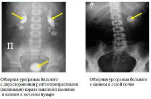
- Survey urography of the kidneys. A simple diagnostic technique, which consists of a conventional x-ray examination of the organ. Compared to other types of urography, which involve the introduction of contrast, the survey diagnostic method is considered less informative. At the same time, it has a minimal number of side effects. Survey urography is prescribed to obtain a general characteristic of the functioning of the kidneys, as well as to detect large and medium-sized stones;
- Retrograde urography. This diagnostic technique is performed under anesthesia. The administration of a contrast agent is carried out after catheterization of the ureter and bladder. Urogram images allow you to obtain detailed information about the condition of all urinary organs;
- Intravenous urography of the kidneys. Before an X-ray examination is performed, the patient is first injected with a contrast agent. More often it is an iodine solution in glucose or water. After a certain time, a series of images are taken that allow you to visualize the condition and structure of the kidneys.
Urography is a method for examining the kidneys, based on the administration of contrast followed by X-ray examination. Preparation for the procedure consists of eliminating possible contraindications and bowel movements.
Taking into account the goals and objectives of the X-ray contrast study, as well as the patient’s condition, the following types of intravenous urography are distinguished:
- Infusion It involves the introduction of a radiopaque contrast agent through a catheter. Usually prescribed to patients who are unable to move independently;
- Excretory urography of the kidneys. Performed to study the excretory function of the kidneys by assessing the rate of removal of contrast. To do this, photographs are taken after a strictly set time;
- Compression urography. The procedure involves clamping the ureter through the abdominal cavity. Thanks to this, the photo is clearer. The disadvantage of the study is that the procedure is highly painful.