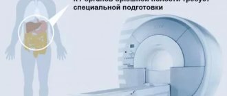
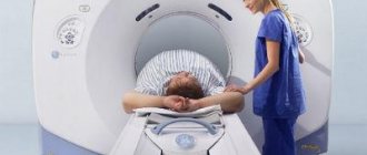
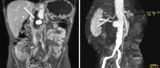
MRI of the abdominal cavity is a completely non-contact method for diagnosing diseases of the digestive tract, and with extended examination, also the urinary system. Using tomography, it is possible to visualize soft tissues, organs, and systems in high resolution. Due to the detail and high quality of the image, MRI of the abdominal cavity is considered the gold standard for diagnosis. Non-invasiveness, complete safety, and the ability to undergo examination repeatedly make tomography one of the main ways of examining patients.
Which authorities check?
Magnetic resonance imaging allows you to determine pathologies of the structures of the digestive tract. The following internal organs are visualized:
- Pancreas.
- Spleen.
- Liver.
- Stomach.
- Gallbladder.
- Various parts of the intestine.
Despite its high information content, tomography allows one to evaluate only the structural features of the digestive tract. The study makes it possible to diagnose the disease or confirm the doctor’s suspicions. However, to obtain more information about the functional state of the gastrointestinal tract structures, additional diagnostics may be required. For example, scintigraphy, studying the functioning of organs after the administration of a radioisotope drug, etc. The choice of examination tactics is decided by the doctor after assessing the situation.
Alternative Methods
In cases where, for one reason or another, it is impossible to perform an MRI, alternative research methods are used to diagnose diseases. These include:
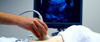
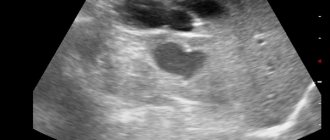
- Ultrasonography . A fairly informative method that allows you to diagnose a variety of pathologies of the abdominal organs (spleen, liver, etc.);
- X-ray examination of the gastrointestinal tract using barium sulfate . During the study, the patient drinks a contrast suspension, and its passage is monitored during fluoroscopy;
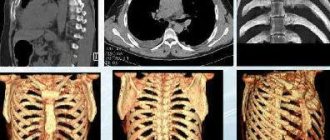
- computed tomography (CT) of the abdominal cavity . This is one of the most informative methods for diagnosing numerous diseases of the hollow organs of the abdominal cavity, which include the stomach and intestines;
- Irrigoscopy . This radiological diagnostic method is used to detect pathology of the large intestine;
- Intravenous cholangiocholecystography . Allows you to identify diseases of the biliary tract. The patient is given a contrast solution intravenously, and then a series of x-rays are taken;
- Diagnostic laparoscopy . Used to identify certain pathologies of the abdominal organs;
- Targeted biopsy under X-ray or ultrasound control . The need to use this method arises when a malignant process is suspected.
Indications for MRI of the abdominal cavity
Tomography of the abdominal organs is carried out in the presence of symptoms that are typical of a pathological process from the structures of the digestive tract. Or when other diagnostic methods did not provide an accurate answer to the question of the origin and type of the disease. Specific indications for diagnosis include:
- Pain in the abdomen. If localized on the left, it may be a manifestation of pancreatitis or gastric pathologies. On the right, under the ribs - almost always indicate liver damage. It is impossible to make a diagnosis based on sensations alone. For verification purposes, magnetic resonance imaging is used.
- Digestive disorders.
- Dyspeptic phenomena. Including heartburn, belching, indigestion, increased intestinal gas formation, diarrhea, constipation, alternating stool disorders. Examination of the tissues of the digestive system may provide an answer to the question of the origin of such symptoms.
- Malaise, strange weakness without signs of damage to the central nervous system and in the absence of manifestations of an infectious disease. Especially if other suspicious signs are detected in parallel, indicating potential damage to the digestive tract.
- The results of other studies are not fully understood. If ultrasound, CT, scintigraphy, or x-ray do not give a clear answer to the question about the type of pathological process, it makes sense to undergo a survey tomography or MRI with contrast.
- Preparation for surgical or, less commonly, conservative treatment. The task is to determine the scale of the work to be done and assess the location of the problem area.
- Study of the results of therapy, including conservative therapy.
- Preventive examinations in patients who already have a history of digestive tract disease. If there are risks and dynamic monitoring is required.
Tomography is performed for suspicious symptoms to obtain more information about the condition of the gastrointestinal tract.
Where can I get an abdominal MRI?
At clinic No. 1 in Lyublino, MRI of the abdominal organs has affordable prices. Reception and diagnostics are carried out by qualified specialists using unique high-precision equipment, which completely eliminates negative consequences for the body. Competent therapy based on instrumental examination gives a good treatment result. If magnetic resonance imaging needs to be done urgently, notify the consultant by phone. In addition, you can make an appointment and receive a completely free initial consultation with a general practitioner.
We work daily from 8 to 21 hours. The schedule is convenient for students, employees of institutions and people of retirement age. Getting to the clinic is also not difficult. You only need to spend 5-10 minutes walking from the Volzhskaya and Maryino metro stations. An online consultant works in real time, and pre-registration is by phone or via the Internet.
Make an appointment with a specialist, without queues, at a convenient time
+7
Sign up
Clinic No. 1 is located at (within walking distance from the Volzhskaya and Maryino metro stations).
Moscow, st. Krasnodarskaya, house. 52, bldg. 2
+7
We work on weekdays and weekends from 8.00 to 21.00
Contraindications
For standard tomography, contraindications are minimal:
- The presence of metal structures in the body. Plates, clips, insulin pump.
- Having electronic devices in the body, such as a pacemaker.
- Body weight over 120-130 kilograms. The contraindication is relative; you can undergo the procedure at the doctor’s discretion and with his permission.
- Pregnancy in the first trimester. Also subsequent stages of gestation. An exception is if the study is authorized by the gynecologist who is caring for the expectant mother.
- Fear of confined spaces. The issue is resolved by using a closed-type tomograph with a wide tunnel. If high detail is not required, you can get by with a low-field device rather than a high-field one.
- Hyperkinesis, which does not allow you to lie quietly.
- Mental inadequacy.
Many contraindications are relative and temporary.
Indications for MRI of the abdominal cavity and retroperitoneal space
- Stomach ache;
- inflammatory, purulent-inflammatory diseases (abscesses, pancreatitis, cholecystitis, etc.);
- oncological diseases (primary tumors, metastases);
- suspected organ development abnormalities (for example, abnormalities in the shape and size of the gallbladder);
- injuries and damage to organs, suspicion of internal hemorrhages;
- hereditary metabolic diseases;
- atherosclerosis, accessory vessels, etc.;
- suspicion of diseases of the abdominal organs, with questionable results of other diagnostic methods;
- postoperative observation;
- clarification of data from other studies, etc.
MRI of the abdominal cavity effectively detects malformations and anomalies, as well as other pathological changes in internal organs using magnetic waves, which makes the procedure absolutely safe.
Sometimes the information obtained from a standard MRI is not enough and the doctor requires additional examination using intravenous contrast agents to make an accurate diagnosis. The most common type of contrast agent is gadolinium, which causes virtually no allergic reactions. However, people with kidney disease who need dialysis should tell their doctor before the test.
What does an abdominal MRI show?
Tomography shows the condition of the digestive tract. Based on the diagnostic results, a group of diseases can be detected:
- Gastritis. Inflammation of the gastric mucosa.
- Pancreatitis. Inflammation of the pancreas.
- Hepatitis. Liver damage.
- Cirrhosis of the liver.
- Intestinal disorders.
- Tumors, neoplasms.
- Structural congenital and acquired anomalies of the gastrointestinal tract.
To diagnose neoplastic processes, MRI of the abdominal organs with contrast is prescribed. The technique involves the administration of a contrast agent intravenously or otherwise for some diagnostic modifications (for example, MRI cholangiography). The contrast agent accumulates in the tissues and enhances the picture. The doctor can determine the size, structure of the tumor, and its effect on other tissues.
MRI diagnostic services at CELT
The administration of CELT JSC regularly updates the price list posted on the clinic’s website. However, in order to avoid possible misunderstandings, we ask you to clarify the cost of services by phone: +7
| Service name | Price in rubles |
| MRI of the abdominal organs (liver, gall bladder, pancreas, spleen) | 7 000 |
| MR tomography of the abdominal organs (liver, gall bladder, pancreas, spleen) + MR cholangiography | 8 000 |
| MR imaging of the abdominal organs (liver, gallbladder, pancreas, spleen) + MR imaging of the kidneys and adrenal glands with intravenous administration of contrast agent | 15 000 |
All services
Preparing for an abdominal MRI
Preparation is simple and includes several steps:
- Diet. For two days, you need to give up carbonated drinks, milk, garlic, cabbage - everything that provokes increased formation of intestinal gas. Meals should be light and gentle.
- Within 6-7 hours, food is completely abandoned. The examination is carried out on an empty stomach. You can't eat, you can drink without restrictions.
Preparation for adult patients and children will be the same.
How the research works
The algorithm is like this:
- The patient makes an appointment for an MRI.
- According to the appointment, he arrives at the appointed time.
- Next, you must leave all prohibited items in a specially designated place. This includes metal items, phones, bank cards.
- The subject lies down on the couch.
- You need to lie still and follow the instructions of the doctor's assistant.
- At the end of the procedure, you need to wait for the results.
It will take about 20-40 minutes to decipher. A person receives a radiologist’s report and images in digitized or printed form.
How long does an abdominal MRI take?
The procedure itself takes about 20-40 minutes. If contrast is administered intravenously - 40-60 minutes. All this time you need to lie still. Otherwise, the information content of the study will suffer.
Prices for abdominal MRI in Moscow
The Bibirevo Central Clinic offers everyone an MRI of the abdominal cavity in Moscow. At the Moscow MRI center in North-Eastern Administrative Okrug, a high-field tomograph is at your service, as well as experienced radiologists and specialized specialists who will prescribe the necessary treatment. The price of the study depends on the complexity, the need for contrast and other factors. Call us by phone, describe the situation, and we will advise you on the cost and all questions of interest.
Abdominal MRI cost
| Abdomen (liver, pancreas, spleen, gallbladder) | 7500 rub. | 5250 rub. | sign up |
| IV contrast | 4500 rub. | 3150 rub. | sign up |
| Photos printed on film | 800 rub. | sign up |
| Recording research results on a CD | for free | sign up |
| Interpretation of the results of studies conducted in another diagnostic center | 1550 rub. | 1085 rub. | sign up |
MRI of internal organs
MRI of internal organs is a highly accurate, safe method for assessing the condition of various organs and systems. These include:
- kidneys and adrenal glands,
- liver,
- gallbladder,
- ducts,
- spleen,
- pancreas.
Auxiliary methods are MR-cholipancreatography (study of the ducts) and urography (study of the urinary tract).
Types and costs
| Service | Cost, rub. | Action in the center on Musa Jalil |
| MRI of the female pelvis (uterus, ovaries, vagina, bladder, rectum, pelvic diaphragm, regional lymph nodes and cellular spaces of the pelvis) | 4500 rub. | 3950 rub. |
| MRI of the male pelvis (prostate gland, seminal vesicles, bladder, rectum, pelvic diaphragm, regional lymph nodes and cellular spaces of the pelvis) | 4500 rub. | 3950 rub. |
| MRI of male external genitalia (penis, testicles, inguinal canals, inguinal lymph nodes) with intravenous contrast | 9900 rub. | 8900 rub. |
| COMPLEXES (profitable) | ||
| MRI of the abdominal cavity (liver, 3D - cholangiopancreatography and examination of the bile ducts, gallbladder, pancreas, abdominal aorta, spleen, cellular spaces and abdominal lymph nodes) | 10300 rub. | 8950 rub. |
| MRI of the retroperitoneal space (kidneys, adrenal glands, cellular spaces and lymph nodes of the retroperitoneal space, 3D urography) | 9900 rub. | 8950 rub. |
The prices indicated on the website are not a public offer (according to Article 435-437 of the Civil Code of the Russian Federation). You can find out the exact cost of studies and additional services from the administrators of our MRI centers by calling the numbers listed on the website or using the feedback form.
Indications
Using this method, the following is revealed:
- Formations of malignant, benign nature and metastasis.
- Foreign bodies and stones (including small ones) – calculous cholecystitis, urolithiasis.
- Congenital anatomical features, developmental anomalies.
- Ischemic lesions.
- Internal bleeding, the presence of free fluid and pathological fluid formations in the abdominal cavity and retroperitoneal space.
- Diseases of an inflammatory, dystrophic nature (fatty degeneration, abscesses, etc.).
- Pathologies of the reproductive system, infertility.
- Assessment of the nature of tissue scarring after injuries and surgical interventions.
- Disorders of the vascular system - the presence of blood clots, aneurysms, etc.
- Pathologies of the functioning of lymph nodes, changes in their structure and size.
Contrast is required to differentiate the nature of tumors. MRI of the pelvic organs, among other things, assesses the condition of bone tissue - the hip joint, femoral neck, etc.
Tomography of internal organs is required to assess the dynamics of the condition during prescribed treatment and after surgical interventions.
Contraindications
This method is contraindicated for persons with metal or metal-containing structures and elements, or electronic devices in the body. In addition, the procedure is not recommended for patients with renal failure. The first trimester of pregnancy, lactation period, the presence of allergic reactions are relative contraindications for contrast.
Preparation
MRI of the internal organs of the abdominal cavity and the procedure for examining the retroperitoneal space should be carried out after simple preparation:
- the day before the examination, it is prohibited to eat foods that cause increased gas formation - black bread, cabbage, legumes, fruits, fermented milk products, carbonated drinks;
- when diagnosing the liver, pancreas, spleen, a carbohydrate-free diet is prescribed;
- it is important to refrain from drinking 4-6 hours and from eating 6-8 hours before the examination;
- If, while following a diet, increased gas formation still remains, special medications are prescribed. They reduce the amount of gases, take them on the day of the procedure;
- Also, before doing an MRI of internal organs, they may prescribe antispasmodics.
Otherwise, the preparation is no different from other types of magnetic resonance imaging. It is necessary to remove clothing and other items containing metal, and warn your doctor about possible pregnancy or allergic reactions. This is especially true when it is necessary to perform a pelvic MRI with contrast or other procedures that require contrast.
Process
The procedure lasts from 30 to 60 minutes, with contrast injection – up to 90.
