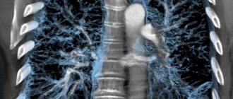
Computed tomography is an external scanning technique that is based on scanning the human body with X-rays. Multiple special sensors determine the density of the radiation flux. They catch the moment when it weakens. The scan results, unlike conventional radiographic images, are processed on a computer. The central processor collects the received information and analyzes it. The output is a photograph with an accurate image in three planes, the step of which is 1 mm.
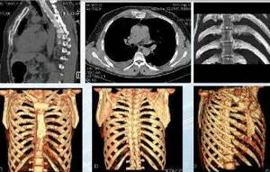
Chest CT is a reliable diagnostic method for organ pathologies, consequences of injuries and diagnosis. The resulting data is interpreted by a radiologist. They are written to disk. The conclusion is also written on paper. Everything is then given to the patient. Now a new type of chest CT scan is gaining popularity, where the step cut is 0.5. It's called multi-computed tomography.
The first experience of use took place in 1971. Subsequently, the devices were improved, and the speed of their distribution increased. Their technical equipment was improved. Modern scanners allow you to take pictures in a fraction of a second.
Why is a chest CT scan done?
This is a complex type of study used in cases where other types of diagnostics have not helped determine the cause of the disease. It is used when it is necessary to confirm or exclude a presumptive diagnosis. The chest is scanned with a CT scan to track pathologies of the intrathoracic organs, bones and vascular system. This helps to accurately identify the affected area and prescribe correct and timely treatment. Since this area of the body contains vital organs, the heart, the lungs, a timely examination often saves the patient’s life.
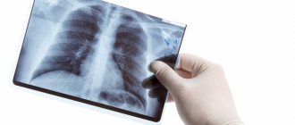
The attending doctor should definitely refer you for an x-ray. Sometimes this is enough for diagnosis.
When is a chest CT scan ordered?
Scanning is used in a wide range of cases:
- various reasons for emergency hospitalization of a person;
- clarification of the diagnosis identified on the basis of standard radiography or fluorography;
- for chronic pathologies in the lungs;
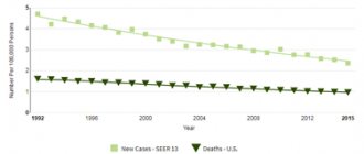
- oncological diseases (since tomography is quite sensitive, it is able to recognize the disease even at the earliest stage, show the location of tumors, confirm the presence or absence of metastases);
- any neoplasms (shows what kind of tumor, nature and malignancy);
- suspicion of pneumonia or inflammation of the pulmonary lobes (when there is a painful cough, increased body temperature, shortness of breath);
- control of oncology treatment (after medications and chemotherapy, when you need to track the dynamics);
- after injuries and bruises of the sternum (detect the presence of cracks and fractures, ruptures of organs and internal bleeding);
- checking the cardiovascular system (if thrombosis or deposition of cholesterol plaques is suspected);
- diagnosis of the condition of the esophagus, trachea and mediastinum;
- pathologies in the thoracic spine;
- cutting, stabbing or squeezing pain behind the sternum;
- if a child inhales or swallows a small object.
Are there any risks with a chest CT scan?
- The patient may ask their healthcare provider about the amount of radiation used during a CT scan. You should tell your doctor about previous CT scans and other types of x-rays. Risks of radiation exposure may be associated with the total number of x-ray examinations or procedures over a long period of time.
- Pregnancy is a contraindication for CT scanning. Radiation exposure during pregnancy can cause birth defects. When breastfeeding, it is also necessary to inform your doctor.
- If contrast dye is used, there is a risk of an allergic reaction to the dye. You should tell your doctor if you have ever had a reaction to contrast dye or had kidney problems.
- If a patient has kidney failure or other kidney problems, they should tell their doctor. In some cases, contrast dye can cause kidney failure. This is more likely if the patient has serious kidney problems or is dehydrated.
- If a patient takes a diabetic medicine called metformin with contrast, there is a risk of developing metabolic acidosis. This is a condition in which there is an unsafe change in the pH of the blood. People with kidney disease are more likely to experience kidney damage after exposure to contrast.
- There may be other risks depending on your specific medical problems. Before the procedure, you must make sure that the attending physician is aware of all medical problems.
Several factors may reduce the accuracy of a chest CT scan. These include:
- Barium in the esophagus after a recent barium study
- Chest piercing
- Metal objects in the chest, such as surgical clamps or a pacemaker
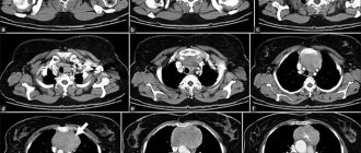
Diseases and disorders in the chest organs, which are determined by CT
What the study shows:
- deviations in the functioning of the pericardium;
- mediastinal abnormalities;
- the appearance of a liquid substance in the pleural area;
- heart and aortic diseases;
- fractures and cracks of the costal arches;
- mid-spine injuries;
- organ perforation;
- internal bleeding;
- pathologies of the joints of the shoulder girdle (when there is pain and impaired mobility);
- chondritis (inflammation of cartilage);
- benign and malignant tumors of organs and bones;
- tuberculosis;
- pneumonia;
- cardiosclerosis of the heart vessels;
- autoimmune diseases;
- heart attacks and strokes;
- lymphogranulomatosis;
- angina pectoris;
- ischemia;
- vascular atherosclerosis;
- diffuse changes in the mammary glands;
- bronchiectasis (the process of irreversible dilatation of the bronchi).
What does diagnostics show?
This method allows you to visualize:
- The presence of effusion (blood, pus, ichor) in the pleural cavities.
- Pneumothorax (air in the pleural cavity).
- Infectious diseases - pleurisy, mediastinitis, pneumonia, tuberculosis.
- Lung abscess.
- Adhesions in the pleura.
- Thrombosis.
- Cysts.
- Lymphogranulomatosis and other systemic diseases.
- Bronchiectasis.
- Cancer of the mediastinum, lungs, chest wall.
- Emphysema.
- Aortic aneurysm.
- Vascular pathologies, for example, malformation, narrowing of the lumen.
- Interstitial lung diseases and disseminated processes.
- Pulmonary embolism.
- Heart attack.
- Enlarged lymph nodes.
- Lymphadenopathy.
- Congenital and acquired anomalies such as emphysematous, paralytic, rachitic, pectus excavatum, scaphoid or kyphoscoliotic chest. As well as fusion or bifurcation of ribs.
The introduction of contrast improves the quality of visualization. This substance can accumulate in pathological soft tissues, allowing the disease to be detected at a very early stage. Bolus administration of the substance improves information content in the study of diseases of the vascular system. It also shows:
- Borders, shape and size of pathological foci.
- Disruption of the blood-brain barrier.
- Condition of vascular grafts and anastomoses.
Preparing for a computer test
It is not prescribed to everyone, as it has absolute and relative contraindications. It is strictly prohibited for people with absolute health conditions, as it can cause harm to health.
These include:
- patients who have pacemakers, neurostimulators, pacemakers and other types of pacemakers installed;
- pregnant women (especially in the early stages);
- children as long as they are under 14 years of age (due to the intensity of radiation exposure. Permitted only in cases where there is a threat to the child’s life);
- those suffering from claustrophobia (fear of narrow and closed spaces, since a panic attack may begin in the tomograph and the scanning will have to be stopped. In some cases, diagnostics are carried out under sedation or anesthesia);
- if there are metal objects in the body (implants, plates, pins);
- presence of vascular stents, clips;
- diagnoses of mental disorders (in which a person behaves inappropriately);
- high body weight (over 130 kg);
- the presence of hyperkinesis and convulsions that cannot be controlled with sedatives;
- barium suspension in the digestive system (may distort the quality of images).
Foreign objects in the body distort the image or interfere with its detailed examination.
Make sure you are not included in the group of prohibiting factors.
Relative prohibitions imply a temporary restriction on research. Permission must be given by a doctor. Among them:
- problems with the thyroid gland;
- patients with an allergy to a contrast agent (during examination with contrast);
- cancer of the circulatory system;
- severe diabetes mellitus;
- pathologically complex forms of asthma;
- severe myocardial dysfunction;
- insufficiency of the kidneys (in case of administration of contrast).
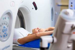
No preparation is required for a CT scan. If the diagnosis is planned without contrast, there are no special preparations for it. A medical specialist will instruct you about the features and relieve unnecessary worry and anxiety. This is very important for those who came to the procedure for the first time. Bring loose clothing. Sometimes clinics provide a disposable shirt. All metal objects on the body, including jewelry and piercings, must be left in the changing room. When using intravenous contrast, take the last meal 4 hours before the procedure. You may experience unpleasant symptoms such as fever, itching, mild shortness of breath, and rapid heartbeat. This is the body's reaction to the drug. Its manifestations will quickly disappear. In some cases, this will take a day. Afterwards, you should drink more clean water so that the substance is eliminated from the body as quickly as possible. The whole procedure will take a few minutes. In some cases where contrast is used, it can take up to an hour. You will have to lie on your back on a mobile couch. The body will be secured with straps. After which it will slide into the cylindrical apparatus. You cannot move during the scan. In the next office, the doctor will monitor the entire process. Interaction with it is carried out via voice communication. Listen to the staff's instructions. You may be asked to roll onto your side or hold your breath. The device is noisy, so you can use earplugs or headphones.
When everything is over, the results can be collected on the same day. They will be recorded on a disc, which your personal doctor will be able to view when you come to see him. In addition, the radiologist will attach the data on paper.
How is the examination carried out?
During the examination, the patient is positioned horizontally on a special table - he will have to lie still. This is not difficult, given that the research takes very little time. The table is connected to the scanning elements of the tomograph, which looks like a ring. The scanner rotates and emits X-rays. Due to differences in tissue density, it becomes possible to image different organs. Typically, the patient is required to take a breath and hold it for a few seconds - at this moment the scan takes place. If the photo is taken, the examination ends. Thus, the entire procedure takes a few minutes.
There is also a computed tomography technique using a contrast agent. The essence of the method is that the patient is injected intravenously with a radiopaque substance, which helps to obtain a clearer image. Before a CT scan with contrast, additional blood tests are prescribed. There are no unpleasant sensations during the examination.
Advantages
Application experience shows that the method is safe, since the radiation exposure, unlike with an X-ray machine, is small. It is allowed to be carried out once or twice during the year. If absolutely necessary, repeat three times with a period of four weeks is possible. Among the advantages is also the duration of the procedure. It is less than with MRI. The price for a chest CT scan is about the same. Compared to X-rays, scanograms can be reconstructed. Tissue differentiation is better. The method is distinguished by high sensitivity.
Contraindications
This is a radiation study, so the limitation for use in women is:
- Pregnancy at any stage. Ionizing radiation is primarily dangerous for rapidly dividing fetal cells. It can cause developmental abnormalities and severe pathologies in a child.
- Lactation. Contrast agents pass into breast milk. If tomography is prescribed for vital reasons, it is recommended to interrupt breastfeeding for 48 hours.
Common contraindications for both men and women are:
- The patient's body weight exceeds the load capacity of the tomograph table.
- Childhood. If the benefit of diagnosis outweighs the possible risk, a referral for examination is issued by doctors after a consultation.
- Liver, kidney and heart failure. These pathologies make it difficult to remove radiopaque contrast from the body.
- Allergy to iodine. It is included in most contrast agents.
- Decompensated diabetes mellitus.
- Thyroid diseases.
- Bronchial asthma in severe form.
- Hyperkinesis, in which the patient is unable to remain motionless for a long time.
- Psychoneurological diseases in the acute phase.
If you have claustrophobia and panic attacks, it is better to choose ring tomographs.
How much does a chest CT cost?
In medical clinics in the capital, prices for chest CT scans start at a lower threshold of 3,000 rubles. It all depends on where the specific center is located, as well as the characteristics of the tomograph, the experience and skills of the staff. The better and more advanced the equipment, the higher the cost of the procedure. It is up to you to decide where you want to have your chest CT scan done. To be aware of how much it costs to do a chest CT scan, use our service.
What is the difference between CT and MRI
This is the most common question asked to a radiologist. CT and MRI are diagnostic methods. They have similarities and differences. The similarity lies in the external similarity in the design of the devices themselves - tomographs. In their center there is a tunnel into which the table slides.
The main difference is a completely different principle for obtaining images: CT uses x-rays, MRI uses a magnetic field. Which method is right for you is determined by your doctor, taking into account the specifics of the disease and the purpose of the study.
Where to get a chest CT scan in Moscow
There are dozens of clinics in the capital that have a tomography scanner. We own information about all institutions and have collected only real reviews from visitors to medical centers. The location of institutions and the price list for chest CT scans in Moscow can be viewed on the map on our website yourself. Contact us and we will tell you where to get a chest CT scan. We will answer all your questions, tell you about promotions and discount bonuses. Call the number and our consultants will select the best offer for you free of charge. Both in price and location.
CT diagnostic services at CELT
The administration of CELT JSC regularly updates the price list posted on the clinic’s website. However, in order to avoid possible misunderstandings, we ask you to clarify the cost of services by phone: +7
| Service name | Price in rubles |
| CT scan of the abdominal aorta and its branches and screening examination of the abdominal and pelvic organs | 14 000 |
| CT coronary arteries | 16 000 |
| CT scan of the spine (1st section) | 7 500 |
All services
Make an appointment through the application or by calling +7 +7 We work every day:
- Monday—Friday: 8.00—20.00
- Saturday: 8.00–18.00
- Sunday is a day off
The nearest metro and MCC stations to the clinic:
- Highway of Enthusiasts or Perovo
- Partisan
- Enthusiast Highway
Driving directions
Preparation
No special training required
General requirements for contrast-enhanced CT:
To perform the test, you must have the result of a blood test with you.
for creatinine, the statute of limitations is no more than 30 days from the date of submission of the biomaterial.
Be sure to tell the radiologist before the examination with contrast:
- Do you have allergies to iodine, medications or food?
- whether you have diabetes, asthma, heart or thyroid disease
How is a CT scan performed?
The examination is usually performed lying on your back, rarely - lying on your side or stomach. To maintain correct or still body position during the examination, you may be asked to place your arms under your body or raise your arms above your head.
If it is necessary to use a contrast agent, it is administered either orally (orally - swallowed) or administered intravenously, depending on the type of study being performed.
You may be asked to hold your breath during the scan. Any slightest movement of the body can lead to artifacts in the images. After the test is completed, you may be asked to wait a short time while the radiologist checks the quality of the image.
Typically, a CT scan takes about 30 minutes. The administration of a contrast agent during the study lasts from 10 to 30 seconds.
What sensations arise during the study.
A CT scan is a painless and quick procedure that is easily tolerated by the patient. Although the scan itself is not painful, there may be some discomfort from having to remain still for several minutes. If you have pain and find it difficult to lie still, your doctor may suggest painkillers. When a contrast agent is administered intravenously, you will feel a rush of warmth and a slight metallic taste in your mouth. These sensations last no more than two minutes.
The contrast agent may taste unpleasant when administered orally, but almost all patients tolerate the procedure well.
After the CT scan, the patient can immediately return to his normal routine.
Indications
- A more detailed study of pathological processes detected during standard chest radiography;
- Diagnosis of the causes of symptoms such as cough, shortness of breath, chest pain, long-term high body temperature, etc.;
- The need to exclude oncological diseases, such as a primary tumor of the lungs and mediastinal organs, secondary damage to the lungs in the presence of a tumor of another location, damage to the lymph nodes of the chest and mediastinum;
- Assessing the effectiveness of cancer treatment results, including after radiation or chemotherapy; determining the presence of free fluid in the pleural cavity;
- Assessment of the condition of the lungs, blood vessels, bone structure (ribs, spine) for injuries of varying severity;
Benefits and risks of CT
Advantages
The results of computed tomography allow doctors to make the most accurate, correct diagnosis and timely prescribe treatment. Computed tomography is a non-invasive, painless diagnostic method. Carrying out CT scans very often reduces the required number of studies to a minimum and eliminates the advisability of using invasive techniques.
CT scan:
— allows you to study the state of the bone structure, soft tissues, blood vessels and individual organs layer by layer; - often used in emergency situations, for post-traumatic disorders, for suspected damage to internal organs, bleeding; — can be used if the patient has implanted medical devices of any kind; — used for control during biopsy of organs and pathological areas of the human body. X-rays from a CT scan have no immediate side effects.
Estimated risks and contraindications
The effective dose of radiation from computed tomography is minimal, however, it is always present. The radiation dose is usually specified in the study protocol. In the interests of accurate diagnosis, especially in acute cases, this risk is mitigated. Women are required to inform the referring physician and radiologist about the possibility of pregnancy. Because of the potential risk to the baby, CT scans are not recommended for pregnant women unless there are specific medical indications. It is extremely rare, but allergic reactions to a contrast agent that contains iodine do occur. In such cases, PATERO CLINIC has all the necessary means to stop such reactions.
Manufacturers of intravenous contrast media do not recommend that mothers breastfeed for 24 to 48 hours after receiving or administering contrast media.