Every person wants to have beautiful and straight teeth. Modern dentistry offers many options for eliminating dental and bite defects: from polymer coating to installing removable and fixed dentures. Magnetic resonance imaging (MRI) doctors often encounter patients who have crowns, braces, or dental implants in their mouths. Can such patients undergo an MRI procedure? To answer this question, we should delve a little deeper into the principle of MRI and the magnetic properties of some substances.
MRI is based on the phenomenon of magnetic resonance of hydrogen protons in water molecules under the influence of a magnetic field. After the cessation of magnetic influence, the protons relax and emit magnetic resonance pulses, which are detected by the sensitive detectors of the tomograph. The computer processes the received pulses and converts them into a digital image.
When exposed to a magnetic field, some substances and bodies exhibit magnetic properties. These substances are called magnets. There are three types of magnetic materials (according to their magnetic susceptibility and degree of response to an external magnetic field):
- paramagnetic substances are substances with weak magnetic susceptibility and are weakly attracted to a magnet. Without exposure to a magnetic field they do not magnetize. These include oxygen, nitric oxide, aluminum, sodium, magnesium, tungsten, chromium, platinum;
- Diamagnets are also weakly magnetic substances, but they are repelled by a magnet. Without exposure to a magnetic field they do not magnetize. These include nitrogen, carbon dioxide, water, copper, silver, gold, zinc;
- Ferromagnets are highly magnetic substances. They have residual magnetization. Under the influence of a magnetic field they can shift and heat up. These include iron, nickel, cobalt, and some silver alloys.
Whether it is possible to do an MRI on patients who have objects made of metals in their mouth depends on what type of magnet they are.
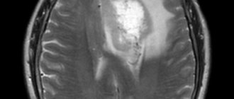
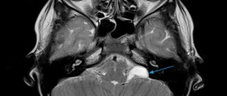
How MRI works
The final result of an MRI examination is an image of the human body “from the inside”, created using magnetic waves. A magnetic field is formed in the device, in which the patient is placed, and the computer records the return signal emanating from the molecules of his body.

The resulting image is not just a photograph. Magnetic pulses create a three-dimensional layer-by-layer image, that is, the body can be viewed at different depths and from different angles. The method allows you to obtain accurate information:
- about the location of neoplasms, even small ones;
- about micro-strokes and hemorrhages;
- vascular deformation, aneurysm and multiple sclerosis, invisible to other types of scanning;
- intervertebral hernias not detected on other devices.
When is CT scanning necessary in dentistry in general?
In dentistry, the use of computed tomography is justified for diagnosing the maxillofacial apparatus as a whole and its individual sections. Applicable in the following cases:
- to identify tooth retention, abnormal bite development, deviations in the location of teeth,
- detection of tumors (even small ones) in soft tissues,
- assessing the condition and volume of bone tissue,
- determining the localization of foci of inflammation and injury,
- studying the entire maxillofacial system before carrying out major surgical interventions to accurately determine the location of nerves, vessels, and important organs. These include implantation, prosthetics, and orthodontic treatment.
Effect of MRI procedure on implants
Since the examination is based on the use of magnetic waves, many patients have a logical question: is it possible to do MRI with dental implants? These concerns are understandable, but not always justified.
In dentistry, as in any other area of medicine, prosthetic technology is constantly evolving, new materials and techniques are appearing. So, if previously dental crowns were made of an alloy of copper and gold, today you will be offered metal ceramics or titanium alloy.
What is the difference? Old copper-gold technology produced distortions in MRI images
, but did not affect the health or condition of the patient during the procedure. Today in dentistry, titanium with small admixtures of other elements is widely used, which makes the prosthesis lighter and more durable. By its nature, titanium is chemically inert, therefore it does not oxidize, does not emit harmful substances and does not react to magnetic waves, which means it does not distort images. All this applies not only to teeth, but also to prosthetic bones and joints.
Metal-ceramics during MRI also does not affect either the state of health or the diagnostic result.
But, in addition to the implant itself, there are also pins, plates and screws on which it is attached. They may be the problem. Implant fastening parts are made from various ferromagnets and special iron alloys
, which have positive magnetic susceptibility. Exposure to a magnetic field can produce distorted results and affect the patient’s well-being during the procedure.
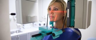
How to do a 3D examination
A dental 3D tomograph is a vertical open platform, in the upper part of which there are rotating blades.
The patient will:
- Stand in the center of this apparatus so as to place your chin on a special stand.
- Lightly bite down on a small dental plate with your teeth and grip it with your teeth.
- Remain still while the machine rotates.
The entire procedure takes no more than 40 seconds for a full examination, 10 seconds for examining the area of several teeth.
The quality of the images will depend on the patient's immobility during scanning.
How do iron parts of implants affect different areas during MRI?
Regardless of the area of examination, before the procedure it is necessary to remove all metal objects:
- buttons,
- rivets,
- hooks,
- lightning,
- buckles,
- keys,
- coins,
- keychains,
- decorations,
- watch,
- Cell phones,
- magnetic media (cassettes, floppy disks),
- credit cards.
In addition, it is necessary to warn medical personnel about the presence of implants or prostheses in the body.
, especially those containing metals. During an MRI of the head and cervical spine, the iron alloys in the denture fixtures can heat up and move. This can lead not only to distorted diagnostic results, but also to discomfort or possible injury to the subject. However, such an outcome is only possible if there are large fragments of metal in the body, and the mass of the pin or plate is negligible. Therefore, even with a large number of implants, the occurrence of such a situation is almost impossible. And certainly, the presence of an implant in the body is not a reason to refuse an MRI.
The presence of metal fasteners in dental implants does not have any effect on other types of MRI - bones, joints, thoracic and lumbar spine. Direct MRI diagnostics of teeth is rarely prescribed; it is difficult to “get close” to the jaw using this tomograph.
And MRIs of the limbs, lower back and spine are without consequences. Dental diagnostics using this method is rarely prescribed - it is difficult to “get close” to the jaw using MRI.
Is it harmful to undergo a CT scan and to what extent?
The radiation exposure of dental tomographs is about 50–60 μSv (microsieverts), which, according to WHO standards, allows CT examinations to be carried out about 20 times a year (no more than 1000 μSv). This is a safe diagnostic method. For comparison, a person in ordinary life receives about 400 μSv per year from food and water, and from concrete walls, sidewalks and roads we receive about 30 μSv per year. But for pregnant women and nursing mothers, X-ray examination is carried out strictly according to indications - however, dental implantation is prohibited during these periods of life.
Myths about the MRI procedure
What not to believe
when going for an MRI:
During the procedure, the implants heat up and move.
The specific weight of the metal part of the implant fastening is negligible, which means there will be no defects in the images or discomfort for the patient. But it is still worth warning medical personnel if it consists of zirconium dioxide, pure ceramics or expensive alloys, there are no contraindications for MRI, but if it is metal ceramics, it is necessary to clarify what metals and impurities the implant consists of. If the composition contains a lot of ferromagnetic metals, the image quality will most likely be reduced and it will be impossible to evaluate the structures behind the structure.
You should not do an MRI during pregnancy.
This is only partially true. In the first trimester of pregnancy, the laying of tissues and vital organs of the child occurs, so any physical, chemical and biological effects should be excluded. Therefore, MRI during this period is performed only for emergency indications. However, in the later stages, diagnosis makes it possible to determine the position of the child, identify a narrow pelvis in a pregnant woman and enable the obstetrician to prepare for the correct delivery.
Going through an MRI with prosthetics is painful.
No, this is an absolutely painless and safe procedure. You can undergo MRI even with titanium plates; they are not affected by the magnetic field and cannot cause harm. The procedure can only be denied if a steel plate is implanted in the patient's body, since it heats up and moves under the influence of a magnetic field, which can injure the patient.
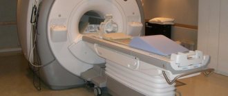
Computed tomography – what is it and why is it necessary in dentistry
Dental computed tomography is the process of obtaining a layer-by-layer three-dimensional image, which is formed using a special apparatus - a tomograph - and makes it possible to accurately assess the anatomical features of the structure of the oral cavity and adjacent areas.
New teeth in 1 day - All-ON-4 - 180,000 rub.
All inclusive!
3D modeling of the structure with a prosthesis, implantation of 4 Osstem implants, installation of a fixed prosthesis on the same day. Free consultation with an implantologist +7 (495) 215-52-31 or write to us
In dentistry and implantology in particular, CT has been used relatively recently. At the same time, the method has become a priority [1] in comparison with other currently existing clinical and laboratory diagnostic methods due to its informativeness, ease of use and quick receipt of high-quality results.
The image - tomogram - provides a detailed image of the teeth, roots and root canals, allows you to accurately determine the volume of bone tissue, identify atrophy of the bone necessary for installing an implant, simulate the installed implant in the bone structure, and also calculate its location and direction in the jaw.
In dentistry, in addition to CT, two other types of X-ray examinations are also used. This is an x-ray - a targeted image of 1-2 teeth, and an orthopantomogram (OPTG) - a flat panoramic image of the jaws.
What could be the real reason for refusing an MRI?
- Claustrophobia. The patient is placed in a closed apparatus for a short time, which can provoke a panic attack. But here it all depends on the degree of claustrophobia and the type of examination. In addition, each device has an “alarm” button, when pressed, the procedure immediately stops and medical personnel approach the patient.
- An installed pacemaker may come into dangerous resonance with magnetic waves and stop working.
- Before starting the examination, the hearing aid must be removed, since the magnet in the hearing aid may be damaged under the influence of a magnetic field. And the presence of middle and inner ear implants is an absolute contraindication, since they often contain metal alloys with strong magnetic properties.
- An insulin pump is an absolute contraindication for MRI.
Before the procedure, you must tell your doctor
about everything that may affect your health and the diagnostic result. You can’t hush something up for fear that an MRI won’t be done. As a rule, any problem can be solved, and in the case of metal prostheses and implants in the patient’s body, the doctor will simply adjust the tomograph so that, taking into account the location and composition of the prosthesis, the device will produce the highest quality clear image. If there are absolute contraindications to MRI, the doctor will select the most informative and permitted type of examination in your case.
If the patient “forgets” about the fake ideal smile, the pictures will turn out blurry, and he will have to undergo an MRI again, having honestly admitted all the interventions in the body.
If you are planning to undergo MRI diagnostics from qualified specialists, using modern expert-class equipment in comfortable conditions and in the shortest possible time, call the phone number listed on the website, or leave a request in the feedback form. Medical specialists will answer your questions and conduct all necessary examinations within one business day. Let's take care of your health together!
Types of three-dimensional research using tomographs
Tomographs have evolved since 1974, when the first device appeared, developed by scientists Allan Cormack and Godfrey Hounsfield, who received the Nobel Prize for its invention. Modern dental devices are the fifth generation of tomographs. They are divided into three types:
- sequential: consists of layer-by-layer scanning of an area with a step of at least 1 mm. The gaps between steps are not covered, but are simulated by the computer based on available data (interpolation), which makes the result not very accurate. This method has practically been driven out of practice,
- spiral: the method is so called due to the movement of the emitter in a tight spiral around the area of interest,
- cone beam: The x-ray beam is delivered in a cone shape and is emitted continuously, there are no steps. The latter is used so far only for diagnosing the maxillofacial area and appeared already in the 21st century.
An even more accurate device used for dental implantation is a multislice computed tomograph (MSCT). In it, the detectors are located not in one row, but in several: if the first double-spiral (double-slice) tomographs appeared in 1992, now there are already 640-slice tomographs with two X-ray tubes. All information can be obtained in half a turn, and for a full turn it doubles, making the result even more accurate.
Such systems are not available in all dentistry and are used in complex cases for precise diagnostics (for zygomatic implantation, for example) in specialized centers. What CT scan is needed for implantation? For dental practice in all its diversity, a cone-beam examination is quite sufficient, but sometimes implantologists refer patients to MSCT. Which CT scan should you choose? Here everything will depend on individual indications - therefore, before undergoing the examination, it is better to consult a doctor (visit the clinic in person or address the problem online).
Why is a CT scan needed for implantation, and an OPTG image is not enough?
An orthopantomogram (OPTG) is a flat scanned image of both jaws, which is also widely used in dentistry. But for implantation, especially when it comes to restoring all teeth and using immediate loading methods, OPTG data is not enough. This is due to significantly less detail of the structures, the inability to consider sections of sections and planes other than the frontal one. And when assessing areas of bone atrophy or the presence of tumors, this is the most important information that cannot be missed.
Simply put, for a complete picture and analysis, the image needs to be “rotated”, examined from different angles, and volumes examined, which a flat OPTG does not allow - CT is an order of magnitude more informative. This is a more professional and modern type of jaw examination, indispensable when an image in several planes is required, which is especially important before full dental implantation.
Computed tomography for dental implantation is performed both before the installation of implants and after surgery. Initially, CT is needed to create a 3D image as part of preparation and diagnosis. Subsequently, for the use of X-Guide digital navigation during implant installation, as well as for analyzing the work performed. During annual examinations in the clinic, after dental implantation, CT scans are also performed to assess the condition of the bone tissue near the implants.
Validity period of CT result
How long does a CT scan last before dental implantation? Here, the validity period of the examination is longer than that of a blood test, for example. But how long jaw tomography lasts will depend on the rate of bone tissue atrophy in the patient, the presence of inflammation and neoplasms under the roots of the remaining teeth. It is desirable that a minimum amount of time pass between the CT scan and the implantation itself - literally 1-2 weeks (which are needed for preparation and planning).
DENTAL PROSTHESIS WITH 4 OSSTEM IMPLANTS FOR 1 JAW - RUB 130,000.
Implantation even with bone tissue atrophy. Work guarantee! Save RUR 20,000.
on promotion >> Free consultation with an implantologist +7 (495) 215-52-31 or write to us
If you undergo a tomography and do not install an implant in the near future, then within 3-6 months the bone in the area of the extracted tooth will sag by several millimeters. Then, according to the existing plan, it will not be possible to place the implant perfectly, and complications may even begin. Therefore, a fresh tomography and new planning will be required.
Advantages of computed tomography over other methods
- diagnostic accuracy: the image allows you to obtain an unlimited number of highly detailed sections to calculate the height and thickness of the bone tissue required for installation and fixation of a dental implant,
- formation of a 3D model of the jaw on a scale of 1:1, which makes it possible to study all structures in the most thorough manner,
- safety, painlessness, non-invasiveness of the method,
- the possibility of high-quality endodontic treatment of teeth, detection of hidden foci of inflammation in soft tissues,
- saving time: one image is enough for in-depth study, since the finished image can be stored in a computer database for an unlimited time,
- guarantee of results: a three-dimensional model increases the information content of the study several times, thanks to which the doctor has the opportunity to build a treatment plan most effectively,
- the versatility of using a three-dimensional image, which can be used as a model for planning an operation, modeling a prosthesis, as a simulator for working out a surgical intervention, and more.
Computed tomography of the jaw is indispensable in 3D dental implantation. This method involves modeling the step-by-step process of implant installation on a computer and gives the patient the opportunity to evaluate the expected result before the operation begins.
For more information about preparation for implantation and the use of computed tomography for this, read the material “Consultation, diagnostics and tests.”.