- What is a biopsy for cancer?
- When is a biopsy ordered?
- How the procedure is performed
- How the material is examined
- Preparing for the study
- Contraindications for biopsy
- Advantages of punch or needle biopsy
- Cancer biopsy in oncology
Trillions of cells function in the human body every day.
Each of them has its own development cycle. They usually grow and divide. When they age or are damaged for some reason, they naturally die off so that their place can be taken by equally new ones. When cancer develops, this orderly process is disrupted. Cells become abnormal, old or damaged cells continue to function, and new ones are formed even when they are not needed. For some reason, the immune system does not have time to react and block such division. Thus, additional cells multiply without stopping and combine to form tumors.
Some types of cancer are malignant formations; they represent a certain mass of tissue that can grow into neighboring organs or put pressure on them, which gives clinical manifestations. Also, as cells grow, they can separate from the primary tumor, spread through the lymphatic or blood vessels and form new foci - nearby or distant metastases.
Unlike malignant tumors, benign tumors do not spread or invade nearby tissues. However, they can reach significant sizes. After their removal, relapses may develop.
What is a biopsy for cancer?
To confirm the diagnosis of cancer, it is necessary to do an analysis for the presence of cancer cells that differ from normal ones in structure, degree of differentiation and size.
Cell biopsy is the basic test for suspected cancer. It involves removing a tissue sample for study. For such analysis, it is possible to remove a small fragment with a needle or surgically.
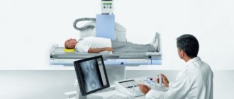
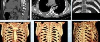
Most biopsies are performed on an outpatient basis, without special preparation. This is usually done under the guidance of ultrasound, x-ray, CT or MRI. Such visualization makes the study targeted and safe.
Meaning of Methods
A puncture differs from a biopsy in that the first is one of the methods for performing the second. Both of these terms do not denote research, but a method of obtaining material for further study.
Based on the results obtained, the patient is given an accurate diagnosis and treatment is prescribed. The patient’s life may depend on the timeliness of the examination.
If the doctor considers it necessary to perform a biopsy, then there is no other way to clarify the nature of the detected pathological formation. How the biopsy will be performed is determined on an individual basis.
The points are sent for cytological examination, which provides information about the cellular structure of the neoplasm. Biopsies are subjected to histological examination, which allows us to learn everything about the morphology of the tissue.
When is a biopsy ordered?
The procedure is indicated when a suspicious formation is detected. It allows you to determine the degree of malignancy, nature and structure. In some cases, it helps to identify other conditions: infections, inflammatory and autoimmune disorders.
It is possible to examine almost any organ or tissue:
- Abdomen. The sample is collected percutaneously by needle (under ultrasound or CT guidance), surgically using a laparoscope, or during open surgery.
- Bones. A biopsy is used to diagnose cancer or infection in the bones. Also performed through the skin with a needle or surgically.
- Bone marrow. The procedure diagnoses malignant neoplasms of the blood system. A small sample of bone and bone marrow is removed using a needle during a core biopsy.
- Breast. It is possible to take a biopsy in several ways: puncture, stereotactic and others.
- Endometrium. The study is prescribed to find the cause of abnormal uterine bleeding, to study the mucous layer of the uterus and to diagnose cancer. It is carried out during diagnostic curettage or using a needle.
- Kidneys. Used to study the condition of an organ with renal failure, inflammation, or if a malignant tumor is suspected. Most often this is a needle biopsy, which is performed using ultrasound or CT.
- Liver. Hepatitis C, cirrhosis, infections and cancer are diagnosed. A needle biopsy is performed through a catheter or during surgery.
- Lungs and mediastinum. The procedure can be performed using bronchoscopy, percutaneously or surgically.
- The lymph nodes. Their examination is carried out during each operation to remove a tumor, and a needle biopsy is also possible.
- Muscles. Diagnosis of infections, connective tissue and vascular diseases is carried out. A needle is used or the sample is surgically removed.
- Leather. A sample of altered tissue is examined. A skin biopsy can be performed by scraping, using a scalpel, or other means.
- Testicles. They are studied to identify the nature of the neoplasm and the causes of male infertility. Most often, tissue collection is performed surgically.
- Thyroid. The biopsy is performed using an ultrasound-guided needle.
Types and characteristics of biopsy
This procedure belongs to the category of diagnostic operations, during which the doctor, using special instruments, removes a small amount of tissue from the tumor for further analysis in the laboratory. The vast majority of cancers are most accurately diagnosed by biopsy. But, in addition to diagnosing cancer, this procedure is also advisable for dystrophy or an inflammatory process.
Biopsy, depending on the method of tissue sampling, is divided into the following types:
- Excision. When tissue is collected, all pathological formations are removed.
- Incisional. A small portion of tissue is removed from the formation.
- Puncture. A small portion of tissue is removed using a syringe.
- Aspiration. Instead of tissue, a small volume of liquid is withdrawn using a syringe.
- Endoscopic. An endoscope is used to collect tissue (for example, when examining the stomach).
How the procedure is performed
Biopsy differs in the methods of collecting material (biopsy):
- Puncture. The material is taken using a hollow surgical needle directly from the lesion. Suitable for biological fluids or cell collection. A less traumatic modification is possible - fine-needle biopsy.
- Aspiration. The collection takes place using a special vacuum device.
- Imprint smears or scrapings of material from the mucous membrane.
- Endoscopic. The sample is taken using special endoscopic equipment. Used to study the gastrointestinal tract, uterus and other organs.
- Sighting. Pathological tissue is examined after collection with special forceps.
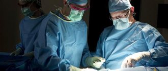
- Excision. A surgical method of completely removing the affected organ or just the tumor.
- Incisional. During surgery, part of the tumor is removed.
- Stereotactic. Suitable for collecting material from hard-to-reach areas, the process is controlled using visualization.
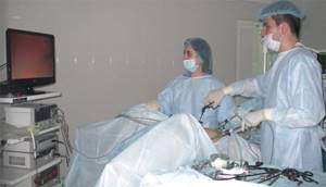
- Laparoscopic. The procedure is performed during diagnostic laparoscopy (examination of the abdominal cavity) or thoracoscopy (chest cavity).
The biopsy usually takes place on an outpatient basis under local anesthesia. Sometimes performed during surgery (intraoperative). Its results may influence further patient management tactics.
How is a biopsy performed?
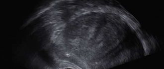
The patient is in a lying position during the manipulation. First, the doctor performs anesthesia. In most cases, local anesthesia is sufficient. The specialist performs a control ultrasound and determines the exact location of the tumor. After the anesthesia takes effect, the doctor will collect tissue from the pathological area. Most often, the patient does not experience pain, but may feel slight pressure, burning, and tingling. When the material is collected, the doctor removes the needle and treats the puncture site with an anesthetic. In some cases, it is recommended to apply cold for 30-40 minutes.
How the material is examined
Once the tissue is collected, it is sent to a laboratory for analysis. There, a histological (if possible) and cytological examination is carried out.
- Histology. After special processing with a microtome (special knife), very thin sections of tissue are made, which, after staining, are examined under a microscope. The structure of the tumor is studied, the presence of pathologically altered cells that absorb dyes more intensely than normal cells is determined.
- Cytology. Smears containing cellular material are examined under a microscope. The size, degree of differentiation and other characteristics of the cells are determined. This is a less informative study than a histological one, since the cells do not always fall into the field of view.
Peculiarities
All cells of the human body have a certain structure. With the development of malignant neoplasms, the structure of cells is disrupted. These changes can be detected under a microscope. This means that a biopsy can accurately confirm or refute the presence of cancer.
The study is accessible and can be carried out at any age. There are no contraindications for it. It is important that the result is obtained within a few days, which allows minimizing the psychological stress in patients associated with long waits.
Biopsy is an invasive procedure. This means that local anesthesia must be used when collecting biomaterial. This does not exclude difficulties in extracting tissue samples.
When performing a biopsy, complete information about the developing pathology is obtained. The procedure allows you to determine the nature of the tumor and prescribe the correct treatment.
Preparing for the study
Before doing a biopsy, laboratory and instrumental research methods are performed that help in diagnosing the disease.
If an outpatient procedure is planned, then special preparation and hospital stay are not required. The patient is advised not to eat or drink before the test if anesthesia is required.
Anticoagulants and other drugs that can cause bleeding in the postoperative period are temporarily discontinued. The doctor may also prescribe sedatives.
Carrying out an incisional biopsy
When conducting this research method, the doctor removes a small part of the soft tissue from the formation under study. To more accurately determine the condition, sampling is done from the center of the tumor. For this purpose, the progress of the operation is monitored by ultrasound. Incisional biopsy is not a curative procedure, but is classified as a surgical procedure.
Due to significant pain during incisional biopsy, it is advisable to use general anesthesia or local anesthesia. After the operation, stitches are placed on the operated area.
Incisional biopsy makes it possible to conduct an accurate diagnosis in the early stages of the disease (determine the nature of the tumor, its growth rate, etc.). Repeated surgery may be prescribed if it is necessary to monitor the course of therapy (confirm its effectiveness or ineffectiveness). If necessary, doctors may order an urgent incisional biopsy, which is performed during surgery.
Advantages of punch or needle biopsy
This is a reliable method of obtaining tissue samples that helps determine whether a tumor is developing and what its nature is.
A puncture biopsy is less traumatic than surgery, which involves incisions in the skin and local or general anesthesia.
The procedure is generally painless and the results are as precise as surgically removing a tissue sample.
Recovery time is short, patients do not require hospitalization.
Carrying out an excisional biopsy
An excisional biopsy involves complete removal of the lesion. Removal is carried out by making several small incisions using a scalpel. Therefore, unlike the incisional type, excisional biopsy is not only a diagnostic, but also a therapeutic surgical intervention. But before carrying out this procedure, a preliminary puncture examination is required. If the needle examination does not provide accurate information, doctors prescribe an excisional biopsy.
The completely remote education is sent to the laboratory for further research. This helps improve the effectiveness of cancer treatment and identify the causes of a specific tumor.
10.10.2017
Why is a biopsy necessary?
A biopsy is necessary to rule out cancer or other malignancy. Also, biopsy and histological examination provide significant assistance in clarifying the diagnosis. Often, biopsies are performed at certain intervals for precancerous lesions, as an observation. For example, for cervical erosion, annual screening is indicated, and it is also indicated for certain types of intestinal polyps.
The importance of biopsy and histological examination to confirm the diagnosis is invaluable. For example, if colon polyps are identified during fibrocolonoscopy, a biopsy can answer a number of questions, including the following:
- type of polyp (adenomatous, villous, hyperplastic);
- the degree of differentiation of the polyp and the presence of malignancy of the intestinal polyp (i.e. the presence of malignant degeneration).
This information is important from the point of view of treatment and observation tactics, the need for urgent removal of the polyp, and the frequency of observation.
In recent years, with the advent of video colonoscopy and examination of the mucous membrane with multiple magnification and in the i-scan mode, it has become possible to detect the early stages of intestinal cancer that occurs without obvious changes in the intestinal mucosa. In this case, a biopsy of the intestinal mucosa followed by histological examination is of decisive diagnostic importance. Detailed cases of early colorectal cancers have not been previously diagnosed.

A biopsy of the mucous membrane has diagnostic value in ulcerative colitis and Crohn's disease, as well as in identifying precancerous intestinal diseases.
During mammography, a detected mass formation indicates the possibility of developing breast cancer. For diagnosis, a puncture biopsy is performed.
In the presence of chronic hepatitis, a biopsy can provide information about the structure of the liver and the developing cirrhosis of the liver.
Complications after biopsy
Biopsy techniques vary greatly in how difficult the test is to perform and how traumatic it is for the patient. When determining the degree of complexity and trauma of a biopsy, the medical term “invasiveness” is used. “
A minimally invasive biopsy (for example, a biopsy of skin growths) can be performed in a dressing room or in a minor operating room, performed after an injection of local anesthetic for pain relief. After this, the procedure is practically painless and is minimally invasive, i.e., minimally traumatic.
More invasive biopsies are performed in a hospital setting or in a specialized clinic. If the procedure is performed under intravenous anesthesia or sedation, then observation in the clinic or short-term hospitalization will be required after it. The most invasive are surgical biopsies, biopsies of abdominal organs, kidneys, and bone marrow biopsies.
After the biopsy, the patient may feel some pain in the intervention area, so analgesics may be prescribed. A short-term course of antibiotics and hemostatic drugs may also be required.
It is necessary to strictly observe all restrictions prescribed by the doctor (limitation of physical activity, some diet), and also inform the attending physician about the slightest changes in your condition, which include:
- temperature increase;
- blood discharge;
- deterioration of general condition (dizziness, fainting, palpitations, general weakness);
- swelling and increased pain after the biopsy;
- nausea;
- vomit.