What happens to the uterus?
During pregnancy, the uterus increases in volume and size many times – 500 times or more, gaining weight from 50 grams to a kilogram. Under pressure from the baby's body weight, as well as with the development of the placenta, the uterus changes its shape and stretches. And immediately after childbirth, it begins to contract back, most intensely in the first days.
Simultaneously with the change in shape, the uterus returns to its anatomically correct place in the woman’s pelvis, because in the last stages of pregnancy it occupied all the space in the abdominal cavity.
And on a pelvic ultrasound immediately after childbirth, it is necessary to evaluate the weight of the uterus: it is decreasing. The shape gradually changes from round to pear-shaped, as it was before pregnancy and the birth process itself.
Advantages of laparoscopic reconstructive metroplasty
- Low-invasiveness - thanks to the use of video endoscopic equipment, only the scar is excised, the surrounding tissues are minimally affected
- No blood loss
- Reliable suturing - thanks to the author’s technique used, pregnancy and childbirth naturally become possible in the future
- The use of anti-adhesive barriers during surgery on the uterine scar reduces the risk of formation of adhesions
- Short rehabilitation period and minimal hospitalization period
- Fast recovery after
- Excellent cosmetic result - only three barely noticeable marks remain on the skin.
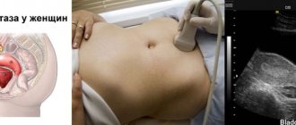
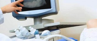
How an ultrasound examination is performed after childbirth
After labor ends, there is no more amniotic fluid left in the uterus. Therefore, before the ultrasound, which is performed in the maternity hospital, the doctor recommends filling the bladder properly in order to slightly elevate the uterus and improve visualization of the organ for a more accurate assessment by a specialist. To sufficiently fill the bladder, you need to drink about one and a half to two liters of warm water. If there is a reason for an urgent examination, and there is no way to wait, the bladder will be filled with fluid through a special catheter.
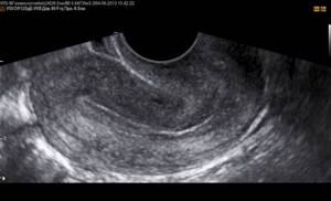
Ultrasound examination in the immediate postpartum period is carried out by installing a sensor on the abdominal wall: the uterus is still very large, transvaginal ultrasound (accessed through the vagina) will not show an objective and accurate picture.
The examination procedure takes about thirty minutes. The woman lies on her back.
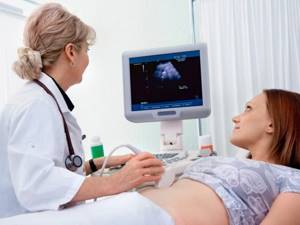
How is ultrasound performed?
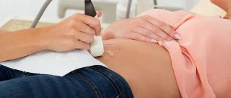
To check for scar tissue after a cesarean section, ultrasound is done in two ways. In most cases, the condition of the uterus is checked through the lower abdomen (abdominal).
How the clinic diagnoses the uterus with ultrasound:
- The patient covers the couch with a towel. She lies on her back, exposing her stomach.
- A special gel is applied to the lower abdomen.
- The doctor moves the sensor over the skin and looks at the image that appears on the screen.
- After the procedure, the woman removes the remaining gel from her stomach.
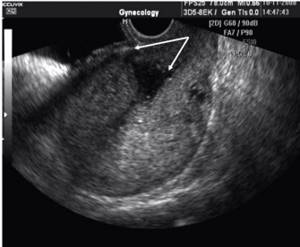
Sometimes the uterus is examined through the vagina (transvaginally). In this case, a condom is put on the device.
To watch a video review from the chief physician of the medical center, Dr. Nikolaev, on the topic:
What will an ultrasound of the uterus show after childbirth?
Diagnosis of the condition of the uterus, as well as the pelvic organs, after childbirth is performed to detect possible disorders that can lead to complications. The doctor analyzes and studies the woman’s health status according to several parameters:
- the size of the uterus, how it contracts, where and how it is located at the time of examination;
- whether there are any formations or fragments in the cavity inside the uterus (for example, blood clots, parts of the fetal membrane or remaining placenta may be found);
- what is the volume of fluid in the uterus, is it excessive;
- assessment of the endometrium: is there any development of inflammatory phenomena;
- assessment of the suture at the surgical site (if the birth took place by cesarean section);
- general condition of all pelvic organs.
Doctor's comment
Are you worried about the upcoming operation to remove an incompetent scar on the uterus? However, the proprietary technique used in our clinic excludes the development of any complications during the intervention. Thanks to the practiced methods of anesthesia and modern painkillers, the patient does not experience pain either during metroplasty or after removal of the uterine scar. The laparoscopic method used in our clinic also makes it possible to reduce the length of hospitalization to a minimum; after 1.5 months you can return to your usual physical activities. But the most important thing is that after treatment of the uterine scar, it becomes possible to carry and give birth to a child naturally. If you are just planning an examination, in our clinic you can undergo all the necessary procedures, after which treatment tactics will be selected taking into account the characteristics of your body. Do not delay treatment, because correct diagnosis and timely correction are the first step to happy motherhood!
Head of the surgical service at SwissClinic Konstantin Viktorovich Puchkov
Ultrasound results after natural childbirth
After the child is born through the natural birth canal, diagnostics of the pelvic condition by ultrasound is prescribed from the second to the fourth day. If the birth was complicated or there is concern that there is a rupture of the uterine wall, an ultrasound will be performed as an emergency. Visually, normally the uterus will have a certain shape, close to spherical. Immediately after childbirth, it will be located in the middle pelvic region. If a woman has had two or more children, or if she has one large baby, the uterus will move slightly toward the lower part of the pelvis.
If you conduct the study several times over a short period of time in the postpartum period, it is possible to monitor how the weight of the contracting uterus decreases and how it narrows. The reduction of the organ and its displacement to the location that was before the birth occurs little by little. The norm for changes is a shift of 1–2 cm every day. In terms of weight, the uterus loses approximately half (about 450–550 g) in the first few days (up to a week). Subsequently, weight loss becomes smoother and more gradual: about 100 g per week, to the standard 95-100 g that was before childbirth.
How dangerous is uterine suture dehiscence?
Expert comment:
In case of divergence, the scar does not burst like a soap bubble, it spreads along the seams, and this process occurs within 4-5 hours. In this case, the pregnant woman experiences aching pain and bleeding. You shouldn’t be afraid of this, because in such a situation the woman has enough time to come to the maternity hospital without panic.
Childbirth with a scar on the uterus can be considered safe, provided that the pregnant woman:
- the placenta is outside the scar;
- fruit weight does not exceed 3600 g;
- having only one cesarean section;
- body mass index does not exceed the norm;
- when suturing, modern absorbable suture material was used;
- there is no anatomical narrowing of the pelvis;
- The thickness of the lower segment of the suture according to ultrasound is at least 3 mm.
Ultrasound results after cesarean section
After a birth performed by cesarean section, the restoration of the reproductive organs takes a long time due to the fact that the mass and overall size of the uterus, taking into account surgical intervention, expands and adds mass by about 40% when compared with natural childbirth. During ultrasound diagnostics, the doctor can see small hematomas, which are foci of hemorrhage in the area of the scar formed at the site of the operation. Such lesions are not dangerous, but they are quite capable of impairing the passage of ultrasound from the sensor to the organ and back. And if the scar swells, then this phenomenon may be a sign of the onset of inflammation in the uterine cavity and endometrium.
After delivery has been performed by cesarean section, the weight of the uterus decreases by approximately 200–250 g during the first postpartum week. The organ usually returns to its original weight values after two months. In terms of the shape of the organ: it acquires the appearance it had before birth no earlier than by the middle or end of the second week after the operation. Standard, basic measurement values (length, width, and anteroposterior dimension) are similarly delayed in parameters when compared with those for vaginal delivery. After the surgical delivery, the doctor will certainly conduct an extremely thorough ultrasound examination of the ovaries, veins and arteries of this area to be sure that their integrity has not been compromised.
Risk factors
Risk factors for the formation of a defective scar on the uterus are:
- corporal caesarean section - an incision along the uterus, CME with opening of the uterine cavity;
- inflammatory processes in the recovery period after surgery;
- a short period (up to 2 years) from the time the scar forms on the uterus until the onset of this pregnancy;
- abortions, curettage in the postoperative period before the onset of this pregnancy;
In order for the uterine scar to be complete, it is necessary to wait a period of time after a cesarean section, CME or uterine perforation. The optimal period of rest is at least 2 years. To avoid curettage of the uterus in the postoperative period, protection is mandatory; this can be either hormonal contraception or mechanical (a condom in combination with spermicides). Early use of intrauterine contraception (IUD) is not recommended.
Diagnostics
If 2 years have already passed since surgery on the uterus and you are planning a pregnancy, it is advisable to make sure that the scar on the uterus is complete before pregnancy occurs.
In non-pregnant women, the consistency of the uterine scar is assessed using the following methods:
Ultrasound hysteroscopy
(a study in which an optical device is inserted into the uterine cavity, which allows you to evaluate the walls of the uterus); In this way, the development and severity of connective tissue in the scar is assessed.
Hysterosalpingography
- X-ray examination of the uterus and fallopian tubes after the introduction of a radiopaque substance into the uterine cavity.
If there are no objective signs of scar failure and at least 2 years have passed since the previous operation on the uterus, a woman with a clear conscience can become pregnant, but we should not forget that during pregnancy it will be necessary to monitor the condition of the scar.
By what signs do doctors judge the inferiority of a scar in pregnant women?
During ultrasound, thinning of the lower uterine segment in the scar area (less than 3 mm), dense inclusions in a significant amount in the scar area are determined, which indicates a connective tissue component in the scar area.
When an obstetrician-gynecologist palpates the area of the scar on the uterus after displacement towards the scar on the skin, the uterus usually contracts evenly with a full scar and unevenly with an incomplete one, forming depressions in the anterior wall of the uterus.
Local (local) pain in the area of the uterine scar is also determined.
Pain may occur in the area of the postoperative scar. If a woman with a uterine scar complains of pain, it is necessary to figure out what is causing it. Most often they are associated with the threat of miscarriage, adhesions in the pelvis or stretching of the scar on the uterus. Pain associated with adhesions in the pelvis goes away with a change in body position. They are not associated with the tone of the uterus and do not go away when taking antispasmodics. Pain associated with the threat of miscarriage occurs when the uterus is tense, which the pregnant woman herself feels. In a relaxed state, these pains go away. The inferiority of the scar is indicated by local pain in the scar area that is not associated with the tone of the uterus and does not go away when taking antispasmodic drugs.
Possible complications
A scar on the uterus can cause some complications during pregnancy. Among them are the threat of termination of pregnancy at different times (occurs in every third pregnant woman) and placental insufficiency when the placenta is attached to the area of the postoperative scar. This condition appears due to the fact that the placenta is attached not in the area of full muscle tissue, but in the area of scar tissue; an insufficient amount of oxygen and nutrients reaches the fetus through the vessels of the placenta. In 20% of women with a scar on the uterus with the placenta attached in the area of the scar (mainly after cesarean section), fetal malnutrition is observed (it lags behind in size and does not correspond to the gestational age). Most often, fetal malnutrition is observed when the scar becomes thinner.
Features of labor management
If a scar is suspected, the pregnant woman should be hospitalized.
If any signs indicate an inferior scar on the uterus, there is no doubt that childbirth should be operative - the doctor only determines the timing of delivery depending on the condition of the fetus and mother. With a full scar, spontaneous birth is permissible if the condition of the mother and fetus is satisfactory, if the birth canal is ready for the upcoming birth (mature cervix) and the pregnant woman agrees to spontaneous birth.
If a uterine scar is formed as a result of a cesarean section in a previous birth, the indications for surgery often remain the same as during the previous birth. This could be, for example, an anatomically narrow pelvis, cicatricial deformities of the vagina and cervix, etc. Often, there are indications for surgery specifically during a given pregnancy, regardless of a previous cesarean section, for example, placenta previa, breech presentation of the fetus (in this case, the pelvic end is present at the exit from the uterus), clinically narrow pelvis with a large fetus, etc. In such cases In cases, without a doubt, despite the consistency of the scar, a caesarean section is performed (it would be carried out even if the woman did not have a scar on the uterus).
The absolute indications for repeat caesarean section are:
- A scar on the uterus after a corporal cesarean section (in this case it is located along the body of the uterus).
- Scar after two or more operations.
- Scar failure, determined by symptoms and ultrasound data.
- Location of the placenta in the area of the uterine scar. If the placenta is located in the area of a postoperative scar, then its elements are deeply embedded in the muscular layer of the uterus, which increases the risk of uterine rupture when it contracts and stretches.
Spontaneous births with a strong scar are carried out in an obstetric hospital, where round-the-clock highly qualified surgical care is available, and there are anesthesiological and neonatal services. The possibility of spontaneous childbirth is finally determined at the end of pregnancy. Childbirth is carried out with constant cardiac monitoring (fetal cardiac activity is monitored for timely detection of fetal oxygen deficiency).
If women with a strong scar on the uterus during childbirth experience weak labor forces, then a caesarean section is performed.
Often women, after giving birth by cesarean section, ask the question: how many more times can you give birth? Often, already during a repeat cesarean section, the question of sterilization (tubal ligation) arises, since the risk of scar deficiency increases with each pregnancy. Most often, already at the third caesarean section, a woman is offered sterilization, but without her consent the doctor has no right to take such a step.
Our comment.
A scar on the uterus is a good reason for concern, but not a fatal situation for the pregnant woman and the child. Vaginal birth is possible for most pregnant women with a scar on the uterus after a previous cesarean section or conservative myomectomy, performed not by laparoscopic access, but by conventional surgery. This problem is quite widely discussed in scientific circles in our country and abroad. At the World Congress of Obstetricians and Gynecologists in Kuala Lumpur (Malaysia, 2006), a report was made from the UK on 50% of spontaneous births among 170 pregnant women with a uterine scar. In our country, the leader in the management of natural childbirth after a previous cesarean section is the Moscow Regional Research Institute of Obstetrics and Gynecology (MONIIAG), headed by Corresponding Member of the Russian Academy of Medical Sciences, Professor V.I. Krasnopolsky, where 60.2% of natural births occur in women with a scar on the uterus, 51.7% – in women after in vitro fertilization and 66.6% – in women with a breech presentation of the fetus (from a report at the 10th Russian Forum “Mother and Child” in 2009). In comparison, in Canada, 100% of pregnant women with breech presentation undergo a caesarean section.
Of course, it is necessary to prepare more carefully for childbirth in such a situation. This preparation largely consists not so much in learning any techniques or actions during childbirth, but in trying to understand the internal cause of the mental conflict that arose during the previous pregnancy and did not allow one to complete a simple woman’s task - to give birth to a child. Let me remind you that absolute indications for caesarean section account for no more than 1-3% of all operations performed. You can often hear the phrase that doctors “forced” you. But if a woman consciously or subconsciously refuses to give birth, the child must be saved! And doctors are forced to insist. In our practice, there were different situations when it was possible to give birth, and when we had to perform a repeat cesarean section - about half and half. There was a case of three births after the first cesarean section and only one case of spontaneous birth after two cesarean sections. But this is so-called casuistry. When there is a serious intention to give birth on her own after a previous cesarean section, the woman needs a lot of work on herself, which we can help do in our classes and consultations with a psychologist. Despite the fact that childbirth with a uterine scar is often more difficult, many women succeed. Most of them note a noticeable difference between their second child and a Caesarean in terms of their life-affirming attitude and independence in decision-making. And this means that “fires, waters and copper pipes” are so necessary for the formation of a courageous and strong-willed character of the future citizen of Russia.
What are the complications?
Conducting a pelvic ultrasound almost immediately after childbirth ensures the detection of some possible problems, which, if not eliminated in time, are quite capable of developing into serious complications requiring treatment. The most common abnormalities that a specialist can see during an ultrasound procedure include:
- the presence of residual blood clots that have coagulated in the uterine cavity, or fragments of the placenta. If enough such fragments have accumulated, they interfere with the outflow of normal postpartum secretions, which leads to inflammatory diseases or even bleeding from the uterus. In order to remove debris from the uterus, the doctor will usually prescribe vacuum aspiration (that is, removal) of the contents of the cavity;
- insufficient activity of uterine muscle contractions. The doctor will suspect this condition when the organ measurements do not correspond to standard values. Prescribing certain medications will help correct the situation;
- inflammatory phenomena of the mucous layer of the uterine cavity (so-called endometritis). The basis for the development of the disease, as a rule, is the addition of a bacterial infection. Pathological microorganisms enter the organ cavity from the genitals if shortly before birth the vaginal microflora was disturbed or its sanitation (cleansing) was not carried out.
Monitoring the condition of the uterine scar
After a woman is discharged from the maternity hospital with a uterine scar, the gynecologist will monitor the condition of her scar for a whole year.
To do this, the doctor may prescribe:
- Ultrasound: to assess the shape, length and thickness of the scar, the possible presence of cracks or tears in it;
- X-ray (hysterography): to more accurately assess the structure of the scar;
- hysteroscopy: the uterine cavity is examined with a hysteroscope to see the color of the scar and evaluate the blood network in its tissues;
- MRI: to understand how strong the muscle tissue of the scar is.
Ultrasound of the uterus after discharge from the hospital
The new mother must undergo a repeat or so-called control examination at her place of residence, in strict accordance with the doctor’s recommendations upon discharge. However, emergency testing may be required. Indications for it will be the following conditions and signs:
- a large amount of discharge with a sharp, sour or unpleasant odor from the vagina;
- sudden bleeding;
- pain, discomfort in the pelvic area;
- the occurrence of suppuration and/or swelling of the scar after cesarean section;
- prolonged fever not associated with a cold.

It is important to know that if an ultrasound examination was not performed while the woman was in the maternity ward, you should consult a doctor for consultation and conduct an ultrasound yourself, even if the alarming symptoms do not bother you.
How much does the examination cost?
A referral for a uterine examination is given by a gynecologist or family doctor. If prescribed by a doctor, an ultrasound examination is performed free of charge in a clinic or antenatal clinic.
In paid clinics, the procedure will cost 800–2500 thousand rubles. How much an ultrasound will cost depends on the pricing policy of the clinic and the city.
An ultrasound after cesarean section should be done if any suspicious symptoms occur. This harmless examination will help avoid many dangerous complications.
Share your experience of ultrasound examinations after cesarean section, this information will be useful for women facing the same problems. Repost on social media. networks.
Advantages of performing ultrasound in our clinic
Our Yuno clinic is distinguished not only by highly qualified specialists and modern equipment, but also by such advantages as:
- 1. Location in the city center
- 2. Diagnostic procedures and therapeutic therapy are possible
- 3. Availability of a day hospital
- 4. Low prices for all services
Other services of our clinic:
- — Ultrasound of internal organs
- - Ultrasound of the uterus
- — Office hysteroscopy
- — Ultrasound of the heart
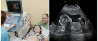
- — Ultrasound by week of pregnancy
- — Treatment of erosion
- — Treatment of urinary incontinence in women
- — Treatment for lack of orgasm
- — Androgyne in gynecology
How to speed up scar healing?
Expert comment:
This can be done with the help of stem cells: research shows that the introduction of mesenchymal stem cells safely speeds up scar healing. At the same time, the cells themselves are completely eliminated from the woman’s body after 2-3 months.
Expert comment:
For expectant mothers who have already had one elective cesarean section, the chances of giving birth spontaneously with a uterine scar are close to 100%. At our National Medical Research Center for Obstetrics, Gynecology and Perinatology. IN AND. Kulakov, pregnant women with a scar on the uterus are not hospitalized in advance - we always wait for the woman to develop independent labor. We say to such a pregnant woman: “Come to us exactly at 40 weeks.” Thus, we give the woman a chance to have contractions without stimulation, without opening the amniotic sac. If at 40 weeks of pregnancy contractions still have not begun and water has not broken out, we perform a repeat cesarean section.
Signs of lochiometra
Delayed discharge of discharge is often diagnosed 5-7 days after birth - the woman notes that discharge from the uterus has become scanty or has disappeared. At the same time, the following clinical signs are observed:
- increase in the size of the uterus;
- cramping pain in the lower abdomen;
- cloudy vaginal discharge with an unpleasant odor.
There are also signs of lochiometra, indicating a deterioration in the general condition:
- increase in body temperature to 38-40 degrees Celsius;
- heartbeat;
- lethargy.