The intestine, consisting of 2 sections (thin and thick), plays an important role in digestion. Violation of intestinal functions affects the condition of the entire body.
It is important to diagnose intestinal anomalies, infectious and parasitic diseases, and tumors at the earliest stages, which will enable the patient to receive timely treatment.
MRI allows early diagnosis.
Typically, an MRI of the intestine is not prescribed separately, but as part of a comprehensive magnetic resonance examination of the abdominal organs and the space behind the peritoneum.
Anatomy of the gastrointestinal tract
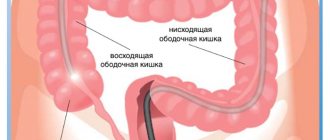
The stomach and intestines are organs of the digestive system that provide the body with proteins, fats, carbohydrates and other nutrients. These are hollow organs with a thin wall - digestion occurs in the lumen of the gastrointestinal tract, during which food is broken down into easily digestible components. The movement of the food bolus is facilitated by constant wave-like movements of the middle wall of the stomach and intestines, consisting of smooth muscle fibers. This phenomenon is called peristalsis. Peristalsis is of great importance in magnetic resonance imaging of the stomach and intestines, as it affects the quality of the resulting images (see preparation for magnetic resonance imaging of the abdominal organs).
The advantages of this technique include:
- Spatial reconstruction, clarifying the location of healthy tissue areas and those in which there are pathological changes.
- No invasive interventions.
- No pain.
- Possibility of reliable assessment of the condition of the intestinal walls and adjacent structures.
This procedure has only one drawback - intestinal peristalsis negatively affects the quality of images. This can make diagnosis difficult when the pathological focus is small.
Diseases of the gastrointestinal tract
Diseases of the stomach and intestines are important for a person’s overall health and well-being. All of them lead to a decrease in quality of life. Constipation, diarrhea, abdominal pain, indigestion and other symptoms of stomach and intestinal diseases can be debilitating, leading to exhaustion and nutritional deficiencies.
Timely diagnosis of diseases of the digestive system is a prerequisite for their successful treatment. However, in reality, most patients seek medical help late, with severe violations of the function and anatomy of the stomach and intestines.
Reasons for late presentation include discomfort and pain associated with fibrogastroduodenoscopy or colonoscopy. MRI cannot completely replace FGDS, since this method is the “gold standard”, but even a single magnetic resonance imaging may be sufficient for timely diagnosis.
Indications and contraindications for MRI of the stomach and intestines
MR imaging can be used in the diagnosis of the following gastrointestinal diseases:
- Complications of gastric and duodenal ulcers - perforation, penetration of ulcers, formation of fistulas and fistulas;
- Polyps of the stomach and intestines;
- Diverticula of the stomach and intestines;
- Intestinal abscesses;
- Inflammatory diseases - Crohn's disease and ulcerative colitis;
- Intestinal necrosis;
- Benign and malignant tumors of the stomach and intestines;
- Determining the stage of stomach and intestinal cancer;
- Preparation for surgical treatment, planning of surgical interventions;
- Assessing the effectiveness of the therapy;
- Anomalies in the development of the gastrointestinal tract.
Acute surgical diseases of the gastrointestinal tract, such as appendicitis or perforated gastric ulcers, are rarely diagnosed using MR imaging. Most often, computed tomography is used for this purpose, as a more accessible and faster method of diagnosing the causes of “acute abdomen”.
What symptoms may be an indication for MR imaging of the gastrointestinal tract:
- Abdominal pain of various localization and nature;
- Bloating, nausea, vomiting;
- Sudden weight loss, lack of appetite, aversion to meat foods;
- Constant constipation followed by diarrhea;
- Volumetric formations of the abdomen, which are determined visually or by touch.
MRI examination of the intestines and stomach is contraindicated:
- Persons whose bodies contain electronic devices - cochlear implants, neurostimulators, cardioverter-defibrillators, heart pacemakers, etc.;
- Persons with foreign bodies made of metals - these are fragments, particles of steel or iron stuck in the eye, medical devices (vascular stents, clips) made of stainless steel;
- Patients of early preschool age, infants;
- Obese people weighing more than 130 kg and having a body girth of more than 150 cm;
- Women in the early stages (up to 12 weeks) of pregnancy.
Preparing for MRI of the stomach and intestines
As stated earlier, gastric and intestinal motility can affect the quality of MRI images. Patient immobility is a prerequisite for obtaining clear and detailed images. Since the intestines and stomach are in constant motion, it can be difficult to obtain high-quality images - with intense peristalsis, they can be blurred, making diagnosis difficult.
Proper preparation for the examination helps reduce peristalsis and increase the quality of images. To do this you need:
- 2-3 days before the scheduled appointment, avoid certain types of food that increase the formation of gases in the intestines, which also contribute to increased peristalsis. These are products such as confectionery, white bread, alcoholic drinks, legumes, fresh vegetables and fruits, dairy, carbonated drinks;
- 8-12 hours before diagnosis (usually in the evening before the morning examination or in the morning if the examination is carried out in the evening), it is necessary to take an antispasmodic (no-shpu) and a carminative (espumisan, simethicone) in the dose recommended by the instructions for use of the drug. These medications relax the intestines and stomach, reduce the severity of peristalsis, and promote the elimination of gases;
- It is not recommended to smoke before the examination - nicotine increases peristalsis, do enemas, take laxatives;
- The examination itself is carried out on an empty stomach - you cannot eat 6-8 hours before the examination (you can drink water).
IMPORTANT! Magnetic fields generated during the examination can attract and heat metal objects and damage electronic devices. It is strictly prohibited to undergo examination with jewelry, piercings, watches, mobile phones, plastic cards, gadgets, etc. in your pockets or on your body. Failure to follow this rule may result in burns and injury.
Is it possible to do an MRI of the stomach and intestines at the same time?
In fact, MRI of the stomach and intestines is included in one standard examination of the abdominal organs, since the field of view of the equipment covers the entire abdomen. This is one of the advantages of magnetic resonance imaging, which takes images not of individual organs, but of the entire body as a whole. The equipment receives a series of layer-by-layer images of the human body in several planes with a step of 1-2 mm. In this way, a comprehensive examination of all organs of the abdominal cavity is performed, and not just the stomach and intestines, which cannot but affect the accuracy and efficiency of diagnosis.
General characteristics of the examination
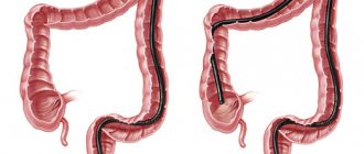
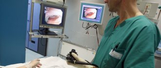
The intestine is the longest organ and the most labor-intensive to examine. Various methods are used to diagnose intestinal diseases. It depends on the part of the intestine and the alleged illness. The most common method is colonoscopy. With its help, you can study the intestines literally centimeters at a time. However, this procedure is quite unpleasant, requires careful preparation and is not always possible. It is not able to cover areas of the small intestine and is contraindicated in a number of diseases. Due to its pain, many patients are looking for alternative methods of examination and ask the question: “Do they do MRI of the intestine and rectum?” Knowing about the safety and effectiveness of the study, it is often preferred. Intestinal MRI is also actively used as an additional diagnostic method.
Do they do MRI of the stomach and intestines with contrast?
Magnetic resonance imaging can be performed with contrast. This technique allows you to increase the information content and sensitivity of the method to some gastrointestinal diseases. Contrast agents for intravenous administration can accumulate only in those tissues that are affected by an inflammatory or tumor process. The affected areas of the intestines or stomach will be highlighted in the images, making diagnosis easier.
In some cases, the use of contrast during MRI helps to clarify the preliminary diagnosis, confirming the results of the biopsy with high accuracy. The reason is that the dynamics of absorption and release of the contrast agent is directly dependent on the type of pathological process and is a constant indicator for a particular disease.
results
After computer processing of the received signals, digital images appear on the screen in 3 projections. After the procedure is completed, the doctor carefully analyzes the images of the intestines and formulates a conclusion. Some clinics provide MRI results the next day.
MRI, given its safety, can be performed repeatedly without any restrictions. Such a need may arise after surgical (or other) treatment in order to monitor the dynamics of the process and the effectiveness of treatment.
How is MRI of the gastrointestinal tract performed?
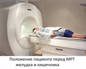
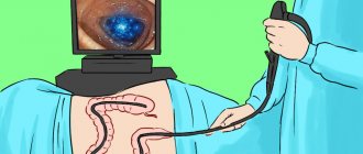
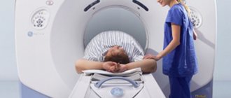
Magnetic resonance imaging of the gastrointestinal tract is an extremely simple, painless and safe procedure. All that is required of the patient is to lie still and follow the operator’s instructions exactly.
Before starting the procedure, you will be asked to put on headphones - they will protect you from the rather loud noise coming from the equipment. They also transmit operator instructions and music to help you relax and make waiting less tiring. The duration of the study is about 40 minutes (for studies with contrast - 55-60 minutes).
After completing the examination, you need to wait for the images to be decrypted. In our center it takes no more than 30 minutes. The images themselves on a CD, together with a specialist’s report, must be handed over to your attending physician. In addition, images can be obtained on film or a flash drive if you pay a little extra - CDs are not always convenient for storing research results.
What does MRI of the stomach and intestines show?
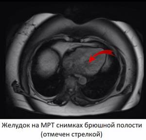
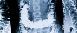
With proper preparation for the study, MRI images of the abdominal cavity show the stomach and intestines along its entire length. When decoding images, the doctor pays attention to the thickness of the wall of hollow organs, the presence of outgrowths, protrusions in the mucous membrane directed towards the lumen, nodes, pockets (diverticula) and neoplasms in the wall of the intestine and stomach. Normally, the mucous membrane should maintain a folded structure, and the large intestine retains haustration. The patency of the intestine, the presence of pathological narrowings and dilations in its lumen are examined.
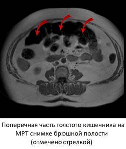
When using contrast, pay attention to the local accumulation of the drug in the tissues of the mucous membrane (a sign of inflammation). In the presence of neoplasms, the dynamics of accumulation and washout of contrast are studied in more detail. Normally, contrast accumulates evenly in all tissues, which is why there are no areas of its accumulation in the images.
Allowed foods and drinks
During preparation for an abdominal MRI, foods are baked in the oven, boiled, or steamed. The diet may include:
- Veal or beef, turkey or rabbit (optimally in the form of minced meat);
- Low-fat fish;
- Buckwheat or rice, oatmeal (it is better to exclude semolina);
- Fruits, vegetables, heat-treated and pureed;
- Vegetable oils;
- Omelettes;
- Dry types of cookies.
Honey is allowed, but in small quantities. Wholemeal bran bread, watermelon, and parsley are acceptable. Rosehip tincture helps reduce gas formation.
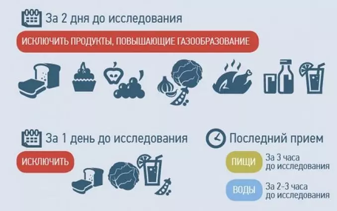
In addition to diet, it is important to maintain a drinking regime. Drinking plenty of fluids helps prevent constipation, but you need to choose the right drinks. These can be unsweetened compotes made from a small amount of berries or fruits, water, herbal tea. Of the juices, varieties that do not contain pulp are allowed; it is better to dilute them with water in a 1:1 ratio.
