Ultrasound of the abdominal cavity Ultrasound of the gallbladder Ultrasound of the kidneys Ultrasound of the bladder Ultrasound of the pelvis Ultrasound of the breast Ultrasound of the thyroid gland Ultrasound of the prostate gland
Today there are many ways to diagnose the disease. But ultrasound examination of organs remains the most popular. The simplicity and relatively low cost of such research help to immediately obtain a clear picture of the processes that occur in the body. And thereby eliminate inflammatory processes in the body. This procedure allows you to easily and painlessly identify the disease and begin adequate treatment. But before carrying out the procedure, it is necessary to prepare the organ for examination. How to do it?
Preparing for an abdominal ultrasound
During an ultrasound examination, internal organs must be open. They should not be obscured by gases and products that are in the intestines. To do this, the intestines need to be prepared.
First of all, within a few days of the planned study, exclude from daily consumption foods that cause bloating. These include beans, peas, brown bread, bananas, and sweets. Fresh vegetables and fruits, especially cabbage, can cause excessive gas formation.
It is also necessary to take espumizan for three days (as prescribed by the doctor). On the day of the study, you will need to increase the dosage to four capsules. They need to be taken once a day, and should not be washed down with water. Abdominal ultrasound is performed on an empty stomach. Before starting this procedure, you should not eat for 6 hours.
A patient suffering from diabetes is allowed a light breakfast.
Which is correct?
So, how to properly drink water before an ultrasound?
For men and women
The size of the bladder in men is slightly larger than in women, therefore, the volume of fluid consumed will be different. Men need to drink 1-1.5 liters of fluid an hour or two before monitoring . After drinking, you must refrain from urinating.
If an hour or two after taking the liquid the patient does not have the urge to deurinate, and the time for the study has not yet arrived, it is recommended to drink another half liter of liquid.
Representatives of the fairer sex are required to drink 1 liter of liquid at least an hour before diagnosis. If the patient's examination coincides with her menstrual period, this is not a reason to refuse the procedure. Menstruation does not interfere with the diagnostic process in any way and does not affect its readings.
Adults need to adhere to a diet (exclude foods that contribute to gas formation from the diet) , refrain from drinking alcohol for a week before the ultrasound, and also notify the attending physician about taking medications taken before the examination.
For children
In children, the size of the bladder varies depending on age, so drinking is prescribed with caution so as not to overload the kidneys.

The volume of fluid consumed will be distributed as follows:
- up to 1 year – feeding after the procedure;
- from 1 to 2 years – as much as they can drink;
- from 3 to 7 years – a glass of liquid;
- from 8 to 11 years – 1.5 cups;
- from 12 to 18 years – 2 glasses.
Drinks should be given to the child 1-1.5 hours before diagnosis. You should avoid consuming soda and other sugary drinks the day before the test.
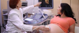
Pregnant
For expectant mothers, what to drink before the procedure depends on at what stage of pregnancy the ultrasound is performed. If this is the first or second trimester, you need to drink 500 ml of liquid 2 hours before the procedure.
Diagnosis carried out in the third trimester does not require special filling of the bladder to avoid additional stress on the pelvic organs. The procedure is carried out strictly on an empty stomach.
You can find out how to drink water before an ultrasound here, and about water consumption in general here.
Preparing for an ultrasound of the gallbladder
Preparation for this study is almost the same as for the study of the abdominal cavity. It is necessary to take for research those products that stimulate the functioning of the gallbladder. You can use full-fat sour cream or purchase a choleretic agent (holiver, corn silk, etc.) at the pharmacy. Children are given dark chocolate instead of sour cream. This ultrasound is done 2 times. First on an empty stomach. And before the second study, they take choleretic products. Therefore, this research takes a lot of time.
What is better to use?
It is best to drink plain water .
It is also allowed to drink unsweetened berry or fruit juices, fruit drinks, compotes or tea.
It is not recommended to drink carbonated drinks and milk, as they stimulate increased gas formation, which negatively affects the reliability of the study results.
Preparing for a pelvic examination
This ultrasound can be performed on any day of the cycle. The exception is the days of menstruation. To clarify the diagnosis, a repeat ultrasound is performed on the day of the cycle, which is prescribed by a specialist. The location of the pelvic organs is the abdominal cavity. They are located deep, making them difficult to explore.
In order to examine the pelvis (uterus, ovaries, fallopian tubes), a special vaginal sensor is used. This research method is called transvaginal. If the girl is a virgin, then this method is not acceptable for her. Transvaginal ultrasound does not require preliminary preparation and is performed through the anterior abdominal wall. Before starting the study, you need to go to the toilet and empty your bladder.
An hour before the appointed time, virgins drink 3-4 glasses of non-carbonated liquid.
What is diagnosed with an ultrasound of the bladder?
If a doctor prescribes an ultrasound examination, this is most likely done to confirm the diagnosis.
Diseases and pathologies that are determined by ultrasound:
- the presence of stones and sand in the kidneys;
- appeared suspensions;
- incorrect location of the organ;
- when determining the residual amount of urine, an increased volume was detected;
- development of tumors and neoplasms;
- change in the shape of the bladder, thickening of its walls;
- echogenic formations;
- acute or chronic cystitis;
- pyelonephritis;
- pyeelectasia;
- hydronephrosis.
These results are not satisfactory, so you should not delay treatment and put off going to the doctor until later. This is especially true for those who are planning to have children, since these diseases can be transmitted at the genetic level.
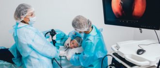
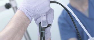
Ultrasound of the prostate gland - Preparation
This study, or TRUS as it is also called, is performed through the rectum. Therefore, it will need to be cleaned. This is done as follows. A couple of hours before the start of the study, the patient is given a microenema. Its volume is 200 ml. To do this, take plain water and use a special bulb to inject it into the rectum. The patient empties within a few minutes. It is necessary to fill the bladder by taking liquid an hour before the procedure. If the patient is unable to tolerate it, you can go to the toilet. But then you will need to drink some liquid again.
Ultrasound cost
| Duplex study of the arteries of the upper or lower extremities | 3000 | rub |
| Duplex examination of the veins of the upper or lower extremities | 3000 | rub |
| Duplex examination of veins and arteries of the lower extremities | 4900 | rub |
| Duplex examination of veins and arteries of the upper extremities | 4900 | rub |
| Duplex examination of the portal system | 3500 | rub |
| Duplex scanning of brachiocephalic and transcranial vessels | 5200 | rub |
| Duplex scanning of brachiocephalic vessels | 3000 | rub |
| Duplex scanning of hemorrhoids | 1900 | rub |
| Duplex scanning of the renal arteries | 3100 | rub |
| Comprehensive examination of the gastrointestinal tract (ultrasound) | 5200 | rub |
| Neurosonography | 3100 | rub |
| Stress echocardiography | 4200 | rub |
| Transcranial duplex scanning of blood vessels | 3100 | rub |
| TRUS of the prostate gland + bladder + scrotum (without CD) | 3400 | rub |
| TRUS of the prostate gland + bladder + scrotum (with color circulation) | 3600 | rub |
| Ultrasound of the abdominal cavity (to exclude free fluid) | 1600 | rub |
| Ultrasound of the abdominal aorta | 3000 | rub |
| Ultrasound of the maxillary sinuses | 1900 | rub |
| Ultrasound of the eyeball and paraorbital space with color circulation | 1900 | rub |
| Ultrasound of the stomach | 1650 | rub |
| Ultrasound of the gallbladder | 1600 | rub |
| Ultrasound of the retroperitoneum | 2200 | rub |
| Ultrasound examination of the salivary glands | 1800 | rub |
| Ultrasound examination of gallbladder function (before food load and after 30-40 minutes) | 1800 | rub |
| Ultrasound of complex MPS in women TA, without CDK (kidneys, ureters, bladder) | 2800 | rub |
| Ultrasound of complex MPS in women with TA with CD (kidneys, ureters, bladder) | 2900 | rub |
| Ultrasound of complex MPS in men TA without CDK (kidneys, ureters, bladder, residual urine, prostate gland) | 3500 | rub |
| Ultrasound of complex MPS in men TA and TRUS without color circulation (kidneys, ureters, bladder, residual urine, prostate gland) | 3800 | rub |
| Ultrasound of complex MPS in men TA and TRUS with color circulation (kidneys, ureters, bladder, residual urine, prostate gland) | 4000 | rub |
| Ultrasound of complex MPS in men TA with CDK (kidneys, ureters, bladder, residual urine, prostate gland) | 3800 | rub |
| Comprehensive ultrasound in men of the TA (bladder, prostate gland, residual urine) with color circulation | 2500 | rub |
| Comprehensive ultrasound in men TA (bladder, prostate gland, residual urine) without color circulation | 2100 | rub |
| Ultrasound of large joints (1 joint) | 2000 | rub |
| Ultrasound of large joints (2 joints of the same name) | 2900 | rub |
| Ultrasound of lymph nodes (group 1) | 1900 | rub |
| Ultrasound of any part of the large or small intestine | 1450 | rub |
| Ultrasound of small joints (up to 2 joints) | 1800 | rub |
| Ultrasound of the mammary glands with regional lymph nodes (without colorectal circulation) | 2200 | rub |
| Ultrasound of the mammary glands with regional lymph nodes (with color circulation) | 2400 | rub |
| Ultrasound of the bladder (without colorectal dosage) | 1700 | rub |
| Ultrasound of the bladder (with color circulation) | 1900 | rub |
| Ultrasound of the bladder + prostate gland TA (with color circulation) | 2400 | rub |
| Ultrasound of the bladder + prostate gland TA (in men) without color circulation | 2200 | rub |
| Ultrasound of the scrotum (without color circulation) | 1600 | rub |
| Ultrasound of the scrotum (with color circulation) | 1900 | rub |
| Ultrasound of the scrotum and spermatic cord (without color circulation) | 2000 | rub |
| Ultrasound of the scrotum and spermatic cord with color circulation | 2000 | rub |
| Ultrasound of the adrenal glands (without color circulation) | 1700 | rub |
| Ultrasound of the adrenal glands (with color circulation) | 1900 | rub |
| Ultrasound OBP + Kidneys | 2900 | rub |
| Ultrasound of the abdominal organs (liver, gall bladder, pancreas, spleen) (without colorectal dosage) | 2300 | rub |
| Ultrasound of the abdominal organs (liver, gall bladder, pancreas, spleen) with colorectal dosage | 2500 | rub |
| Ultrasound of the liver | 1600 | rub |
| Ultrasound of the liver with colorectal dosage | 1800 | rub |
| Ultrasound of the liver + gallbladder without color circulation | 2050 | rub |
| Ultrasound of the liver + gallbladder with color circulation | 2250 | rub |
| Ultrasound of pleural cavities | 1800 | rub |
| Ultrasound of superficial structures (soft tissues) without color circulation | 1800 | rub |
| Ultrasound of superficial structures (soft tissues) with color circulation | 2000 | rub |
| Ultrasound of the pancreas | 1500 | rub |
| Ultrasound of the pancreas with color circulation | 1600 | rub |
| Ultrasound of the spine (1st section) without color circulation | 2200 | rub |
| Ultrasound of the spine (1st section) with color circulation | 2400 | rub |
| Ultrasound of the penis (without color circulation) | 2200 | rub |
| Ultrasound of the penis (with color flow) | 2400 | rub |
| Ultrasound of the kidneys (without colorectal dosage) | 1800 | rub |
| Ultrasound of the kidneys (with color flow) | 1900 | rub |
| Ultrasound of the kidneys and bladder (without colorectal dosage) | 2400 | rub |
| Ultrasound of the kidneys and bladder (with colorectal dosage) | 2600 | rub |
| Ultrasound of the kidneys and ureters | 2100 | rub |
| Ultrasound of the kidneys and adrenal glands | 2200 | rub |
| Ultrasound of the prostate gland (PA) with a full bladder (without CDB) | 1400 | rub |
| Ultrasound of the prostate gland (PA) with a full bladder (with color circulation) | 1500 | rub |
| Ultrasound of the prostate gland (TRUS) (without colorectal dosage) | 1900 | rub |
| Ultrasound of the prostate gland (TRUS) (with color doppler) | 2300 | rub |
| Comprehensive ultrasound of the prostate gland (TA and TRUS) without CD | 2400 | rub |
| Comprehensive ultrasound of the prostate gland (TA and TRUS) with color circulation | 2450 | rub |
| Ultrasound of the spleen | 1500 | rub |
| Ultrasound of the spleen with color circulation | 1500 | rub |
| Ultrasound of the heart without Dopplerography | 2700 | rub |
| Ultrasound of the heart with color circulation (Echocardiography) | 3100 | rub |
| Ultrasound of the hip joints (children) | 2200 | rub |
| Ultrasound of the thyroid gland with regional lymph nodes (without CDK) | 2100 | rub |
| Ultrasound of the thyroid gland with regional lymph nodes (with color circulation) | 2300 | rub |
| Ultrasound guidance (1 organ, 1 area) | 1700 | rub |
| Ultrasound mark (marker) 1 study | 1500 | rub |
| Echohydrosalpingoscopy (with consumables) | 9300 | rub |
| Ultrasonic densitometry | 2000 | rub |
| Ultrasound elastography of parenchymal organs (1 organ) | 3500 | rub |
| Ultrasound of the stomach with water-siphon test | 2500 | rub |
| Ultrasound in gynecology | ||
| Volumetric 3D/4D ultrasound during pregnancy 2-3 trimester with recording on a flash drive/disc | 3800 | rub |
| Volumetric 3D/4D ultrasound during pregnancy 2-3 trimester (with recording on a flash drive) with calculations | 4800 | rub |
| Volumetric 3D/4D ultrasound during pregnancy 2-3 trimester | 3300 | rub |
| Volumetric 3D/4D ultrasound for twins in any trimester (optional) | 1300 | rub |
| Dopplerography (assessment of uteroplacental blood flow) | 2100 | rub |
| Dopplerography in gynecology | 1600 | rub |
| Dopplerography (assessment of uteroplacental blood flow) (twins) | 2900 | rub |
| Dopplerography (assessment of uteroplacental blood flow) according to the program | 0,00 | rub |
| Ultrasound monitoring of the luteal phase, category 1 | 2800 | rub |
| Ultrasound monitoring of the luteal phase, category 2 | 3500 | rub |
| Ultrasound monitoring of the luteal phase, category 3 | 4500 | rub |
| Ultrasound monitoring of follicle and/or endometrial growth without stimulation of superovulation, category 1 | 1600 | rub |
| Ultrasound monitoring of follicle and/or endometrial growth without stimulation of superovulation, category 2 | 1800 | rub |
| Ultrasound monitoring of follicle and/or endometrial growth without stimulation of superovulation, category 3 | 2100 | rub |
| Ultrasound in the first trimester of pregnancy transabdominal (without CD) | 2100 | rub |
| Ultrasound in the first trimester of pregnancy transabdominal (without CDK) according to the program | 0,00 | rub |
| Ultrasound in the first trimester of pregnancy transabdominal (with color doppler) | 2500 | rub |
| Ultrasound in the first trimester of pregnancy transabdominal (twins) (without colorectal doppler) | 2900 | rub |
| Ultrasound in the first trimester of pregnancy transabdominal (twins) (with color doppler) | 3600 | rub |
| Ultrasound in the second and third trimesters of pregnancy (twins) transabdominal (without CD) | 3900 | rub |
| Ultrasound in the second and third trimesters of pregnancy (twins) transabdominally (with color dispersion) | 4800 | rub |
| Ultrasound in the second and third trimesters of pregnancy transabdominally (calculation of fetal size) (without color circulation) | 2800 | rub |
| Ultrasound in the second and third trimesters of pregnancy transabdominal (calculation of fetal size) (without color circulation) according to the program | 0,00 | rub |
| Transabdominal ultrasound in the second and third trimesters of pregnancy (calculation of fetal size) (with color doppler) | 4100 | rub |
| Ultrasound of the symphysis pubis | 1900 | rub |
| Ultrasound determination of fetal sex (without calculations) | 1800 | rub |
| Comprehensive ultrasound of the pelvic organs (TA and TVUS) | 2900 | rub |
| Ultrasound of the pelvic organs (PA) without color circulation | 1900 | rub |
| Ultrasound of the pelvic organs (PA) with color doppler | 2050 | rub |
| Ultrasound of the pelvic organs (TVUS) (with color doppler) | 2300 | rub |
| Ultrasound of the pelvic organs (TVUS) without color circulation | 2100 | rub |
| CDC in the second and third trimesters of pregnancy | 1900 | rub |
| Cervicometry | 2200 | rub |
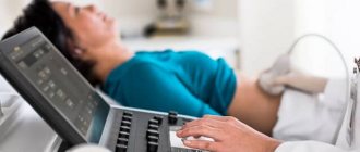
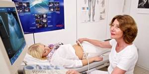
Carrying out the procedure
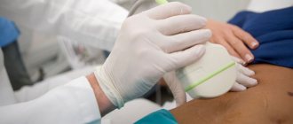
After freeing the lower abdomen from clothing, the patient lies on his back on the couch. The doctor applies a special gel to the area where the ultrasound is performed, and then moves the sensor over the lubricated body, examining the bladder, as well as the organs that lie in close proximity to it. Transrectal ultrasound of the bladder (TRUS) is performed by inserting a sensor into the rectum. This method is used when it is necessary to conduct research on men or girls who have not been sexually active, so as not to break the hymen, as well as patients with contraindications for transabdominal ultrasound. Using this method, it is possible to examine pathological changes in the bladder and prostate in men.
To carry out this diagnosis, it is necessary to clean the rectum. This can be done in the following ways:
- microenema;
- insertion of suppositories with glycerin into the rectum;
- taking a herbal laxative.
Recommended drugs with a long-term laxative effect are phytolax, senade, senadexin, mucofalk, agiolax. They should be taken the night before the examination, as their effect begins within 6-12 hours.
Drugs with a fast-acting laxative effect - fortrans, prelax, magnesium sulfate. These drugs will take effect 15-20 minutes after taking them.
Transvaginal bladder ultrasound (TVUS) is performed by inserting an ultrasound probe into the vagina. It is considered the most accurate and informative method; it is performed on an empty bladder. It can be performed without reference to the menstrual cycle. The only condition for obtaining accurate data is an empty bowel, in which there are no gases.
Transurethral ultrasound (TUUS) is a method in which a sensor is inserted into the urethra. Indicated in diagnosing the connection between diseases of the bladder and urethra. With its help, you can see the extent of damage to the walls of the urethra and the participation of nearby organs in this development. The method is characterized as very accurate. It is performed under anesthesia and there is a high probability of damage to the urethra by the sensor. Due to these factors, it is used very rarely.
In what cases is an ultrasound of the kidneys required?
Various disorders can occur in the urinary system - bladder, kidneys and adrenal glands:
- neoplasms: stones, polyps and cysts;
- inflammatory processes: cystitis and pyelonephritis;
- congenital features and injuries.
Symptoms may include: lower back pain, change in urine color, pain when urinating, swelling, abnormal urine tests.
Ultrasound of the urinary system and kidneys is almost always included in the preoperative examination.
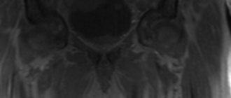
Study results - what ultrasound shows
Ultrasound can be used to guide doctors or surgeons during a procedure, such as a biopsy. It is very important in planning certain types of therapy and surgery, and in determining whether the patient's body is responding to treatment. Ultrasound can be used to check for some types of cancer, including to detect abnormal tumors. The study can help diagnose (find):
- how much urine remains in the patient’s organ after urination;
- the presence of an inflammatory process (for example, cystitis, known to many);
- any obvious abnormalities of the bladder, its size, wall thickness, presence of stones.
Normally, an ultrasound of the bladder shows the physiological location of an organ of normal size, without signs of an inflammatory process or formations, including stones. Ultrasound examinations do not cause pain. Tests that require insertion of a probe may cause slight discomfort. A standard ultrasound examination usually lasts up to half an hour; after this time, the patient receives an image of the internal organ being examined, as well as a specialist’s opinion, after which he is sent to the doctor for a treatment regimen.